PHYSIOLOGY OF THE NERVE
MORPHOLOGYNerve cell represents the building unit of the nervous system with the specialized function of impulse transmission. Typical nerve cell has a cell body (soma) with 5-7 short projections (dendrites) and a longer fibrous axon. The dendrites, receive impulses from other cells and transfer them to the cell body (afferent signals). The axon ends in a number of synaptic knobs (terminal buttons) which stores the neurotransmitter synthesized by the cell body. Axons of some nerve fibers have a myelin sheath, a protein-lipid insulator formed by Schwann cell wrapping around the axon. The sheath envelops the axon except at its ends and periodic constrictions of 1 mm distance called (node of Ranvier), these are called myelinated nerve fibers while those without myelin sheath are called unmyelinated nerve fibers (figure1).
Excitable Tissue: A tissue that responds to stimulation by generating and propagating an Action Potential (AP), it includes nerve and muscle.
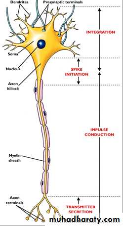
(figure1)
GENESIS OF RESTING MEMBRANE POTENTIAL (RMP)
RMP is an electrical potential exists across the cell membranes with a negative charge inside the cell and the positive charge outside. Resting membrane potential is produced by:1. passive diffusion of Na, and K, ions through leak channels and called a potassium-sodium (K + -Na + )“leak” channel. This channels are more permeable to potassium than to sodium, normally about 100 times this create (-86 mv).
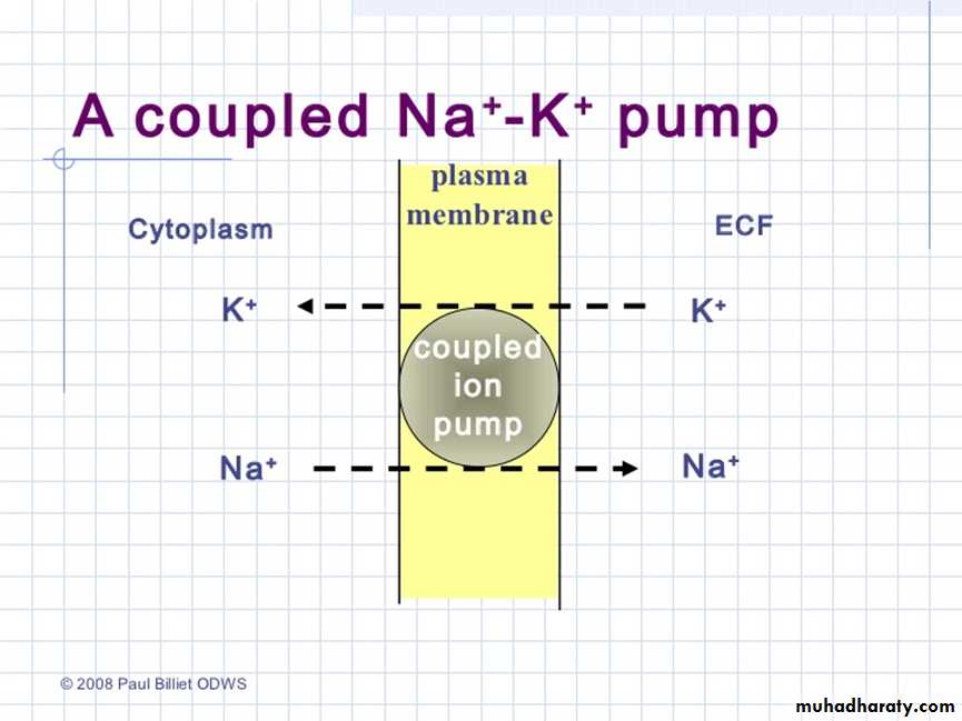
2. Na-K pump: this is an electrogenic pump because more positive charges are pumped to the outside than to the inside (three Na + ions to the outside for each two K + ions to the inside), leaving a net deficit of positive ions on the inside; this causes a negative potential inside the cell membrane. This will add (-4 mv) making a total of (-90 mv) (i.e the potential inside the fiber is 90 millivolts more negative than the potential in the extra-cellular fluid )This -90 mv is the RMP for large neuron (figure 2).
Membrane potential is measured by introducing microelectrode inside the cell with other electrode on the outer surface. The two electrodes are connected to a voltmeter.
(figure 2)
NERVE ACTION POTENTIAL (AP)Nerve signal (impulse) is transmitted by action potential which is defined as a "sudden change from the normal negative resting potential inside the cell to a positive potential (depolarization) followed by rapid return back to the negative potential (repolarization).
Action potential moves along the nerve fiber to its end in a constant rate and amplitude.
Voltage-Gated Sodium Channels (fast channels)
The closure and opening depends on the voltage difference across the membrane. At rest they are closed preventing Na entry. After stimulation the membrane potential becomes less negatives thus opening Na channels for very short time allowing large amounts of Na ions to enter the cell down electrical and chemical gradients thus reversing the membrane potential to positive (depolarization). At the end of depolarization Na channels close (figure 3).Voltage-Gated Potassium Channels
Closed at rest but opened near the end of depolarization allowing large amounts of K to move outside the cell resulting in repolarization (figure 3).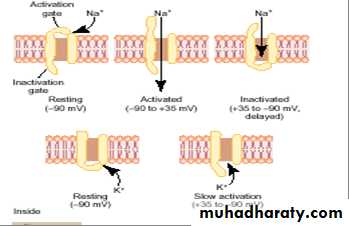
(figure 3)
STIMULUS FOR NERVE1- Chemical: acid, base, strong salt, acetylcholine
2 -Mechanical: crushing, pressure, pricking.
3- Electrical: electrical current.
All these factors increases membrane permeability to Na (leak channels) allowing enough amounts of Na to enter the cell shifting the membrane potential toward the firing level(open voltage gated Na channels) producing an AP.
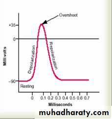
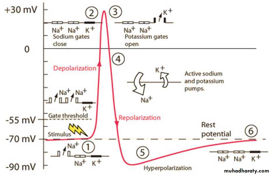
STAGES OF ACTION POTENTIAL
1. Resting stage: The membrane is said to be “polarized” during this stage ,–90 millivolts negative membrane potential is present.
2. Depolarization:
a. increased Na permeability by opening voltage-gated Na channels.
b. inside becomes positive. In large nerve fibers the membrane potential overshoot beyond the zero level and to become somewhat positive
3. Repolarization:
a. closure of voltage-gated Na channels.
b. opening voltage-gated K channels.
c. inside cell returns back to negative.
Action potential lasts for 1 millisecond in large myelinated nerve fiber.(figure 4)
(figure 4)
re-established of RMP by the action of Na-K pump. Activity of this pump is strongly stimulated when there is excess Na inside the cellCATHODE-RAY OSCILLOSCOPE (CRO)
An electrical instrument used to record very small electrical events in millivolt (mv) which occurs very rapidly in millisecond (ms) in the living tissues. It consists of a cathode which emits a beam of electrons directed to strike a florescent face emitting light. A record of AP can be obtained. CRO has two electrodes applied to the nerve, a stimulating electrode and a recording one(figure 5).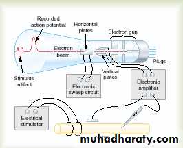
(figure 5)
ACTION POTENTIAL AS RECORDED BY CRO1. latent period: the time taken by an impulse to travel from the stimulating electrode to the recording electrode. From the duration of the latent period the velocity of nerve conduction can be calculated.
2. Depolarization: slow at first but after initial 15 mv of depolarization the rate increases sharply(firing level) due to sudden opening of fast Na channels.
3. Repolarization: this stage starts by closure of fast Na channels and opening of K channels retuning membrane potential back to the polarized state.
4. After depolarization: when the repolarization is 70% completed the rate is decreased due to built up of large amounts of K outside the cell which resist K outflow.
5. After hyperpolarization: when membrane potential reaches the resting level, it becomes somewhat more negative than normal because:
A. continuous K outflow as many K channels remain open after repolarization is completed.
B. the Na-K pump which causes the net movement of positive charges outside the cell (figure 6).
(figure 6).
Intensity of Stimulus: The minimum intensity of stimulus that will just produce a response (AP) is called threshold stimulus. It varies according to the type of axon, therefore different axons within the same nerve trunk has different thresholds.At the level of single axon, any stimulus with subthreshold intensity will not produce an AP. Again, increasing the stimulus intensity above threshold level will produce no change in response, thus the AP of a single nerve axon obey the "all- or- none law" Once an action potential has been elicited at any point on the membrane of a normal fiber, the depolarization process travels over the entire membrane if conditions are right, or it does not travel at all if conditions are not right. This is called the all or none low (all-or-nothing principle).
REFRACTORY PERIOD
When AP is produced, another AP cannot occur as long as the nerve membrane is depolarized. The reason for this is that shortly after the action potential is initiated, the sodium channels become inactivated. The only condition that will allow them to reopen is when the membrane potential return to or near the original resting membrane potential level. Then, within another small
fraction of a second, the gates of the channels open, and a new action potential can be initiated. Refractory period is divided into:
1. Absolute refractory period: the period from firing level until repolarization is about half completed.
2. Relative refractory period: from end of absolute refractory period to the start of after depolarization. Very Stronger stimulus is required to produce a response.(figure 7)
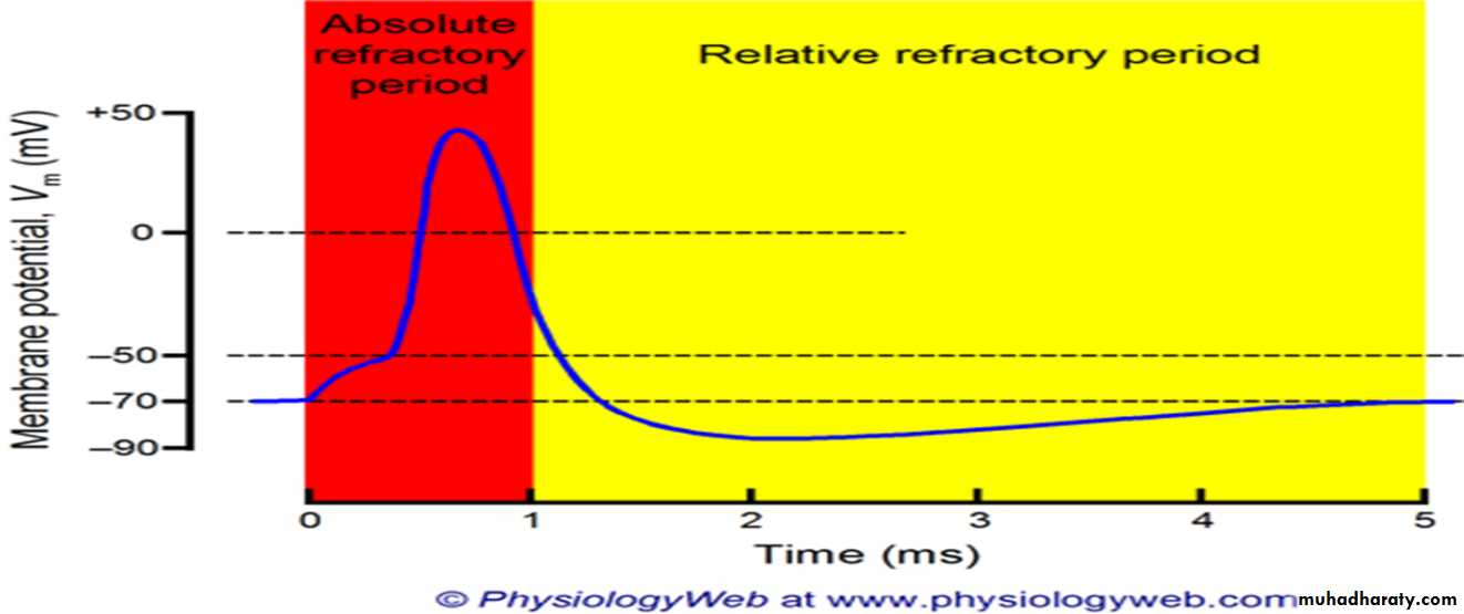
Figure 7
PROPERTIES OF MIXED NERVES
Each peripheral nerve consists of a number of neurons bound together by fibrous sheath(epinurium) The large fibers are myelinated, and the small ones are unmyelinated. The average nerve trunk contains about twice as many unmyelinated fibers as myelinated fibers, therefore the electrical potential record from peripheral nerve have some differences from single axon, these differences are:1-Different neurons have different thresholds:
Subthreshold stimulus produce no response. When threshold stimulus is applied, some neurons with low threshold intensity will respond first producing an AP. As the stimulus intensity is increased (submaximal stimulus), more and more neurons will be brought about into action (recruitment) producing larger AP. The stimulus which excites all neurons is called maximal stimulus. After that, increasing stimulus intensity (supramaximal stimulus) will produce no change in response, therefore mixed nerves don't obey all or none law
2. Different neurons have different speed of conduction giving rise to compound AP on recording.
SUMMATION OF NERVE IMPULSES
Nerve impulses obey all- or- none law, however stronger nerve signals can be obtained by two means:1-Spatial summation: many fibers discharge impulses at the same time
2-Temporal summation: the same fiber discharge impulses rapidly and repeatedly
PROPAGATION OF AP
Nerve cell membranes are polarized at rest with outside positive. When stimulus of enough strength is applied on axon, an AP will be generated at the site of stimulation causing polarity to be reversed (inside becomes positive). Positive charges then move from the adjacent part of membrane to the area of negativity creating a local circuit of current flow between the depolarized area and the adjacent resting area of the membrane, this will decrease the polarity of the adjacent membrane thus activating new Na channels and when the firing level is reached, a new AP develops. This spontaneous sequence of events will move along the unmyelinated axon to its end (figure 8). The moving impulse does not depolarize the area behind it because it is refractory.
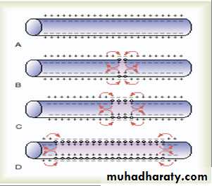
(figure 8)
In myelinated axons, AP will jump from one node of Ranvier to another (myelin acts as an insulator). This type of conduction is called saltatory conduction which is about 50 times faster than the unmyelinated fiber(figure 9). That is, electrical current flows through the surrounding extra-cellular fluid outside the myelin sheath as well as through the axoplasm inside the axon from node to node, exciting successive nodes one after anotherRepolarization starts at the point of original stimulus and spreads in the same direction of depolarization.
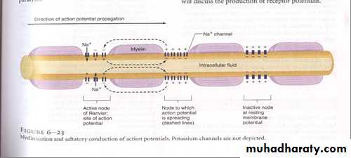
(figure 9)
FACTORS WHICH AFFECT THE CONDUCTION VELOCITY1. Myelination: myelinated nerve is about 50 times faster.
2. Axon diameter: in unmyelinated nerve axon, the conduction velocity is directly proportional to the square root of axon diameter while in the myelinated neuron conduction velocity increases directly with axon diameter, thus myelination saves considerable space in the nervous system.In small unmyelinated nerve axon, the conduction velocity is about 0.5 meter/second while in the largest myelinated nerve axon it is about 100 meter/second.
PLATEAU IN SOME AP
In some excitable tissues, repolarization does not occur immediately after depolarization, instead the membrane potential remains near the peak of spike for many milliseconds(plateau) before repolarization starts, therefore plateau greatly prolongs the period of depolarization(figure 10). This type of AP is seen mainly in cardiac muscle where it lasts for as long as 0.3-0.5 seconds thus prolonging the time for cardiac contraction. Plateau is due to:
1. Opening of slow Ca channels
2. Delayed opening of voltage-gated K channels.
While the usual fast voltage-activated sodium channels opened and causes the spike portion of the action potential.
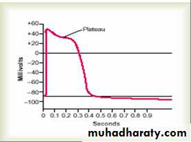
figure 10
SPONTANEOUS RHYTHMICITYSpontaneous discharge occurs normally in cardiac (heart beating) and smooth muscle (intestinal peristalsis) and many neurons in the CNS (rhythmic control of breathing). Spontaneous repetitive discharge occurs due to Na leak during the resting state. The RMP of such cells is about - 60 to -70 mv which is not enough to keep Na or Ca channels closed so there will be Na or Ca leak to inside the cell making the membrane potential less negative, this will cause more channels to open allowing more Na and Ca inflow until the firing level is reached and another AP is produced. At the end of AP, the membrane repolarizes again but shortly then a new AP is generated spontaneously. This cycle is repeated again and again causing the cell to induce rhythmic spontaneous discharge.
The Effect of Calcium Ions on Neuron Excitability
The membrane of most body cells has Ca pumps which deliver Ca ions to the outside the cell (or into the endoplasmic reticulum of the cell) creating a concentration gradient of about 10,000 folds
A decrease Ca ion concentration in extra cellular fluid (hypocalcemia) leads to increased neuron excitability because of the profound effect of extracellular Ca level on the ease of opening the voltage-gated Na channels . When extra cellular Ca falls 30-50% its normal level, it leads to spontaneous discharge in many peripheral nerves causing muscle tetany which could be lethal when involving the respiratory muscles.
FACTORS WHICH INHIBIT NERVE EXCITABILITY
1 .Hypercalcemia(high ECF Ca): excess Ca binds to Na channels so that higher voltage is required to open them.(decrease Na permeability).
2 .Hypokalemia (low ECF K): this decreases membrane potential to a level which favors closure of most Na channels.
3. Local anesthesia: like cocaine, procaine… also by decreasing Na permeability.