1
Fifth stage
Gynecology
Lec-5
أ
سم
اء
السنجري
17/10/2016
Secondary Amenorrhea
The student at the end of this lecture should be able to:
Define secondary amenorrhoea.
Classify the causes of secondary amenorrhoea.
Describe the commonest three cases of secondary amenorrhoea.
Analyze the history and examination of a case of secondary amenorrhoea.
Diagram an outline of a case of secondary amenorrhoea.
Analyze the diagnostic role of progesterone challenge test.
Secondary Amenorrhea
Is cessation of menstruation for 6 consecutive months in a women who has previously had
regular periods , or for 12 months in a women With previous oligomenorrhoea.
WOMEN WITH SECONDARY AMENORRHOEA
z
Must have a patent lower genital tract.
z
Endometrium that have responsive to hormonal stimulation.
z
Ovaries that have responded to pituitary gonadotropin
-Amenorrhea is absence of menstruation, which might be temporary or permanent.
-It may occur as normal physiological condition as before puberty, during pregnancy,
lactation or after menopause.
OR as a feature of a systemic or endocrine disease

2
CLASSIFICATION OF SECONDARY AMENORRHOEA
UTERINE CAUSES: 5%
z
Asherman ‘s syndrome
z
Cervical stenosis :after cone biopsy (require dilatation)
OVARIAN CAUSES:40 %
z
PCOS
z
Premature ovarian failure : genetic, autoimmune, infective , radio/chemotherapy.
HYPOTHALAMIC CAUSES : 35%
(HYPOGONADOTROPHIC HYPOGONADISM)
z
Weight loss
z
Exercise
z
Chronic illness
z
Psychological distress
z
Ideopathic
PITUITARY CAUSES : 19%
z
Hyperprolactinaemia
z
Hypopitutarism(sheehan’s syndrome)
CAUSES OF HYPOTHALAMIC/PITUITARY DAMAGE(HYPOGONADISM)
z
Tumors (craniopharyngiomas, gliomas, germinomas, dermoid cysts)
z
Cranial irradiation
z
Head injuries
z
Tuberculosis
z
Sarcoidosis

3
SYSTEMIC OTHER CAUSES: 1%
z
Debilitating illness
z
Endocrine disorders (thyroid disease , cushing syndrome..)
z
Drugs:COCP,danazol
THE MOST COMMON CAUSES OF SECONDARY AMENORRHOEA
I.
POLYCYSTIC OVARY SYNDROME
II.
PREMATURE OVARIAN FAILURE
III.
HYPERPROLACTINAEMIA
THESE ACCOUNTS FOR 75% OF CASES
MANAGEMENT OF ACASE OF SECONDARY AMENORRHOEA
HISTORY AND EXAMINATION:
Any change in weight (BMI between 20-25 kg/m2)
unusual exercise or stress
ntrauterine instrumentation (pregnancy termination)
drug history
family history of premature menopause
Signs of hyperandrogenism or virilism.
signs of hyperthyroidism or hypothyroidism
signs of cushing’s syndrome
bitemporal hemianopia and visual disturbance
examination of the breast for galactorrhoea
Bimanual pelvic examination.
Always exclude pregnancy in a women of any age in case of secondary amenorrhoea.

4
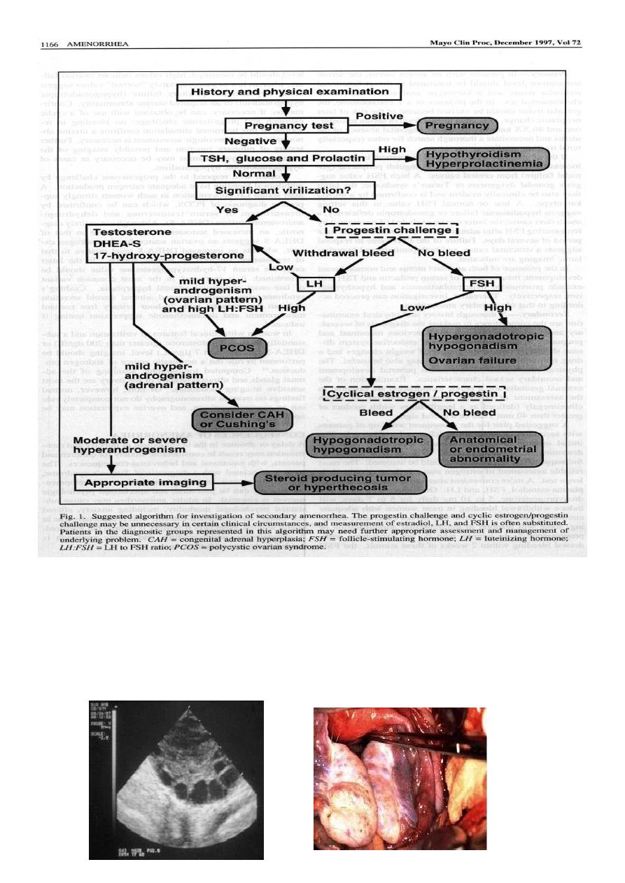
Endocrine Investigation:
Baseline gonadotrophin level (FSH,LH)
FSH and LH >15 IU/l indicate impending ovarian failure (unrelated to preovulatory
surge),their level can differentiate ovarian from hypothalamic causes LH raise alone will
indicate PCOS
Serum prolactin
If >1500 mIU/l indicate pituitary microadenoma .
>5000mIU/l indicate pituitary macroadenoma.
Thyroid function test
Oestrogen state of the endometrium:
By examination of the genital tract or induce withdrawal bleeding by progesterone
administration.
Serum testosterone:
if greater than 5 nmol/l should be investigated to exclude adrenal or ovarian tumors.
24 hour urinary cortisol:
is elevated in cushing syndrome(700 nmol/24 hr)
Other investigation like:
z
Ultrasound : to exclude PCOS, ovarian cyst or tumor,post menstrual endometrial
thickness if >10mm then endometrial biopsy indicated to exclude malignancy.
z
Hystyrosalpingography and hysteroscopy: in cases suspected to have asherman’s
syndrome.
z
CT scan or MRI :hypothalamic tumor, nonfunctioning pituitary tumor compressing
the hypothalamus or a prolactinoma.
z
Skull X ray
z
Karyotype: in patient of premature ovarian failure(<40 years) to exclude sex
chromosomes mosaisim.
z
Auto antibody screen: in patient with POF.

5
PCOS diagnosis
US laproscopic

6
Management of individual causes
Asherman’s syndrome:
Is a condition in which intauterine adhesions prevent normal growth of the endometrium.
Aetiology:
1. Too vigorous endometrial curettage affecting the basalis layer of the endometrium.
2. Episodes of endometritis.
3. Oestrogen deficiency in breast feeding women
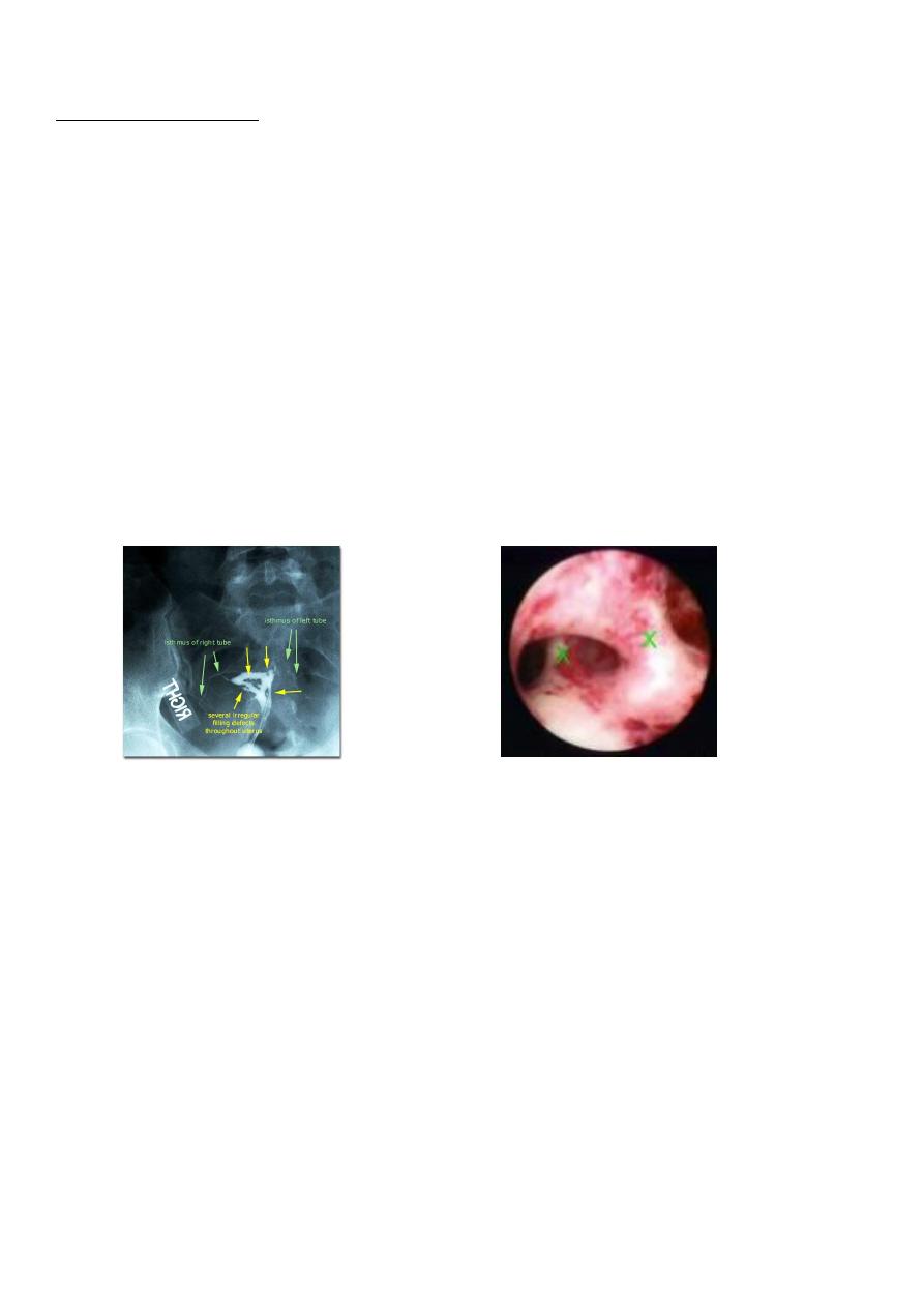
Intrauterine Adhesion
HSG Hysteroscopy
Amenorrhoea is not absolute,withdrawal Bleeding can be induced with oestrogen
/progesterone administration.
Diagnosis: by HSG and or hysteroscopy.
Treatment:
by adhesiolysis followed by 3 months of oestrogen progesterone cyclical therapy.
A foley catheter can be inserted postoperatively for 7-10 days, or IUD inserted for 2-3
months.
The pregnancy rate after treatment depends on initial severity of the adhesions
93% for mild adhesions
57% for sever adhesions

7
Premature ovarian failure (POF):
Is cessation of periods accompanied by raised gonadotrophin level prior to the age of 40
years.
It occurs in 1-5% of female population.
POF have increased risk of:
z
osteoporosis
z
cardiovascular disease
Aetiology:
1. Chromosomal abnormality : it occurs in
70% of cases of primary amenorrhoea
2-5% of cases of secondary amenorrhoea
2. Autoimmune disease.
3. chemo/radiotherapy.
4. Surgery.
Treatment:
Estrogen deficiency : HRT preperation Infertility: in established cases there is resistant to
gondotrophin with absence of ovarian follicle, and reports of pregnancy in treated cases
only indicate fluctuating ovarian function rather than treatment success.
Hyperprolactinaemia:
z
Is the commonest pituitary cause of secondary amenorrhoea.
z
Causes of elevation of serum prolactin:
Mild elevation: -pregnancy(10 fold)
-stress.
-venepuncture.
-postprandial.
-breast examination.

8
Moderate elevation :
-hypothyroidism
-PCOS (up to 2500 Iu/l)
-drugs: dopaminergic antagonist phenothiazines, domperidone,verapamil,
methyldopa,metoclopramide and oestrogen.
Sever elevation :
- prolactin secreting tumor : micro and macroadenoma.
- non-functioning tumor of the hypothalamus or pituitary
Symptoms of hyperprolactinaemia:
z
Amenorhoea: is the bioassay of prolactin excess.
z
Galactorrhoea: in up to 1/3 of patients.
z
Symptoms of hypooestrogenism (amenorrhoea no withdrawal bleeding after
progesterone therapy)
z
Visual disturbances.(bitemrporal hemianopia)
Diagnosis:
z
Prolactin serum assay: ( tumor marker)
z
Skull X ray:
-asymmetrical enlarged pituitary fossa with double contour of it’s floor .
-Erosion of the clinoid processes.
z
Ct-scan or MRI
Treatment of galactorrhea and amenorrhea
1. Observation:
z
periodic observation for patient with galactorrhea who have normal prolactin or
idiopathic elevation.
z
Patient with oligomenorrhea who not desire fertility should be treated with periodic
progesterone if not desire fertility and with COCP,if contraception is required. failure
of progestin to introduce

9
Bleeding is suggestive of hypooestrogenism.
z
Long term treatment with bromocriptine in women with normal estrogen is not
indicated.
z
Observation can be extended to some women with radiologic evidence of pituitary
microadenoma(<=10mm in diameter) because the growth rate in slow,an annual
measurement of serum prolactin is appropriate in patient with normal oestrogen
level.
z
Macroadenoma >=10mm require further evaluation by periodic scanning and
possible treatment.
2. Medical treatment:
z
Patient aim to restore menstrual cyclicity or to prevent osteoporosis COCP are given.
z
Patient require reduction of prolactin and restoration of cyclic physiologic estrogen
secretion require ergot compund:
Bromocriptine:dopamine antagonist:
starting with small dose of 1.25 mg at bed time increased gradually 7.5 mg in divided
Daily doses to initiate response then reduce To a maintenance lower dose .some tolerate
the drug more if given vaginally.
Side effect :
Nausea ,vomiting, headache,
Postural hypotension
Cabergoline :longer acting ,better tolerated,more expensive
Given in 0.25 mg twice weekly,
z
Bromocriptine normalizes the secretion of prolactin in 82% of cases of women with
microadenoma and restore cycle and fertility in more than 90%. It require 6-10 weeks
to restore cycle and 10-16week to establish ovulation. discontinuation of therapy
result into return of hyper-prolactinaemia leading to galactorrhea and amenorrhea.

11
z
Patient with macroadenoma require visual field assesment,other pituitary hormone
assesment,repeat MRI after full therapeutic dose of bromocriptine is
reached.treatment is continued as long as shrinkage of adenoma is required
z
During pregnancy treatment is stopped tumors unlikely to enlarge during
pregnancy,macroadenoma extending beyound the sella tursica require debulking
surgery before pregnancy (15%-30%) risk of enlargement, bromcriptine therapy is
started,patient require repeated visual field assessment once abnormality develops
z
Bromocriptine is instituted or increased for the rest of pregnancy.(there is no
increase in congenital abnormality )
z
Medical treatment is preferred over surgery as recurrence is high: microadenoma
recure30%and macroadenoma recurs in 90%.
3. Surgical : transphenoidal adenectomy
Indicated in cases of:
-Resistance to treatment.
-intolerance to medical treatment.
-supracellar extention not respond to bromocriptine and pregnancy is desired
Radiotherapy: is not desired with modern surgery
A.L.Y
