
Post mortem
• Theory 2h/ week
• Practical General information about P.m and
data collection of some of farm animals
disease

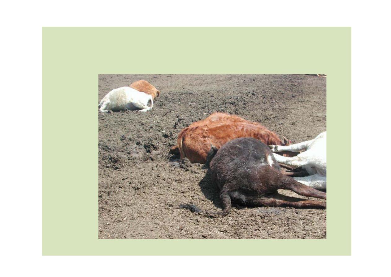
Definition
Necropsy
“seeing a dead body”(Greek)
Definition
Necropsy
“seeing a dead body”(Greek)
Techniques in Necropsy:
Pathway to Knowing “Everything”

Objective: is performing any necropsy is to establish the cause of
illness or death. however, additional information can be gained
that may be of use to other animals in the group E.X: screening
intestinal content for evidence of parasitism
Principles:
The necropsy should be conducted to glean the maximum
information
Examination of one organ should be at the expense of another
All material should be retained until the gross examination is
completed and all samples for analysis have been collected
If the animals presented live, blood samples should be collected
into a range of tubes, with and without of anticoagulants
Necropsy should be performed immediately after death or as
soon thereafter as possible
(why)
Chilling is acceptable when the necropsy has to be delayed
(why)

Safety:
Many dangers must be considered when performing a necropsy but
they fall into two basic categories:
1- Trauma from sharp instruments or bone fragments
2- zoonotic conditions
N.B: the employer must supply adequate protective clothing , safety
equipment and a safe working environment
The Post-Mortem Exam included further things
Cast history
Necropsy
Histopathology
Microbial culture/isolation
Toxicity screening
Gene analysis

Why do a necropsy?
Diagnostic
Prevention
Evaluate health of a population
Educational
Those lesions that don’t fit need to be
explained
Change in management: handling, feeding,
facilities, treatment protocols
Expose all foci of disease/abnormality.
Why do a necropsy?
Diagnostic
Prevention
Evaluate health of a population
Educational
Those lesions that don’t fit need to be
explained
Change in management: handling, feeding,
facilities, treatment protocols
Expose all foci of disease/abnormality.
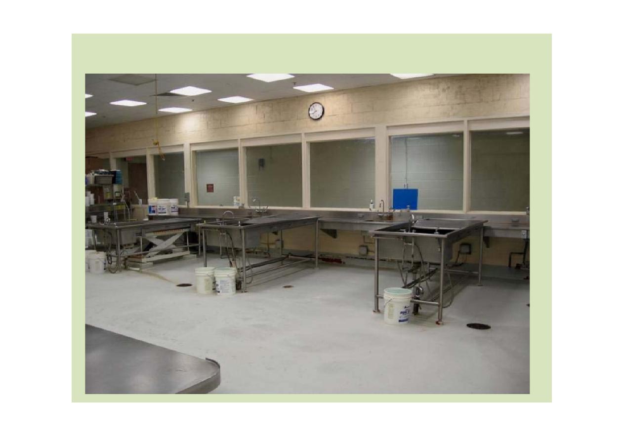
Necropsy Facility
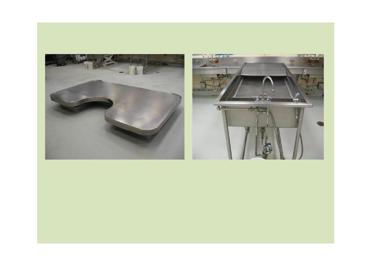
Necropsy Facility

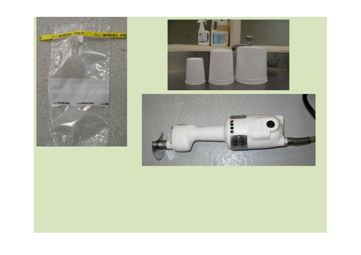
Basic Equipment
will vary with the species, location of cadaver, etc.
CLOTHING:
gloves, boots, coveralls, apron (
)اﻟﻣﺋزر
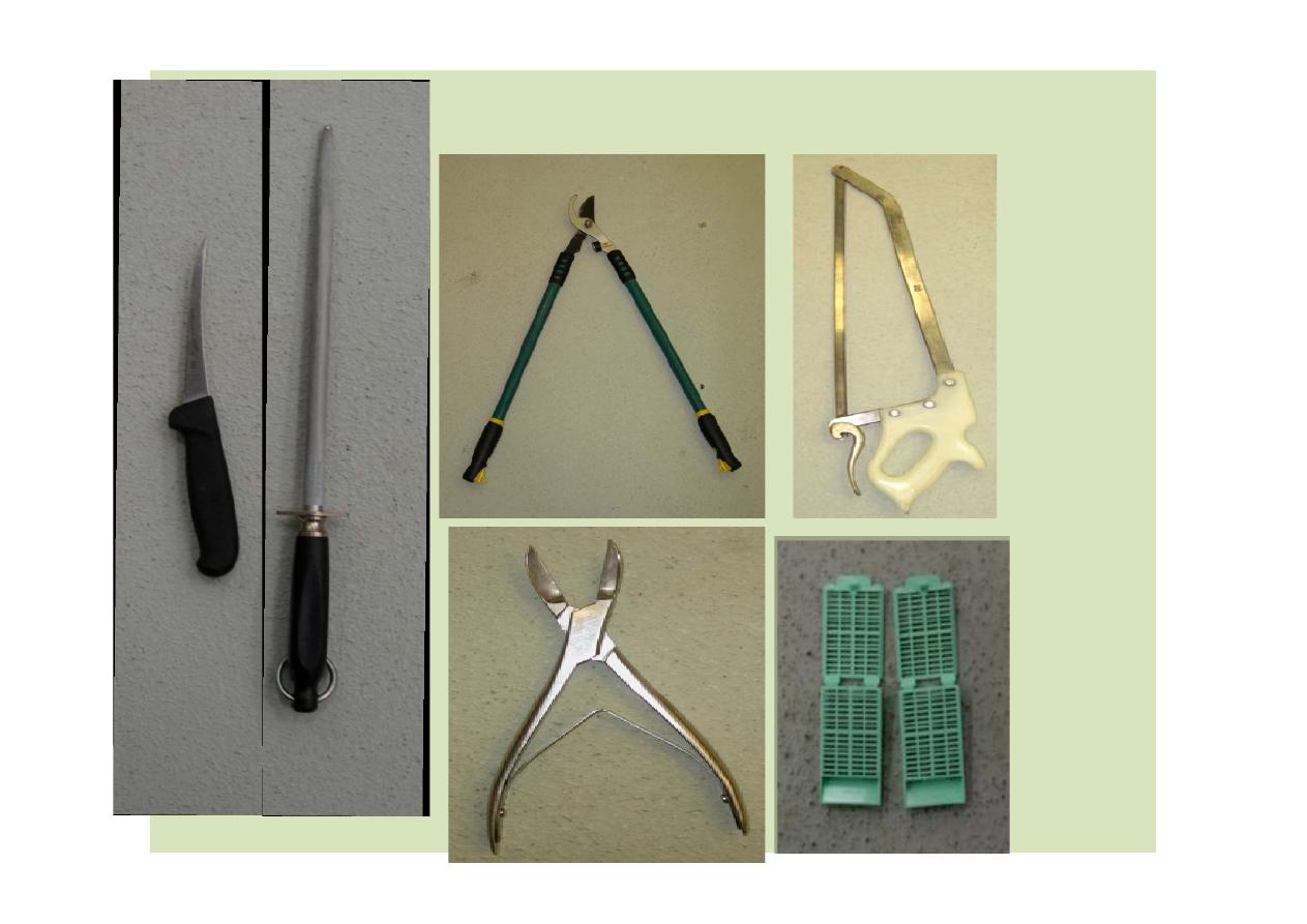
INSTRUMENTS:
Sharp knife and sharpening equip (steel/stone)
Tissue forceps and scissors, saw, cleaver, osteotome, shears, axe,
metric ruler, scale, ruler, Tree limb snips, soap, water, brushes for
cleaning
FIXATIVES & MISC:
fixative and appropriate containers, sterile syringes, needles, swabs,
plastic bags, paper plates, microscope slides, tags (
)ﺑطﺎﻗﺎت اﻟﺑﯾﺎﻧﺎت,
dissecting microscope, photographic capability (not essential)
Basic Equipment
will vary with the species, location of cadaver, etc.
CLOTHING:
gloves, boots, coveralls, apron (
)اﻟﻣﺋزر
INSTRUMENTS:
Sharp knife and sharpening equip (steel/stone)
Tissue forceps and scissors, saw, cleaver, osteotome, shears, axe,
metric ruler, scale, ruler, Tree limb snips, soap, water, brushes for
cleaning
FIXATIVES & MISC:
fixative and appropriate containers, sterile syringes, needles, swabs,
plastic bags, paper plates, microscope slides, tags (
)ﺑطﺎﻗﺎت اﻟﺑﯾﺎﻧﺎت,
dissecting microscope, photographic capability (not essential)
Basic Equipment

Goals of Necropsy in pathology
Data collection
Concise (
رّﻐﺻﻣ) lesions description
Appropriate tissue sampling
Correlate findings with in-life data.

Data Collection
Review history
Systematic
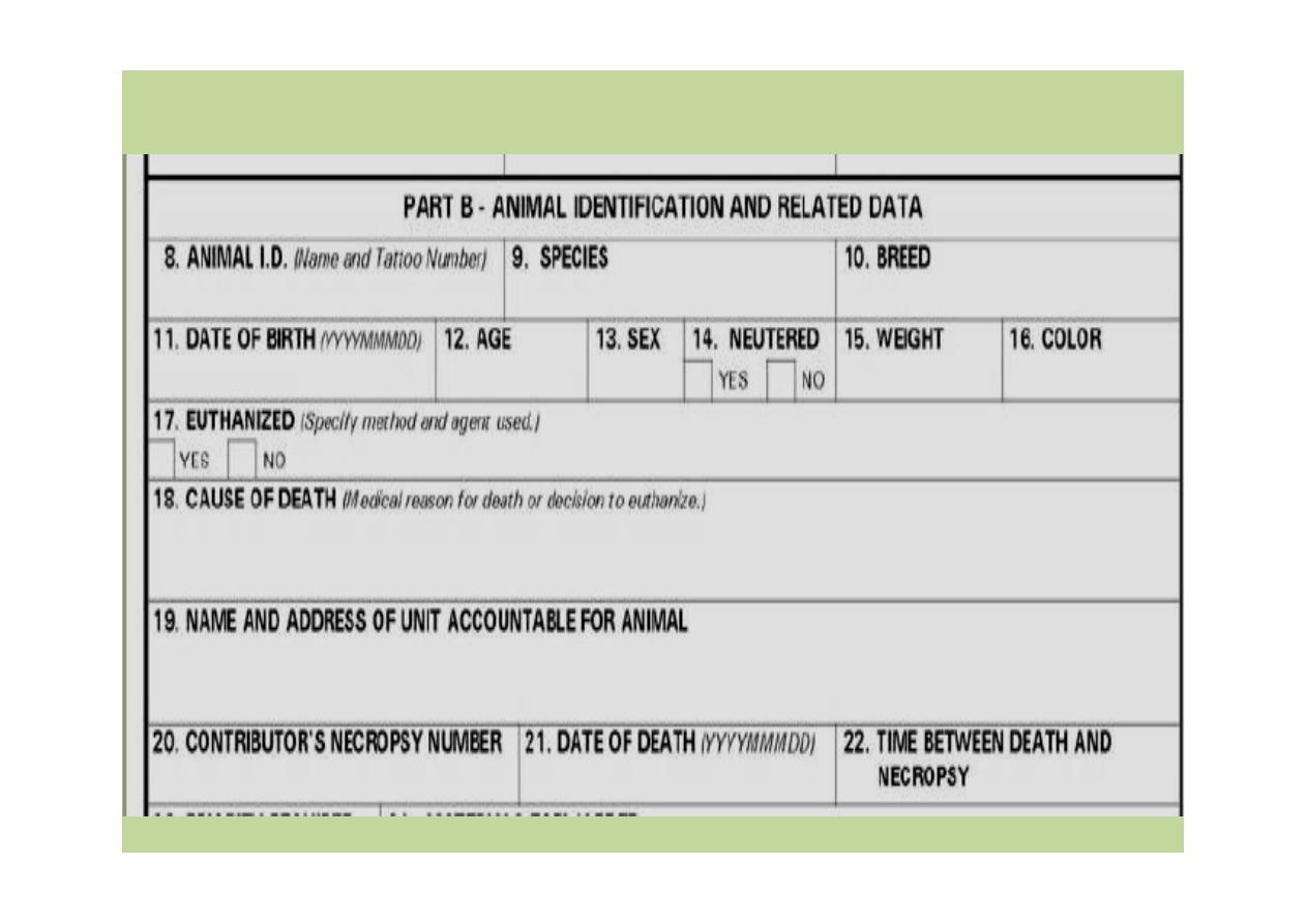
Necropsy record
ID
Date/time of death
Date of necropsy
List of tissues examined
Description of lesions
Data Collection
Review history
Systematic
Necropsy record
ID
Date/time of death
Date of necropsy
List of tissues examined
Description of lesions
Necropsy Record
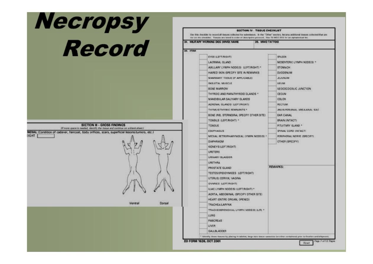
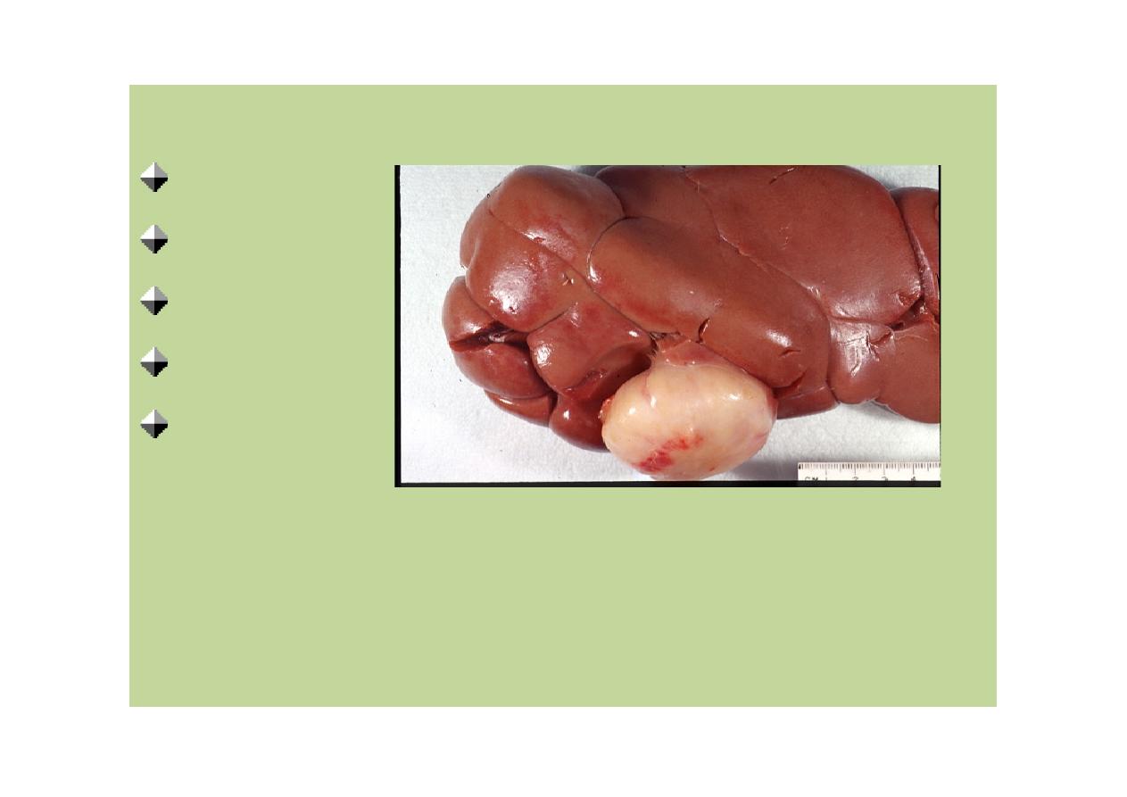
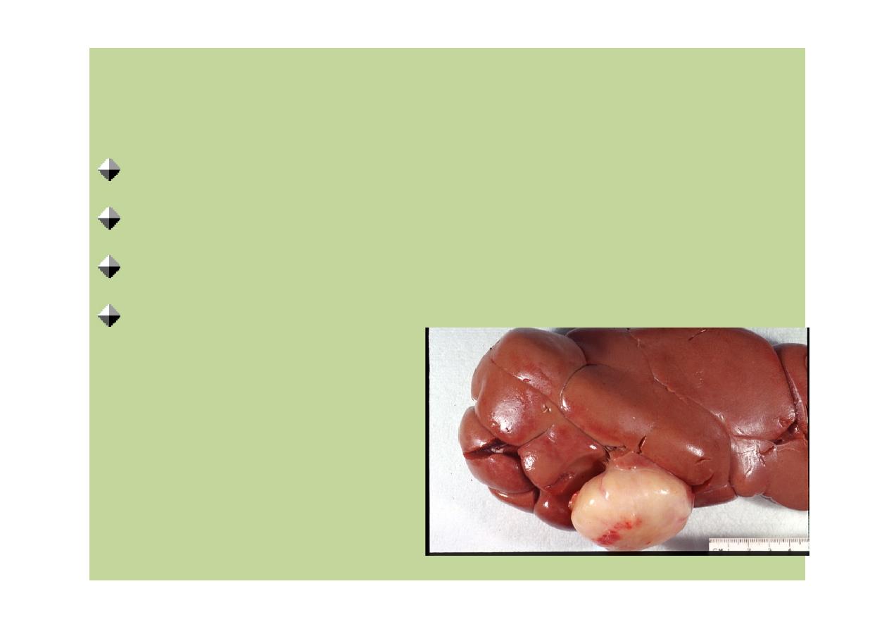
Lesion Description
Tissue
Location
Color
Size
Shape
Lesion Description
Tissue
Location
Color
Size
Shape
Lesion Description
Consistency and texture
Number and extent
Surface appearance
odour
Lesion Description
Consistency and texture
Number and extent
Surface appearance
odour

Tissue Sampling for further using
Cytology
Light microscopy
Microbial isolation
Molecular analysis
Toxicology
Electron microscopy
Tissue Sampling for further using
Cytology
Light microscopy
Microbial isolation
Molecular analysis
Toxicology
Electron microscopy
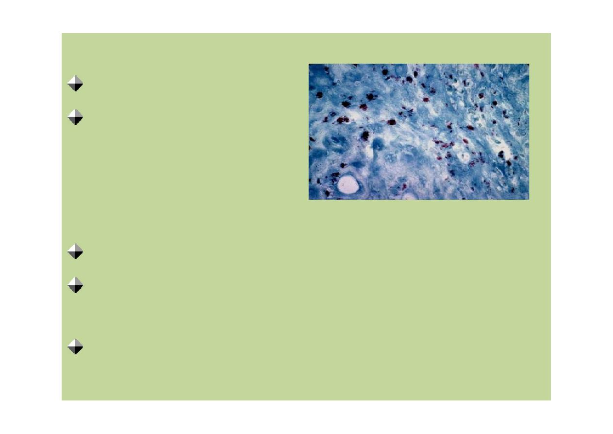
Light Microscopy
Formalin fixed
1cm thick
1:10 of fixative material : tissue ratio
Representative samples should include
junction between normal and abnormal
Histochemistry and immunohistochemistry
Light Microscopy
Formalin fixed
1cm thick
1:10 of fixative material : tissue ratio
Representative samples should include
junction between normal and abnormal
Histochemistry and immunohistochemistry
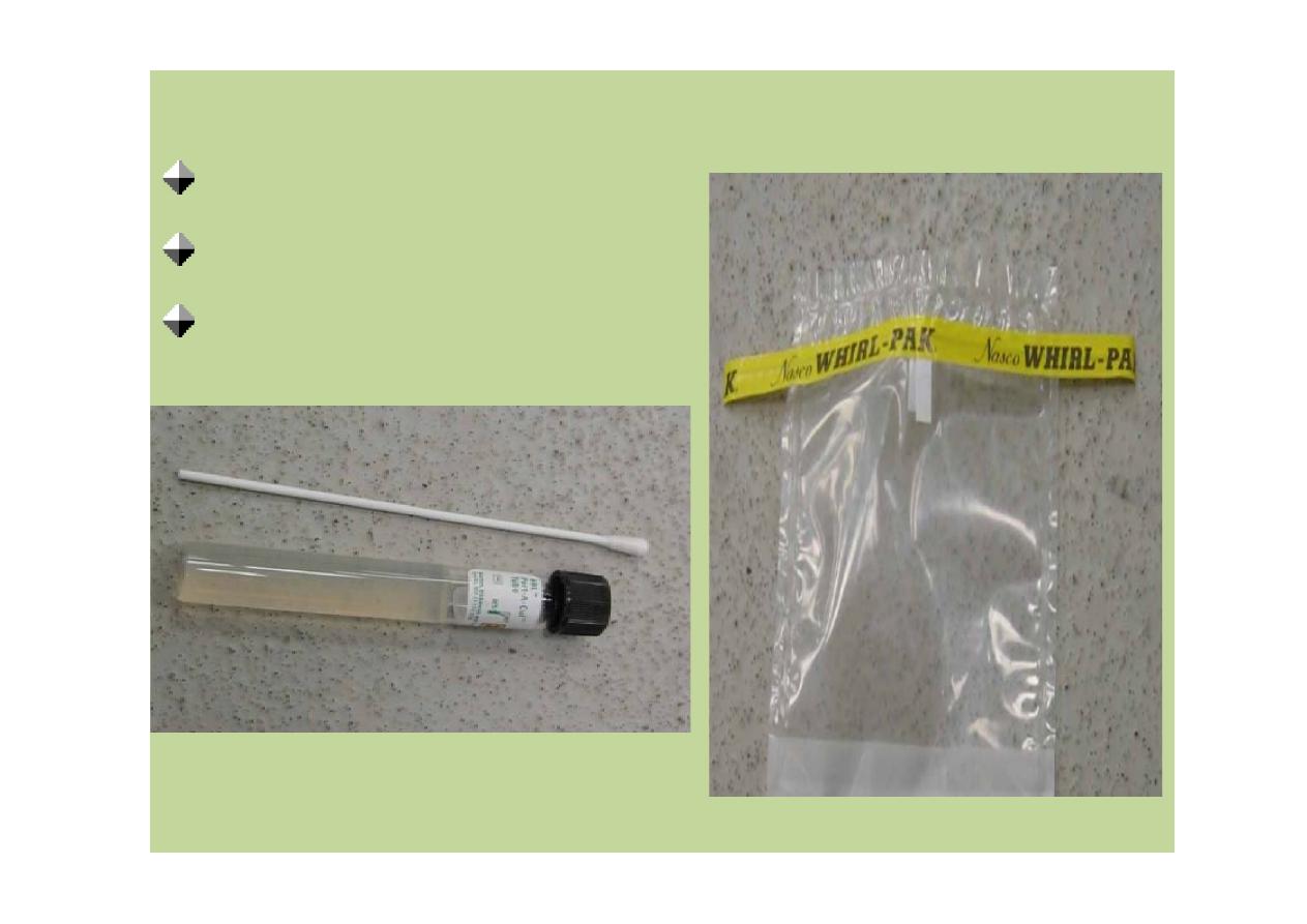
Microbial Isolation
Fresh tissue
Transport media
Frozen tissue (viruses)
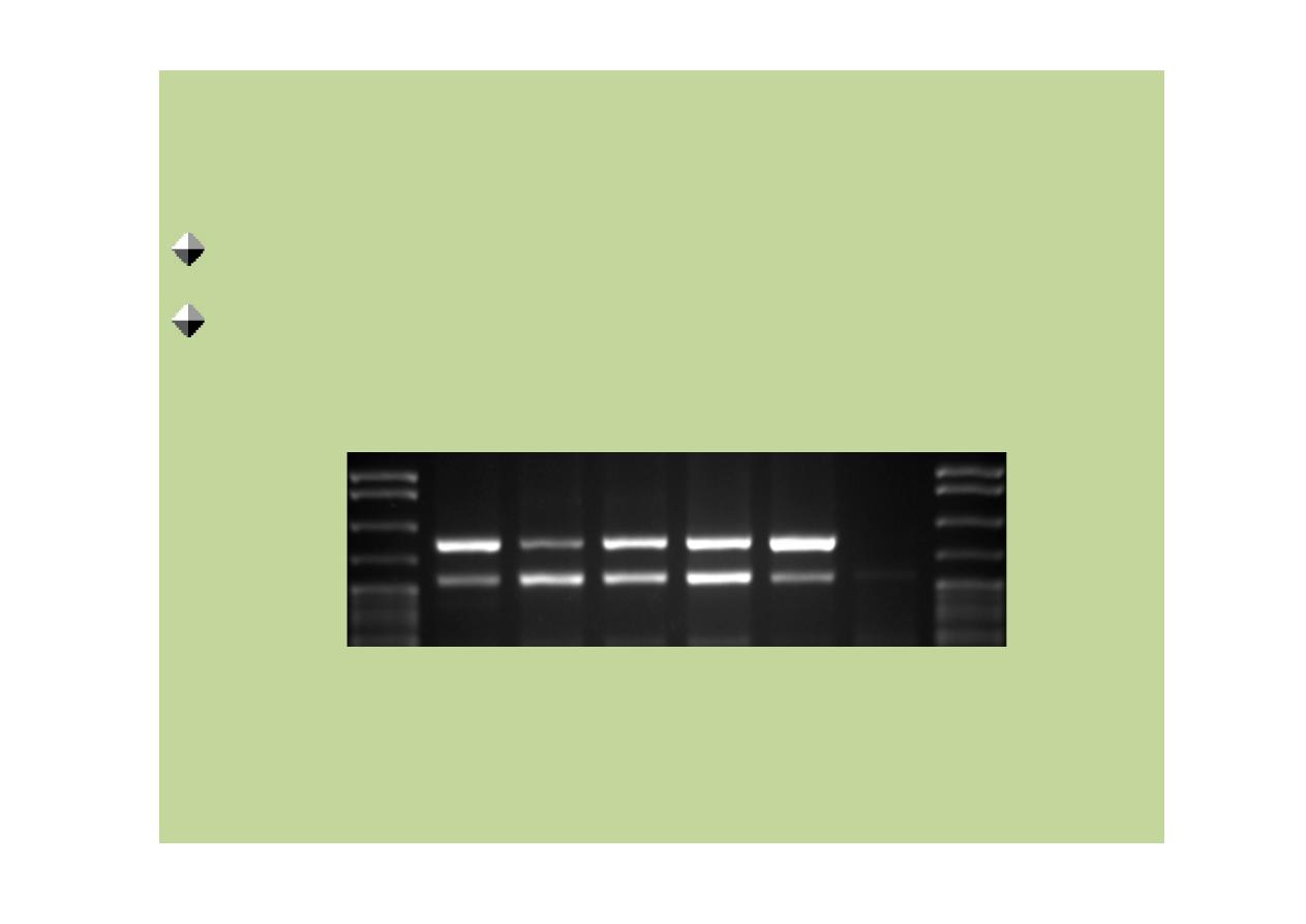
Molecular Analysis
Pathogen identification
Frozen samples are preferred

Toxicology
Fresh or frozen tissue
Liver, kidney, skeletal muscle, fat
Electron Microscopy
1% glutaraldehyde
Tissue perfusion
Paraffin embedded tissue
Toxicology
Fresh or frozen tissue
Liver, kidney, skeletal muscle, fat
Electron Microscopy
1% glutaraldehyde
Tissue perfusion
Paraffin embedded tissue

Summary
Start big; work towards small
Be consistent
Return to the animal’s history
Collect extra tissue if unsure
Do it as often as you can
Summary
Start big; work towards small
Be consistent
Return to the animal’s history
Collect extra tissue if unsure
Do it as often as you can

References
Department of Defence. Veterinary Necropsy Report Checklist and Guidelines
(DD Form 1626). Oct. 2001. 10 June
2008<http://www.dtic.mil/whs/directives/infomgt/forms/eforms/dd1626.pdf>.
Echols, Scott M. Exotic Pet Medicine and Surgery. V.2, Avian Necropsy and
Cytology [1 CD-ROM] : Basic Avian Techniques. Jackson, Wyoming: Teton
Newmedia, 2003.
King, John M. Dr. John M. King's Necropsy Show and Tell. 26 Dec.2007. 10 June
2008 <http://w3.vet.cornell.edu/nst/nst.asp>.
King, John M., Lois Roth-Johnson, and David C. Dodd. The Necropsy Book.
Gurnee, IL: Charles Louis Davis DVM Foundation, 2005.
Munson, Linda. Necropsy of Wild Animals. 10 June
2008<http://www.vetmed.ucdavis.edu/whc/pdfs/necropsy.pdf>.
National Cancer Institute. Registry of Tumours in Lower Animals.30 Sep. 2007. 10
June 2008 <http://www.pathology-registry.org/index_1.asp>.

Rose, Karrie, Scott Newman, and Marcela Uhart. Wild Bird Highly Pathogenic
Avian Influenza Surveillance: Sample Collectionfrom Healthy, Sick and Dead
Birds. Rome, Italy: Food andAgriculture Organization of the United Nations, 2006.
Terrell, Scott P., and Brian A. Stacy. "Reptile Necropsy Techniques."Infectious
Diseases and Pathology of Reptiles. Ed. Elliott R.Jacobson. Boca Raton, FL: Taylor
and Francis, 2007. 219-256.
USDA-APHIS. Animal Health: Collecting Samples for Diagnostic Unknowns. 25
Feb. 2008. 10 June
2008<http://www.aphis.usda.gov/animal_health/lab_info_services/collection_s
ubmission.shtml>.
Woodford, M. H., D. F. Keet, and R. G. Bengis. Post-mortemProcedures for
Wildlife Veterinarians and Field Biologists. Paris, France: Office International des
Epizooties, 2000.
Wyneken, Jeanette. The Anatomy of Sea Turtles. Dec. 2001. 10 June 2008
<http://courses.science.fau.edu/~jwyneken/sta

If you have any comments, criticisms or suggestions about this tutorial
module please let me know.
If you find any errors or typos please let me know too