Muscle
Muscles are responsible for all types of body movement , they contract or shorten and they are the machine of the body.There are more than 600 muscles in our body. Muscles perform many useful functions and help us in doing everything in day to day life. Muscles are classified by three different methods based on different factors:
I. Depending upon the presence or absence of striations
II. Depending upon the control
III. Depending upon the function.
I. DEPENDING UPON STRIATIONS
Depending upon the presence or absence of the cross striations, muscles are divided into two groups:1. Striated muscle
2. Nonstriated muscle.
1. Striated muscle
Striated muscle is the muscle which has a large number of cross striations (transverse lines).Skeletal muscle and cardiac muscle belong to this category.
2. Nonstriated muscle.
The muscle which does not have cross striations is called nonstriated muscle. It is also called plain muscle or smooth muscle. It is found in the wall of the visceral organs.II. DEPENDING UPON CONTROL
Depending upon control, the muscles are classified into two types:
1. Voluntary muscle
2. Involuntary muscle.
1. Voluntary muscle
Voluntary muscle is the muscle that is controlled by the will. Skeletal muscles are the voluntary muscles. These muscles are innervated by somatic nerves.2. Involuntary muscle
The muscle that cannot be controlled by the will is called involuntary muscle. Cardiac muscle and smooth muscle are involuntary muscles. These muscles are innervated by autonomic nerves.III. DEPENDING UPON SITUATION
The muscles are classified into three types depending upon the situation:1. Skeletal muscle
2. Cardiac muscle
3. Smooth muscle.
The functions of muscles
1. Producing movement – facial expressions, movement of food stuffs through digestions and blood through the heart.2. Maintaining posture (skeletal only), very fatigue resistant muscles, ex.erector spinae; and stabilizing joints (holding the skeleton together)
3. Generating heat to maintain body temperature. Muscles make up 40% of body weight and ¾ of energy given off by ATP is heat.
Skeletal Muscle
Skeletal muscle is situated in association with bones forming the skeletal system. The skeletal muscles form 40 to 50% of body mass and they are voluntary and striated. These muscles are supplied by somatic nerves.
The fibers of the skeletal muscles are arranged in parallel.
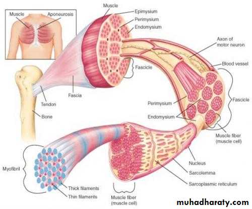
Muscle Mass
The muscle mass (or tissue) is made up of a large number of individual muscle cells or myocytes.The muscle cells are commonly called muscle fibers because these cells are long and slender in appearance. The skeletal muscle fibers are multinucleated and arranged parallel.
The muscle mass is separated from the neighboring tissues by the thick fibrous tissue layer known as fascia. Beneath the fascia, the muscle is covered by a connective tissue sheath called epimysium.
The muscle fibers are arranged in various groups called the bundles or fasciculi. The connective tissue sheath that covers each fasciculus is called perimysium.
Each muscle fiber is covered by the connective tissue layer called the endomysium.
Muscle Fiber
Each muscle fiber is cylindrical in shape. The muscle fibers are attached to bone by a tough cord of connective tissue called tendon.
Each muscle fiber is enclosed by a cell membrane called plasma membrane that lies beneath the endomysium. It is also called sarcolemma. The cytoplasm of the muscle is known as sarcoplasm. Many structures are embedded within the sarcoplasm:
1. Nuclei
2. Myofibril
3. Golgi apparatus
4. Mitochondria
5. Sarcoplasmic reticulum
6. Ribosomes
7. Glycogen droplets
8. Occasional lipid droplets.
`
Myofibril
Myofibrils or myofibrillae are the special structures present only in muscle fibers. These are the fine parallel filaments present in sarcoplasm of the muscle cell. The myofibrils run through the entire length of the muscle fiber.Light microscopic studies show that, each myofibril consists of a number of two alternating bands. The two bands are:
1. Light band or ‘I’ band It is isotropic to polarized light.
2. Dark band or ‘A’ band.
it is anisotropic to polarized light.‘I’ band and ‘A’ band of the adjacent myofibrils are placed side by side and gives the appearance of characteristic cross striations in the muscle fiber.
I band is divided into two portions by a narrow dark line called ‘Z’ line or ‘Z’ disk .
The ‘Z’ line is formed by a protein disk. The portion of myofibril in between two ‘Z’ lines is called Sarcomere is the structural and functional unit of the skeletal muscle.
The sarcomere consists of many thread like structures called myofilaments. Myofilaments are of two types:
1. Actin filaments
2. Myosin filaments.
Actin Filaments
Actin filaments are the thin filaments that extend from either side of the ‘Z’ lines, run across ‘I’ band and enter into ‘A’ band up to ‘H’ zone.
Myosin Filaments
Myosin filaments are thick filaments and are situated in ‘A’ band. Some lateral processes (projections) or cross bridges arise from myosin filaments. These bridges have enlarged structures called myosin heads at their tips. The myosin heads attach themselves to actin filaments.
Extent
Each sarcomere extends between two ‘Z’ lines of myofibril. Thus, each myofibril contains many sarcomeres arranged in series throughout its length. When the muscle is in relaxed state, the average length of each sarcomere is 2-3 microns.
Components
Each myofibril consists of alternate dark ‘A’ band and light ‘I’ band. In the middle of ‘A’ band, there is a light area called ‘H’ zone. In the middle of ‘H’ zone lies the middle part of myosin filament. This is called ‘M’ line. ‘M’ line is formed by myosin binding proteins.

Smooth Muscle
Smooth muscle or visceral muscle is situated in association with viscera. Smooth muscle is nonstriated and involuntary. Because of the absence of cross striations it is called smooth or plain muscle. It is supplied by autonomic nerve fibers.
Structure Of Smooth Muscle
Smooth muscle fibers are fusiform or elongated cells. The nucleus is single and elongated and it is centrally placed. Normally, two or more nucleoli are present in the nucleus. Smooth muscle fibers are covered by connective tissue. But the tendons are absent.Myofibrils and Sarcomere
Well defined myofibrils and sarcomere are absent in smooth muscles. So the alternate dark and light bands are absent.Myofilaments and Contractile Proteins
The contractile proteins in smooth muscle fiber are actin, myosin and tropomyosin. But troponin or troponin like substance is absent.
Thick and thin filaments are present in smooth muscle. However, these filaments are not arranged in orderly fashion as in skeletal muscle.
Dense Bodies
Dense bodies are the special structures of smooth muscle fibers to which the actin and tropomyosin molecules of thin filaments are attached.
Types Of Smooth Muscle Fibers
Smooth muscle fibers are of two types:1. Single unit or visceral smooth muscle fibers
Single unit smooth muscle fibers are the fibers with interconnecting gap junctions. The gap junctions allow rapid spread of action potential throughout the tissue.
The features of single unit smooth muscle fibers:
i. The muscle fibers are arranged in sheets or bundles
ii. The cell membrane of adjacent fibers fuses at many points to form gap junctions.
Through the gap junctions, ions move freely from one cell to the other.
In this way, the visceral smooth muscle resembles cardiac muscle more than the skeletal muscle.
The visceral smooth muscle fibers are in the walls of the organs such as gastrointestinal organs, uterus, ureters, respiratory tract, etc.
2. Multiunit smooth muscle fibers.
The multiunit smooth muscle fibers are the muscle fibers without interconnecting gap junctions.
These smooth muscle fibers resemble the skeletal muscle fibers in many ways. The features of multiunit smooth muscle fibers:
i. The muscle fibers are individual fibers
ii. Each muscle fiber is innervated by a single nerve ending
iii. Each muscle fiber has got an outer membrane made up of glycoprotein, which helps to insulate and separate the muscle fibers from one another
iv. The control of these muscle fibers is mainly by nerve signals
v. These smooth muscle fibers do not exhibit spontaneous contractions. The multiunit muscle fibers are in ciliary muscles of the eye, iris of the eye, nictitating membrane (in cat), arrector pili, and smooth muscles of the blood vessels and urinary bladder.
Cardiac Muscle
Cardiac muscle fibers arranged in a latticework, with the fibers dividing. Cardiac muscle is striated in the same manner as in skeletal muscle. It has typical myofibrils that contain actin and myosin filaments almost identical to those found in skeletal muscle.The dark areas crossing the cardiac muscle fibers are called intercalated discs; they are actually cell membranes that separate individual cardiac muscle cells from one another.
At each intercalated disc the cell membranes fuse with one another that they form permeable “communicating” junctions (gap junctions) that allow rapid diffusion of ions. Therefore, from a functional point of view, ions move with ease in the intracellular fluid along the longitudinal axes of the cardiac muscle fibers so that action potentials travel easily from one cardiac muscle cell to the next, past the intercalated discs. Thus, cardiac muscle is a syncytium of many heart muscle cells in which the cardiac cells are so interconnected that when one of these cells becomes excited, the action potential spreads to all of them, from cell to cell throughout the latticework interconnections.
General Mechanism Of Muscle Contraction
The initiation and execution of muscle contraction occur in the following sequential steps.An action potential travels along a motor nerve to its endings on muscle fibers.
At each ending, the nerve secretes a small amount of the neurotransmitter substance acetylcholine.
The acetylcholine acts on a local area of the muscle fiber membrane to open multiple “acetylcholine-gated” cation channels through protein molecules.
Opening of the acetylcholine-gated channels allows large quantities of sodium ions to diffuse to the interior of the muscle fiber membrane. This causes a local depolarization that in turn leads to opening of voltage-gated sodium channels. This initiates an action potential at the membrane.
The action potential travels along the muscle fiber membrane.
The action potential depolarizes the muscle membrane, and much of the action potential electricity flows through the center of the muscle fiber. Here it causes the sarcoplasmic reticulum to release large quantities of calcium ions that have been stored within this reticulum.
The calcium ions initiate attractive forces between the actin and myosin filaments, causing them to slide alongside each other, which is the contractile process.
After a fraction of a second, the calcium ions are pumped back into the sarcoplasmic reticulum by a Ca++ membrane pump and remain stored in the reticulum until a new muscle action potential comes along; this removal of calcium ions from the myofibrils causes the muscle contraction to cease.
Source Of Reconstitute of ATP
1- The first source of energy that is used to reconstitute the ATP is the substance phosphocreatine, which carries a high-energy phosphate bond similar to the bonds of ATP.
2- The second important source of energy, reconstitute both ATP and phosphocreatine, is “glycolysis” of glycogen previously stored in the muscle cells. Rapid enzymatic breakdown of the glycogen to pyruvic acid and lactic acid liberates energy that is used to convert ADP to ATP; the ATP can then be used directly to energize additional muscle contraction and also to re-form the stores of phosphocreatine.
3- The third and final source of energy is oxidative metabolism. This means combining oxygen with the end products of glycolysis and with various other cellular foodstuffs to liberate ATP.
More than 95 percent of all energy used by the muscles for sustained, long-term contraction is derived from this source.
Sliding Filament Model Of Contraction
Thin filaments slide past the thick ones so that the actin and myosin filaments overlap to a greater degreeIn the relaxed state, thin and thick filaments overlap only slightly
Upon stimulation, myosin heads bind to actin and sliding begins
mechanics of skeletal muscle contraction,
rigor mortis and physiology of cardiac muscleThree basic muscle types are found in the body
Skeletal muscleCardiac muscle
Smooth muscle