Baghdad College of Medicine / 5
th
grade
Student’s Name :
SURGERY
LEC.10
Dr. Ammar Talib
Lec.1
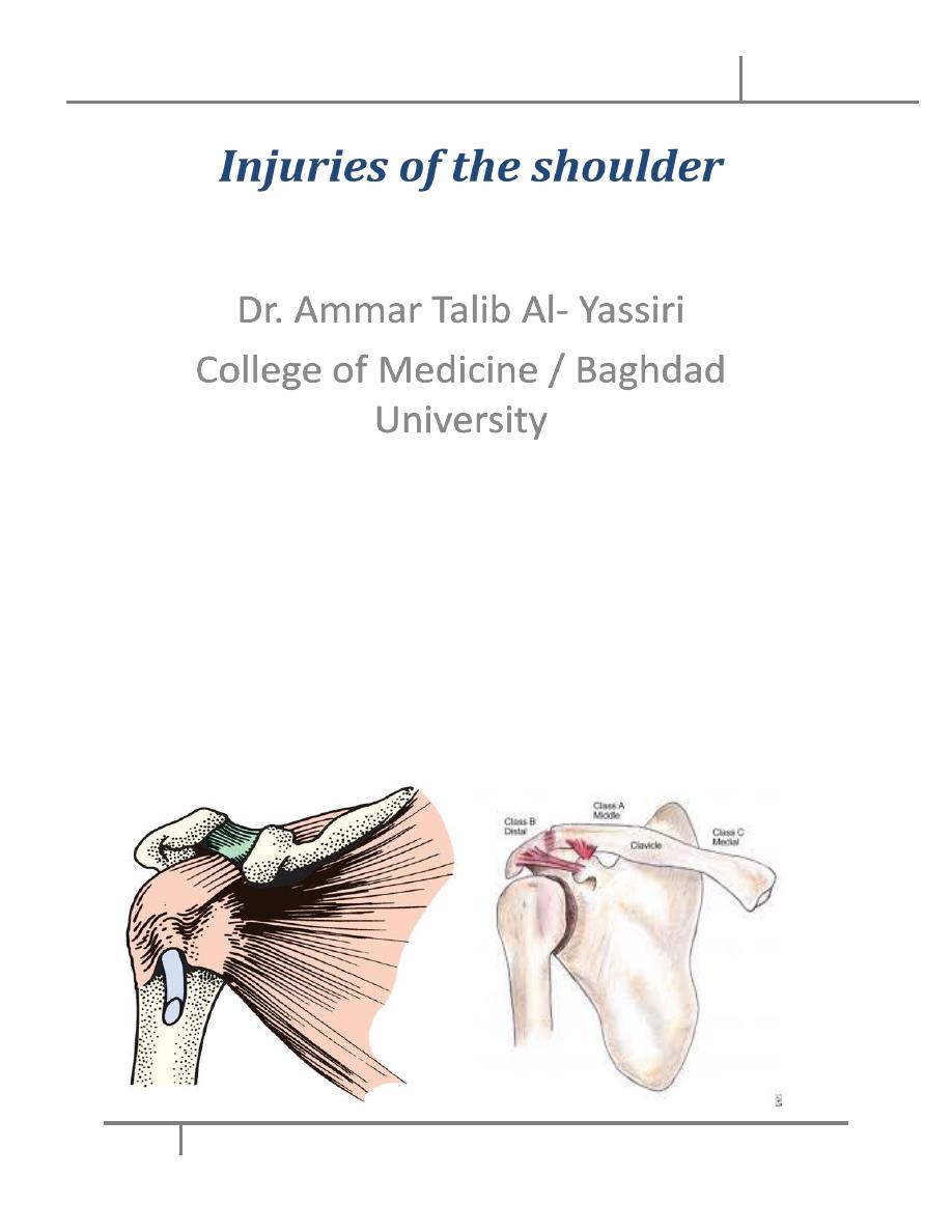
Injuries of the Shoulder
16-10-2016
DONE BY : Mustafa Naser
مكتب اشور لالستنساخ
2016 – 2017
Orthopedics (5)
Injuries of the shoulder Dr. Ammar
16-10-2016
2
@Mustafa Naser 2016-2017
Objectives
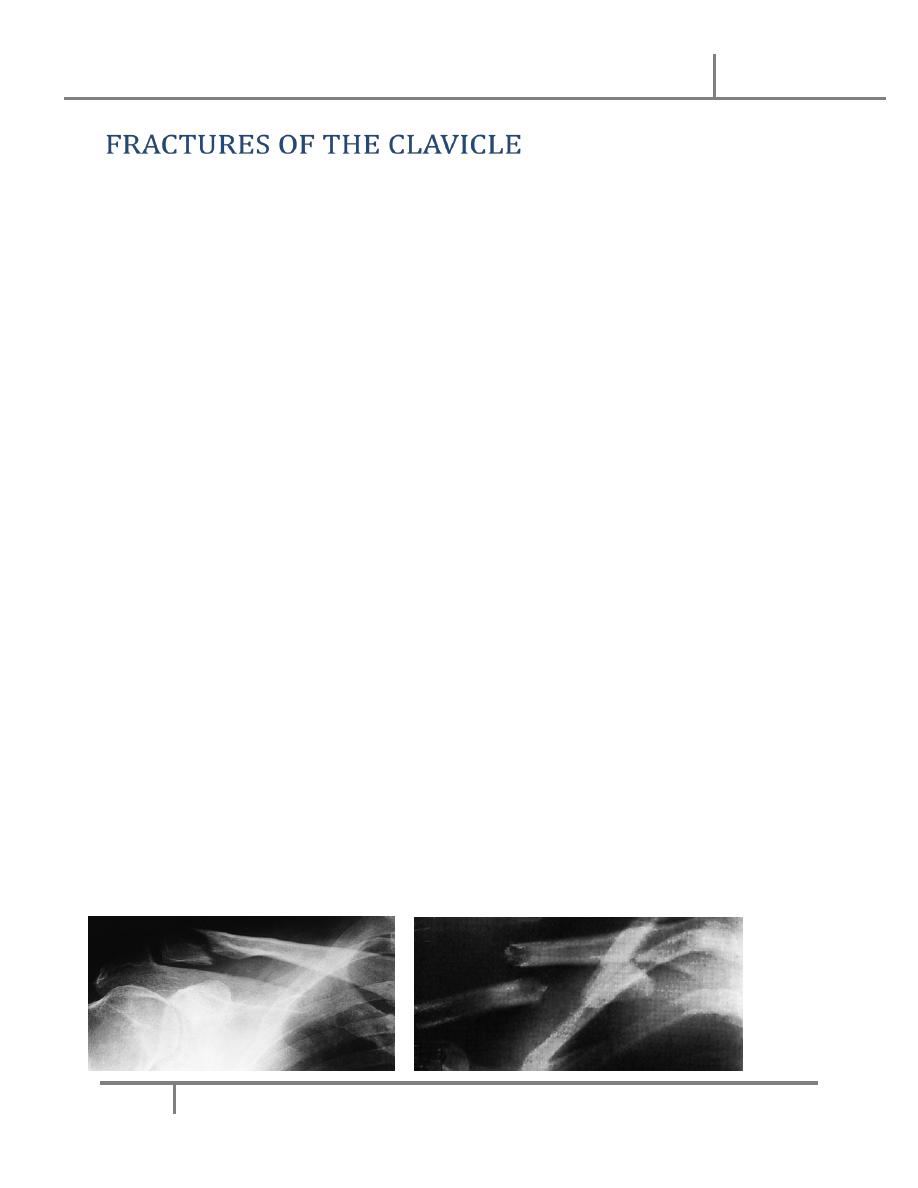
• FRACTURES OF THE CLAVICLE
• FRACTURES OF THE SCAPULA
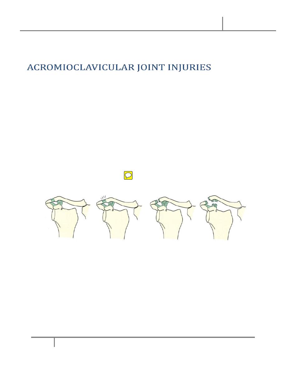
• ACROMIOCLAVICULAR JOINT INJURIES
• STERNOCLAVICULAR DISLOCATIONS
• DISLOCATION OF THE SHOULDER
Injuries of the shoulder Dr. Ammar
16-10-2016
3
@Mustafa Naser 2016-2017
• In children
– fractures easily
– unites rapidly.
• In adults
– much more troublesome
– common.
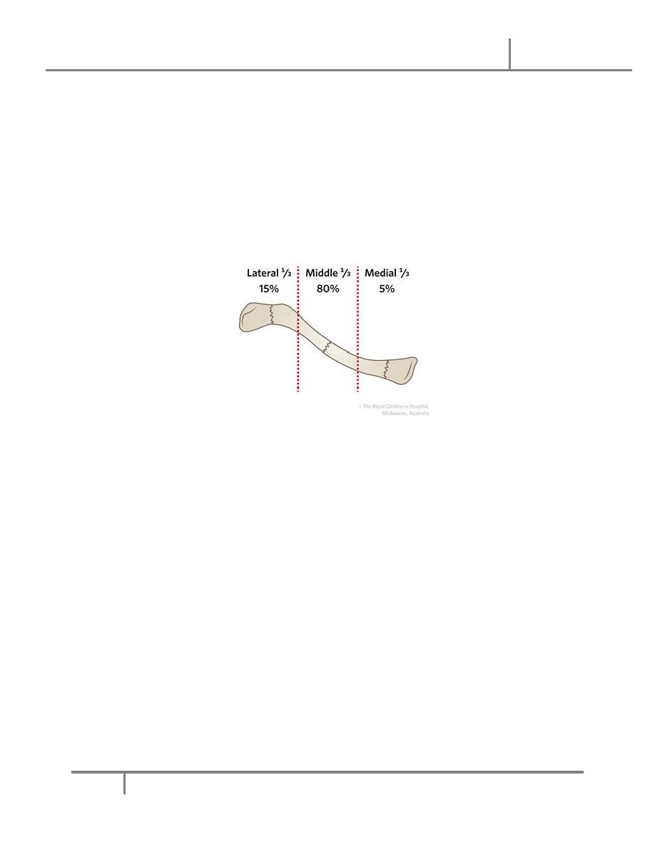
• Fractures of the midshaft > lat. Fractures>med.fractures
Mechanism of injury
• A fall on the shoulder or the outstretched hand
• In the common mid-shaft fracture
– outer fragment is pulled down
– inner half is held up
• In fractures of the outer end
– coracoclavicular ligaments intact little displacement
– Ligments torn or fracture is just medial to these ligaments
severe displacement
Injuries of the shoulder Dr. Ammar
16-10-2016
4
@Mustafa Naser 2016-2017
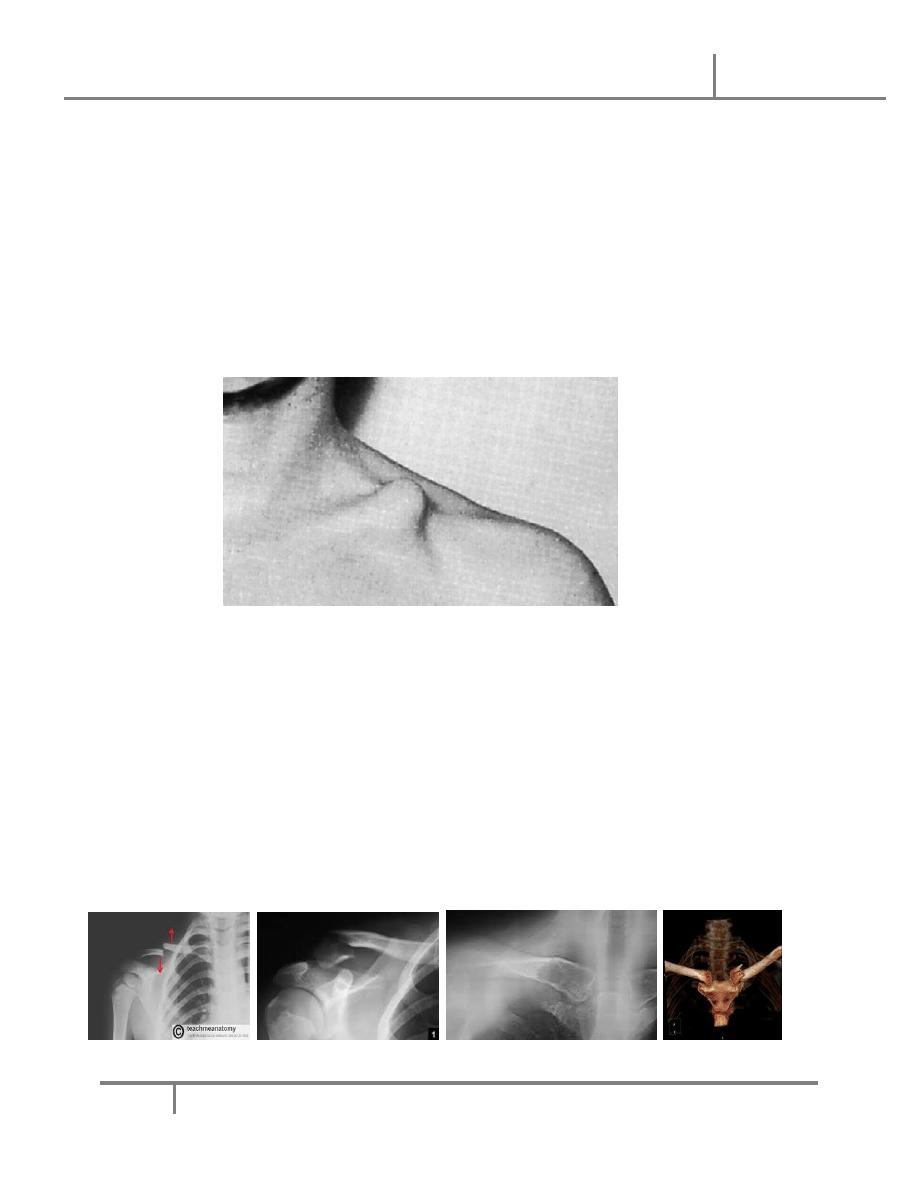
Clinical features:
• The arm is clasped to the chest
• A subcutaneous lump
• it is prudent to feel the pulse and gently to palpate the root of the neck
• Outer third fractures are easily missed or mistaken for acromioclavicular
joint injuries.
Imaging:
• requires at least an AP view and another taken with a 30 degree
cephalic tilt.
• middle third of the bone
• Fractures of the outer third may be missed
• medial third fractures it is also wise to obtain x-rays of the sterno-
clavicular joint
Injuries of the shoulder Dr. Ammar
16-10-2016
5
@Mustafa Naser 2016-2017
Classification:
• Clavicle fractures are usually classified on the basis of their location:
– Group I (middle third fractures),
– Group II (lateral third fractures) and
– Group III (medial third fractures).
Treatment:
• MIDDLE THIRD FRACTURES:Non-operative management (sling for
1-3 WKs)
• LATERAL THIRD FRACTURES
– minimally displaced : sling for 2–3 weeks
– Displaced lateral third fractures: surgery
• MEDIAL THIRD FRACTURES: non operatively
Complications:
• EARLY
– Injury to the vital structures
• a pneumothorax
Injuries of the shoulder Dr. Ammar
16-10-2016
6
@Mustafa Naser 2016-2017
• damage to the subclavian vessels and
• brachial plexus injuries are all very rare
• LATE
– Non-union: Symptomatic non-unions are generally treated with plate
fixation and bone grafting.
– Malunion : All displaced fractures heal in a nonanatomical position
with some shortening and angulation, however most do not produce
symptoms.
– Stiffness of the shoulder : This is common but temporary
Mechanisms of injury
– The body of the scapula crushing force
– The neck of the scapula blow or fall on the shoulder
– The coracoids process fracture across its base or be avulsed at
the tip
– Fracture of the acromion direct force
– Fracture of the glenoid fossa medially directed force or shoulder
dislocation
Clinical features:
• The arm is held immobile
• severe bruising over the scapula or the chest wall.

Injuries of the shoulder Dr. Ammar
16-10-2016
7
@Mustafa Naser 2016-2017
• fractures of the body of the scapula are often associated with severe
injuries to the chest, brachial plexus, spine, abdomen and head.
• Careful neurological and vascular examinations are essential
X-Ray:
• Scapular fractures are difficult to define on plain x-rays because of
the surrounding soft tissues.
– a comminuted fracture of the body of the scapula,
– a fractured scapular neck with the outer fragment pulled
downwards by the weight of the arm.
– Occasionally a crack is seen in the acromion or the coracoid
process.
• CT is useful for demonstrating glenoid fractures or body fractures
Treatment:
• Body fractures: a sling and exercise from the start
• Isolated glenoid neck fractures a sling and exercise from the start
• Intra-articular fractures if displaced, >1/3 of the glenoid surface and
is displaced by >5 mm surgical fixation
• Fractures of the acromion
– Undisplaced fractures are treated non-operatively.
– Displaced fracture require operative intervention to restore the
anatomy.
• Fractures of the coracoid process
– distal to the coracoacromial ligaments (conservative)
– proximal to the ligaments (operative)
Injuries of the shoulder Dr. Ammar
16-10-2016
8
@Mustafa Naser 2016-2017
• Combined fractures ‘floating shoulder’ At least one of the injuries (and
sometimes both) will need operative fixation
• Mechanism of injury: A fall on the shoulder with the arm adducted
• Pathological anatomy and classification:
– Type I
– Type II
– Type III
– Type IV
– Type V
– Type VI
Clinical features:
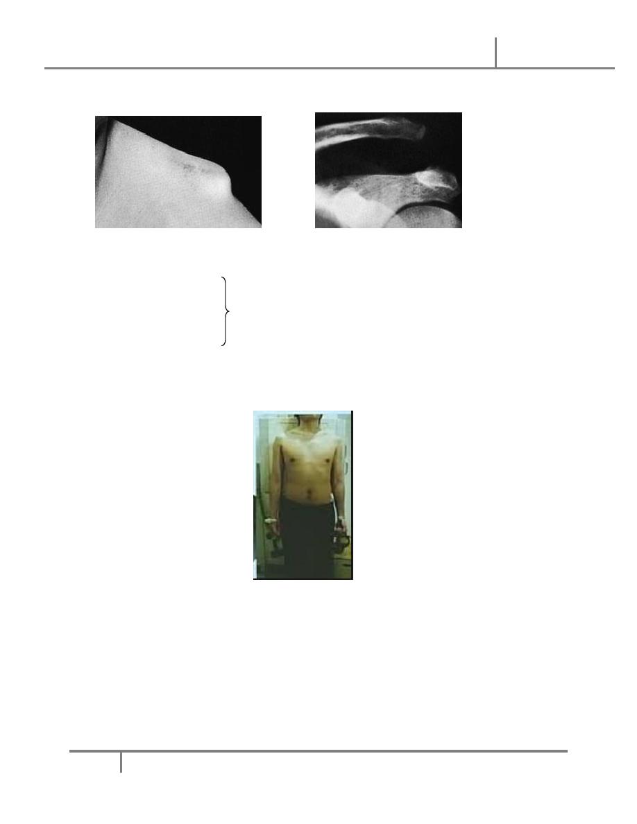
• point to the site of injury
• area may be bruised.
• If there is tenderness but no deformity, the injury is probably a sprain or
a subluxation.
• With dislocation the patient is in severe pain and a prominent ‘step’ can
be seen and felt.
Injuries of the shoulder Dr. Ammar
16-10-2016
9
@Mustafa Naser 2016-2017
• Shoulder movements are limited.
X-ray:
• anteroposterior,
• cephalic tilt and are advisable.
• axillary views
• a stress view is sometimes helpful in distinguishing between a Type II
and Type III injury
Treatment:
• Sprains and subluxations the arm is rested in a sling until pain subsides
(usually no more than a week) and shoulder exercises are then begun.
• conservative treatment for a straight forward Type III injury.
• Operative repair should be considered only for
Injuries of the shoulder Dr. Ammar
16-10-2016
10
@Mustafa Naser 2016-2017
– patients with extreme prominence of the clavicle,
– those with posterior or inferior dislocation of the clavicle and
– those who aim to resume strenuous overarm or overhead activities.
• Mechanism of injury:
– uncommon injury
– lateral compression of the shoulders;
– rarely, it follows a direct blow to the front of the joint.
– Anterior dislocation is much more common than posterior.
– The joint can be sprained, subluxed or dislocated.
Clinical features:
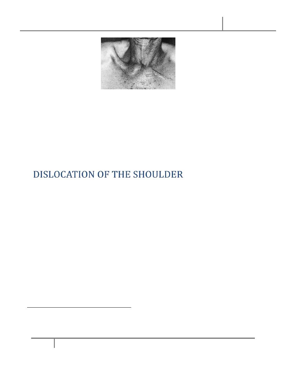
• Anterior dislocation
– prominent bump over the sternoclavicular joint.
– painful
– no cardiothoracic complications.
• Posterior dislocation, though rare, is much more serious.
– Discomfort is marked; there may be pressure on the trachea or
large vessels, causing venous congestion of the neck and arm
and circulation to the arm may be decreased.
• X-Ray: plain x-rays are difficult to interpret. Special oblique views are
helpful and CT is the ideal method.
Injuries of the shoulder Dr. Ammar
16-10-2016
11
@Mustafa Naser 2016-2017
Treatment:
• Anterior dislocation
• Posterior dislocation should be reduced as soon as possible.
• After reduction, the shoulders are braced back with a figure-of-eight
bandage, which is worn for 3 weeks.
• AETIOLOGY: Of the large joints, the shoulder is the one that most
commonly dislocates. This is due to a number of factors:
– the shallowness of the glenoid socket;
– the extraordinary range of movement;
– underlying conditions such as ligamentous laxity or glenoid dysplasia;
and
– the sheer vulnerability of the joint during stressful activities of the upper
limb.
ANTERIOR DISLOCATION
• Mechanism of injury:
– Dislocation is usually caused by a fall on the hand.
Injuries of the shoulder Dr. Ammar
16-10-2016
12
@Mustafa Naser 2016-2017
– The head of the humerus is driven forward, tearing the capsule
and producing avulsion of the glenoid labrum (the Bankart
lesion). Occasionally the posterolateral part of the head is
crushed
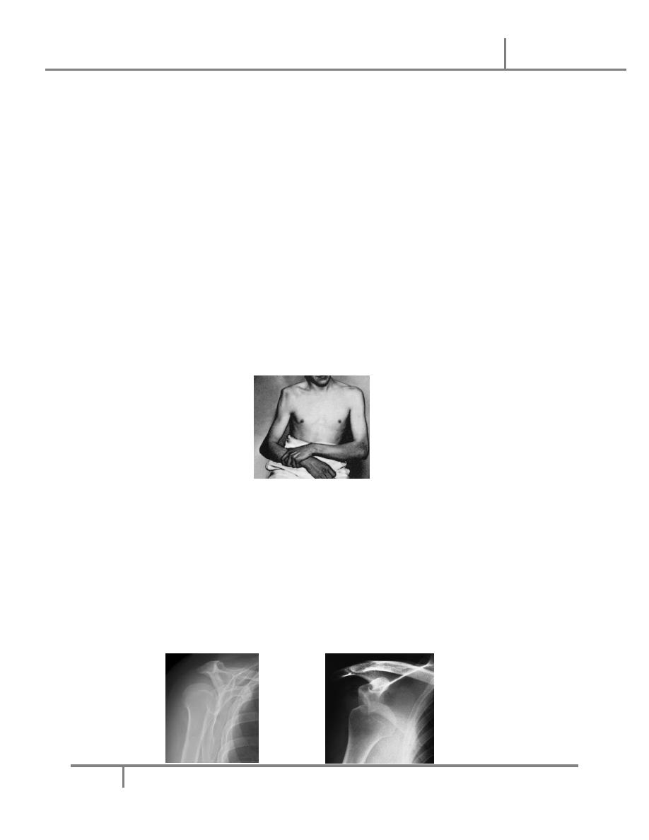
Clinical features:
• Pain is severe.
• The patient supports the arm with the opposite hand and is loath to
permit any kind of examination.
• The lateral outline of the shoulder may be flattened and,
• The arm must always be examined for nerve and vessel injury before
reduction is attempted.
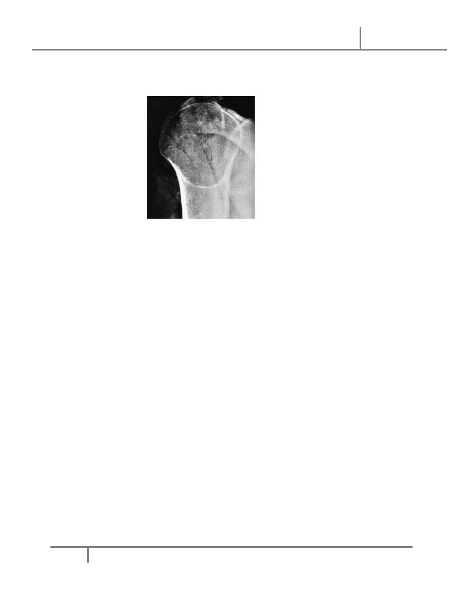
X-Ray:
• The anteroposterior x-ray will show the overlapping shadows of the
humeral head and glenoid fossa, with the head usually lying below and
medial to the socket.
• A lateral view aimed along the blade of the scapula will show the
humeral head out of line with the socket.
Injuries of the shoulder Dr. Ammar
16-10-2016
13
@Mustafa Naser 2016-2017
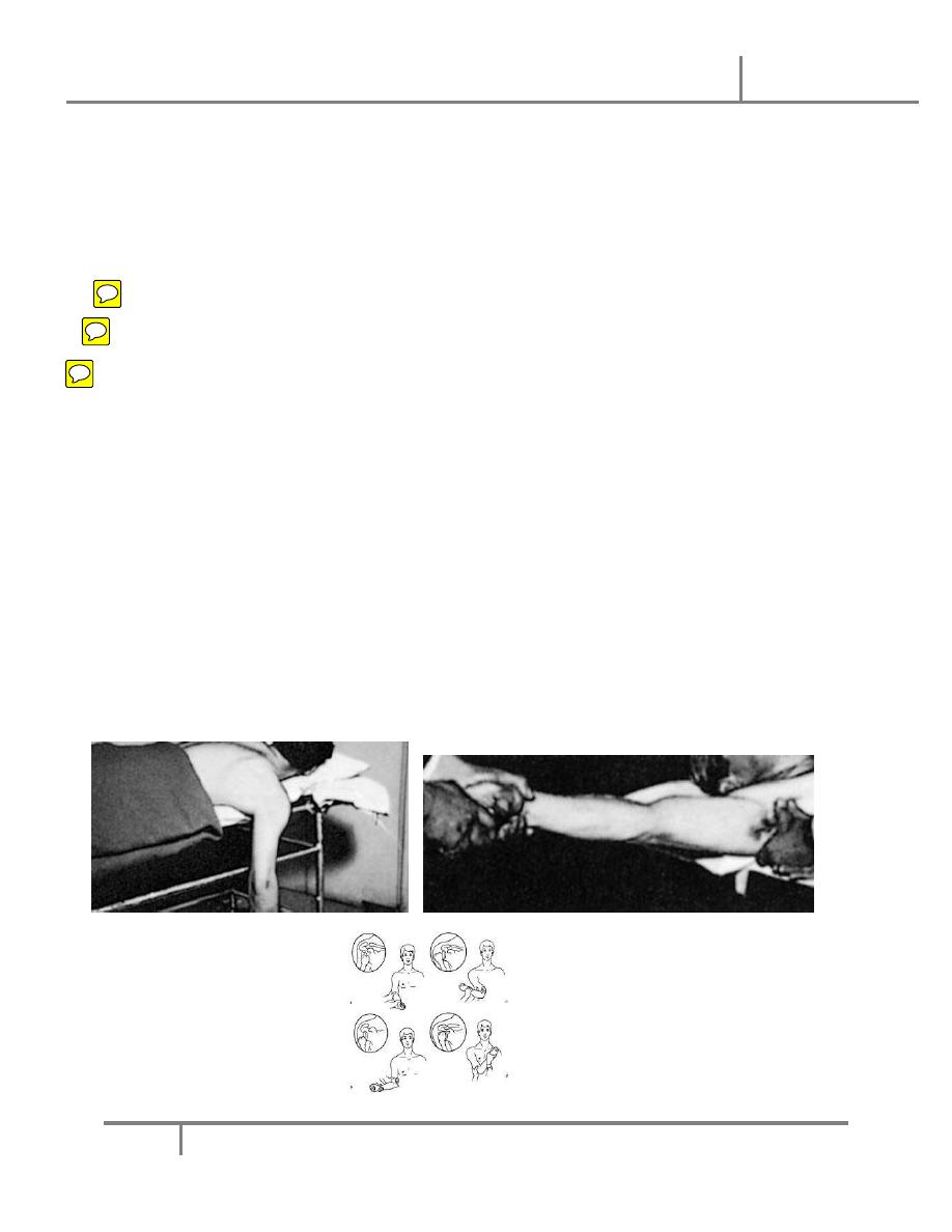
Treatment:
• In a patient who has had previous dislocations, simple traction on the
arm may be successful. Usually, sedation and occasionally general
anaesthesia is required.
• Stimson’s technique,
• Hippocratic method,
• Kocher’s method,
• An x-ray is taken to confirm reduction and exclude a fracture.
• When the patient is fully awake, active abduction is gently tested to
exclude an axillary nerve injury and rotator cuff tear. The median, radial,
ulnar and musculocutaneous nerves are also tested and the pulse is felt.
• The arm is rested in a sling for about three weeks in those under 30
years of age (who are most prone to recurrence) and for only a week in
those over 30 (who are most prone to stiffness).
• Then movements are begun, but combined abduction and lateral
rotation must be avoided for at least 3 weeks.

Injuries of the shoulder Dr. Ammar
16-10-2016
14
@Mustafa Naser 2016-2017
Complications:
• EARLY:
– Rotator cuff tear:
– Nerve injury: The axillary nerve is most commonly injured. Occasionally
the radial nerve, musculocutaneous nerve, median nerve or ulnar nerve
can be injured.
– Vascular injury: The axillary artery may be damaged, particularly in old
patients with fragile vessels.
– Fracture-dislocation:
• LATE
– Shoulder stiffness
– Unreduced dislocation:
– Recurrent dislocation:
POSTERIOR DISLOCATION OF THE SHOULDER
• rare, accounting for < 2 % of all dislocations around the shoulder.
• Mechanism of injury:
– Indirect force producing marked internal rotation and adduction
– Posterior dislocation can also follow a fall on to the flexed, adducted
arm.
•
Clinical features:
The diagnosis is frequently missed
– The arm is held in internal rotation and is locked in that position.
– The front of the shoulder looks flat with a prominent coracoid, but
– swelling may obscure this deformity;
Injuries of the shoulder Dr. Ammar
16-10-2016
15
@Mustafa Naser 2016-2017
– seen from above, however, the posterior displacement is usually
apparent.
•
X-Ray:
– AP film (like an electric light bulb), (the ‘empty glenoid’ sign).
– A lateral film and axillary view is essential; it shows posterior subluxation
or dislocation and sometimes a deep indentation on the anterior aspect
of the humeral head.
•
Treatment:
– The acute dislocation is reduced (usually under general anaesthesia)
– If reduction feels stable the arm is immobilized in a sling; otherwise the
shoulder is held widely abducted and laterally rotated in an airplane type
splint for 3
–6 weeks to allow the posterior capsule to heal in the shortest
position.
– Shoulder movement is regained by active exercises.
•
Complications
– Unreduced dislocation:
Injuries of the shoulder Dr. Ammar
16-10-2016
16
@Mustafa Naser 2016-2017
• At least half the patients with posterior dislocation have ‘unreduced’
lesions when first seen up to two thirds of posteriordislocations are
not recognised initially.
• Typically the patient holds the arm internally rotated; he cannot
abduct the arm more than 70
–80 degrees, and if he lifts the
extended arm forwards he cannot then turn the palm upwards.
• If the patient is young, or is uncomfortable and the dislocation fairly
recent, open reduction is indicated. Late dislocations, especially in
the elderly, are best left, but movement is encouraged.
– Recurrent dislocation or subluxation: Chronic posterior instability of the
shoulder is discussed later
Done By: MustafaNaser