
Temporal, Infratemporal
and Pterygopalatine fossae
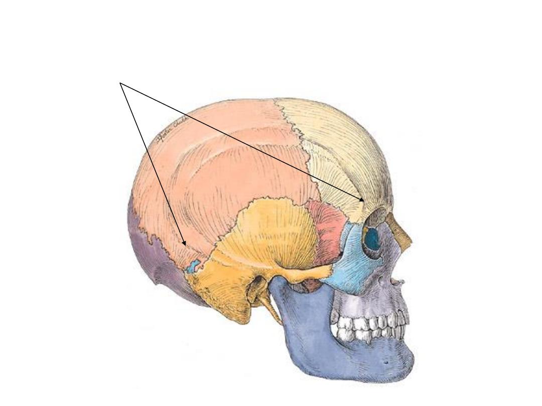
Temporal Fosaa -boundaries

Temporal Fossa - Contents
1. Temporal muscle
2. Temporal fascia (overlies the temporalis muscle)
3. Superficial temporal artery (br. of external
carotid)
4. Superficial temporal vein (unites with the
maxillary vein to form the retromandibular vein)
5. Auriculotemporal nerve (br. of mandibular nerve
which is a br of the trigeminal nerve)
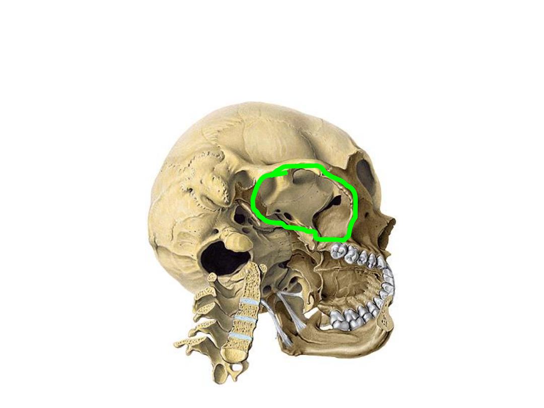
Infratemporal Fossa - boundaries
Boundaries of the infratemporal fossa.
Boundaries:
Lateral=Ramus of Mandible
Anterior=Maxilla
Medial=Lat. Pterygoid Plate
Roof=Sphenoid
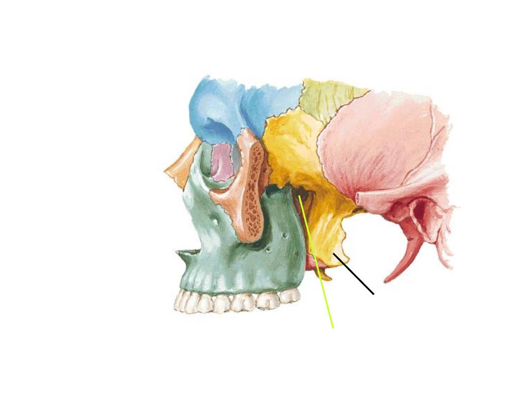
Temporal
Maxilla
Parietal
Sphenoid
Frontal
Z
Lat. Pterygoid Plate
Pterygomaxillary Fissure
Infratemporal Fossa

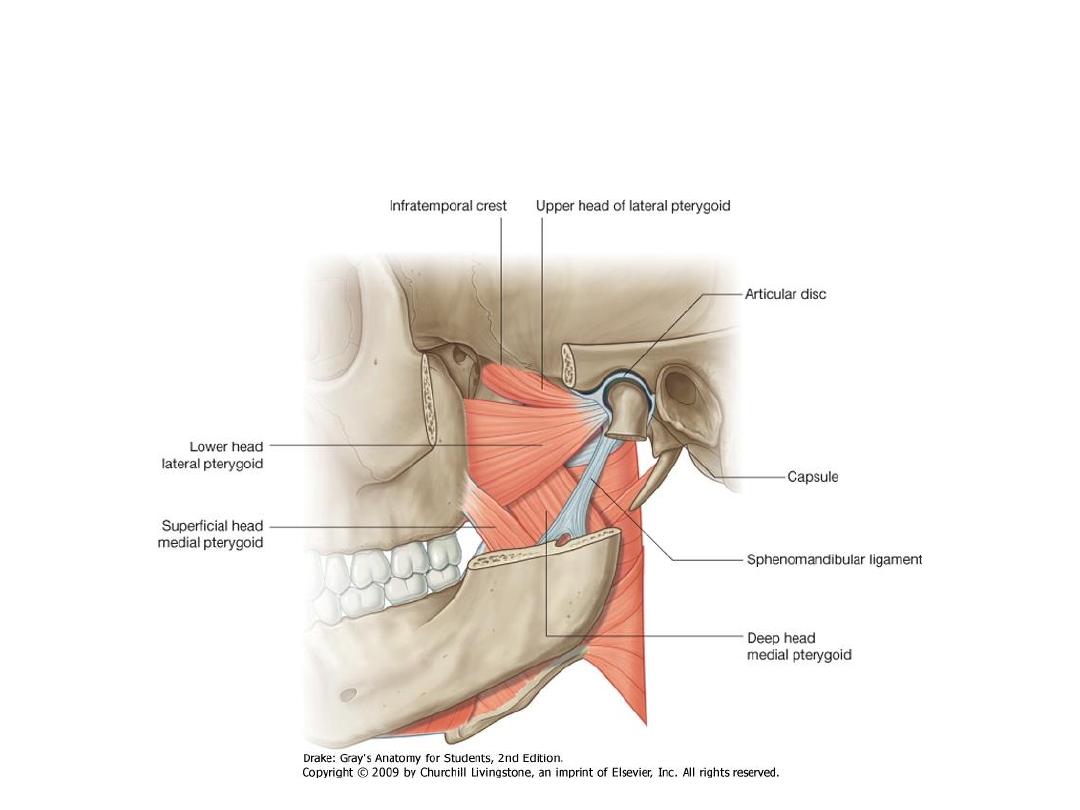
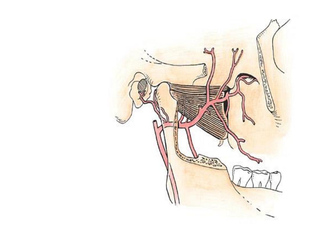
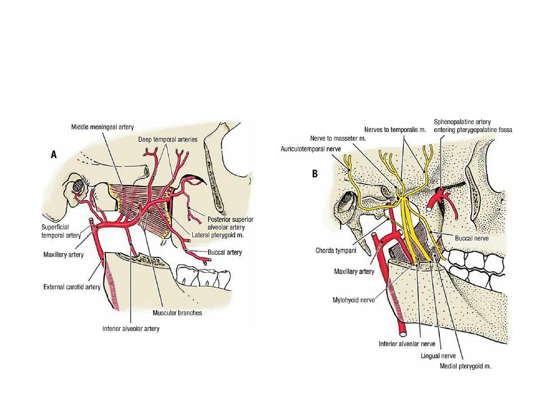
Infratemporal Fossa Contents
• Parotid gland (glenoid process)
• Maxillary artery
• Pterygoid venous plexus
• Otic PS ganglion
• Muscles of mastication
• Mandibular nerve
• Sphenomandibular ligament
Infratemporal Fossa - content
1
st
part
• Deep auricular
• Anterior tympanic
• Middle meningeal
• Accessory meningeal
• Inferior alveolar
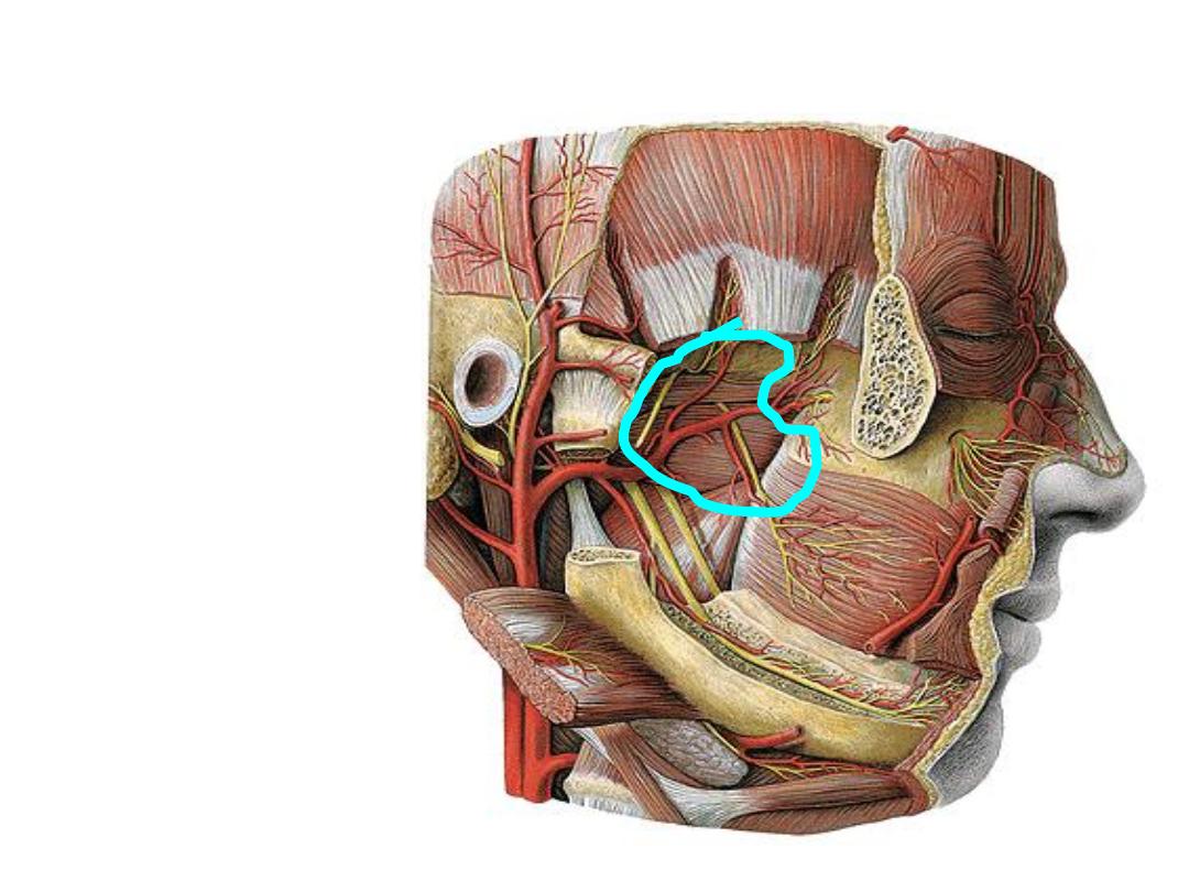
2
nd
part
• Masseteric
• Anterior deep temporal
• Posterior deep temporal
• Pterygoid
• Buccal
3
rd
part
• Posterior superior alveolar
• Infraorbital
• Descending palatine
• Artery of pterygoid canal
• Pharyngeal
• Sphenopalatine

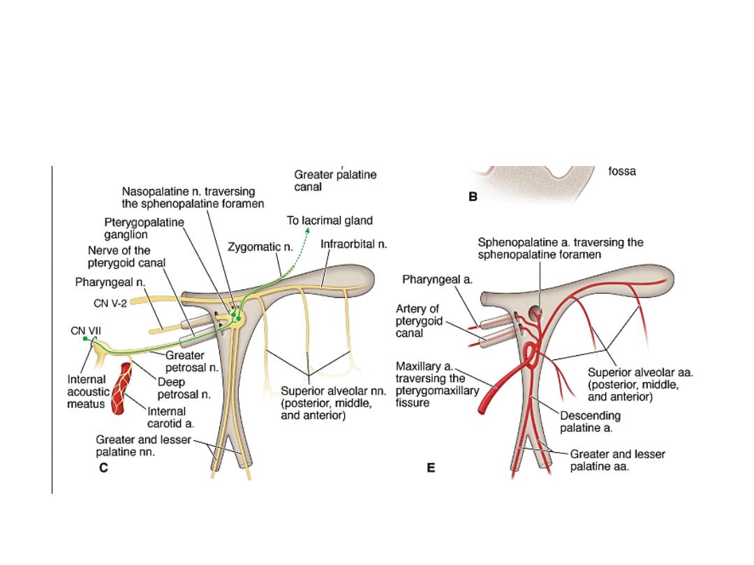
Pterygopalatine fossa – arteries and nerves
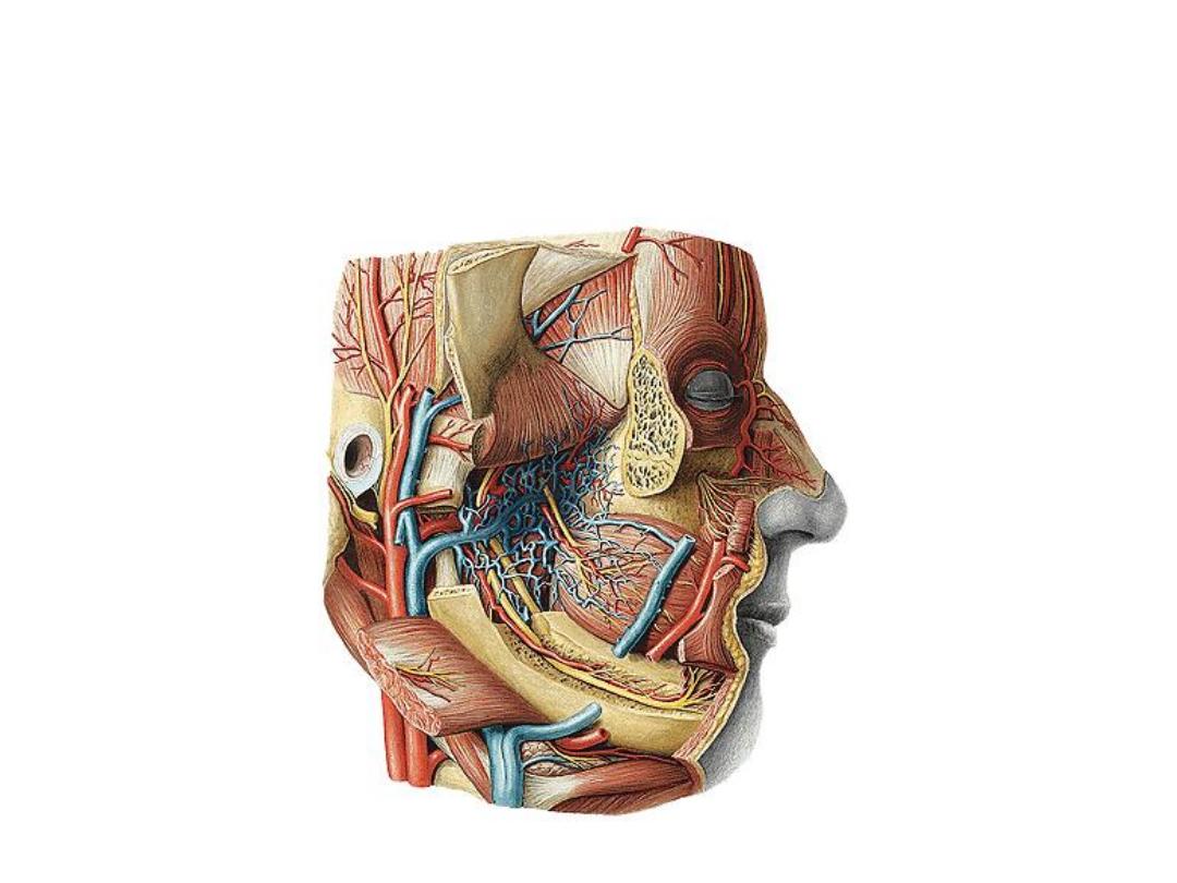
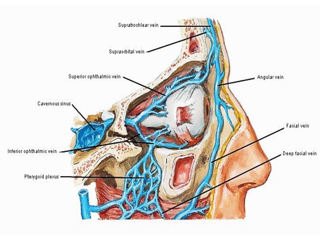
Pterygoid Venous Plexus

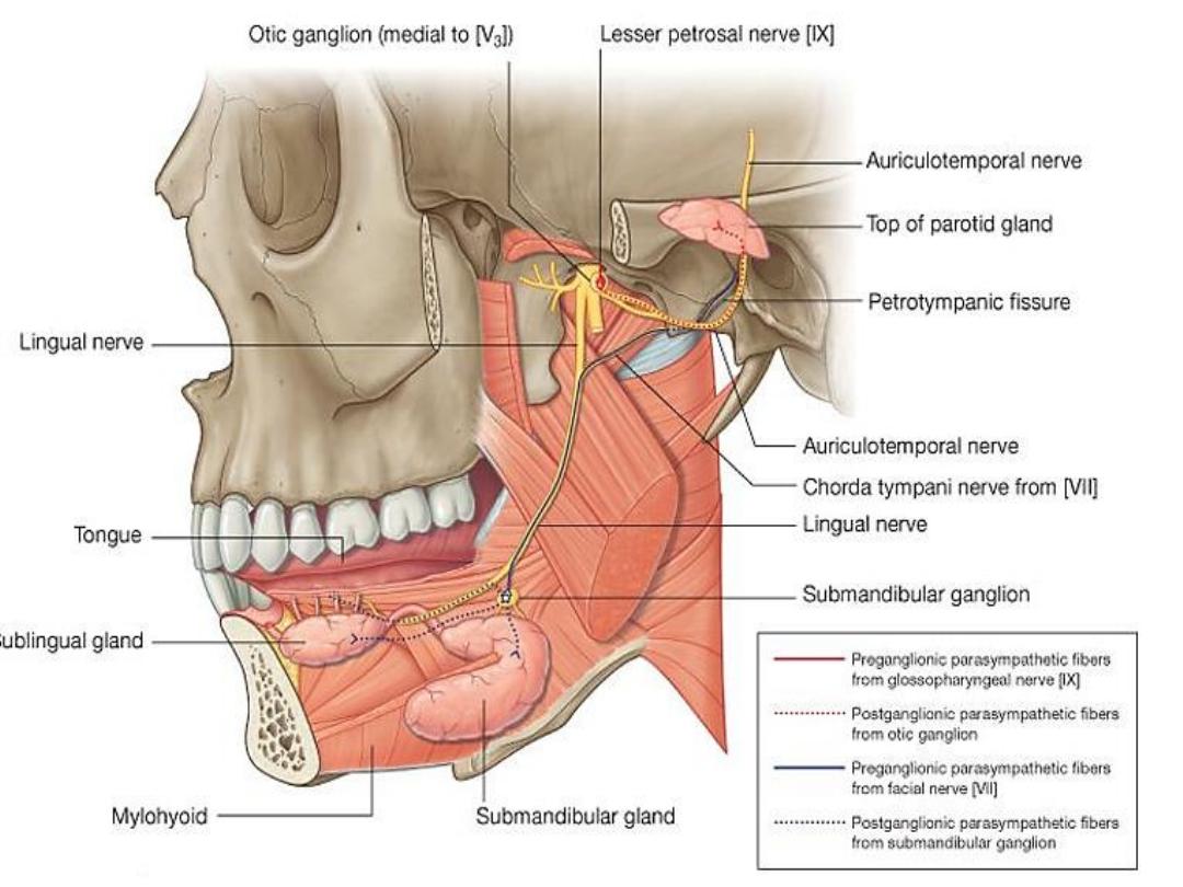
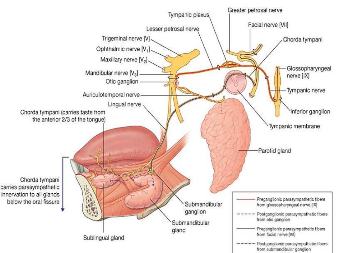
Otic ganglion
(parasympathetic)
Located in the infratemporal fossa, just inferior to
the foramen ovale.
Presynaptic parasympathetic
fibers
, derived mainly from the
glossopharyngeal
nerve
(via the lesser petrosal nerve), synapse in
the otic ganglion.
Postsynaptic parasympathetic fibers
, secretory to
the parotid gland, pass from the otic ganglion to
this gland through the
auriculotemporal nerve
.
glossopharyngeal nerve
(via the lesser petrosal nerve)
auriculotemporal nerve
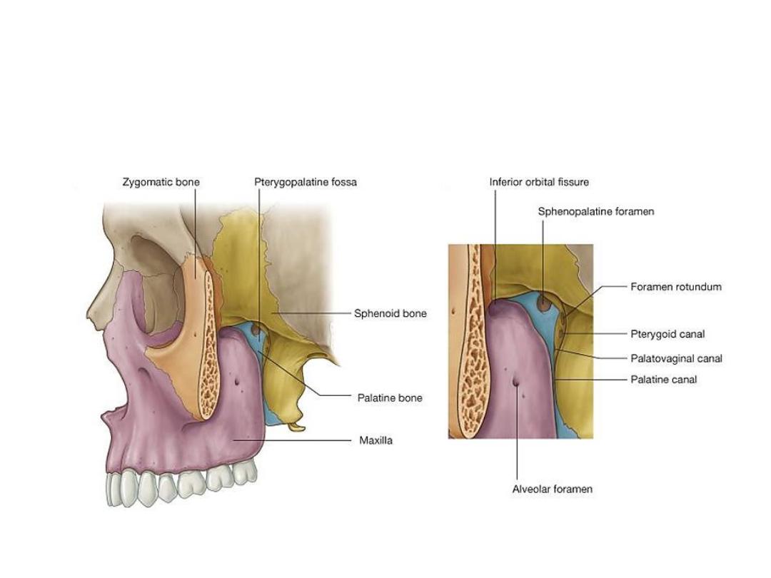
Pterygopalatine fossa
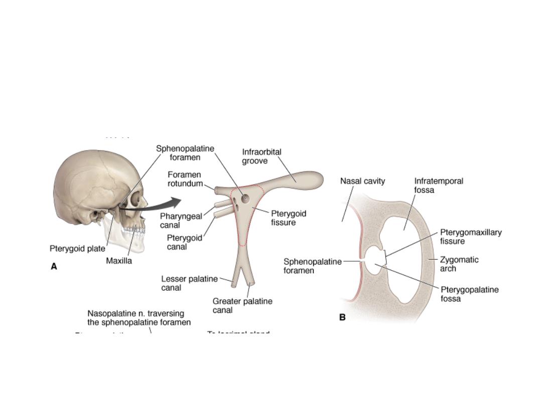
Pterygopalatine fossa – arteries and nerves
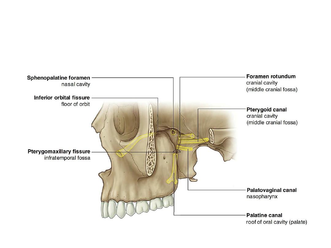
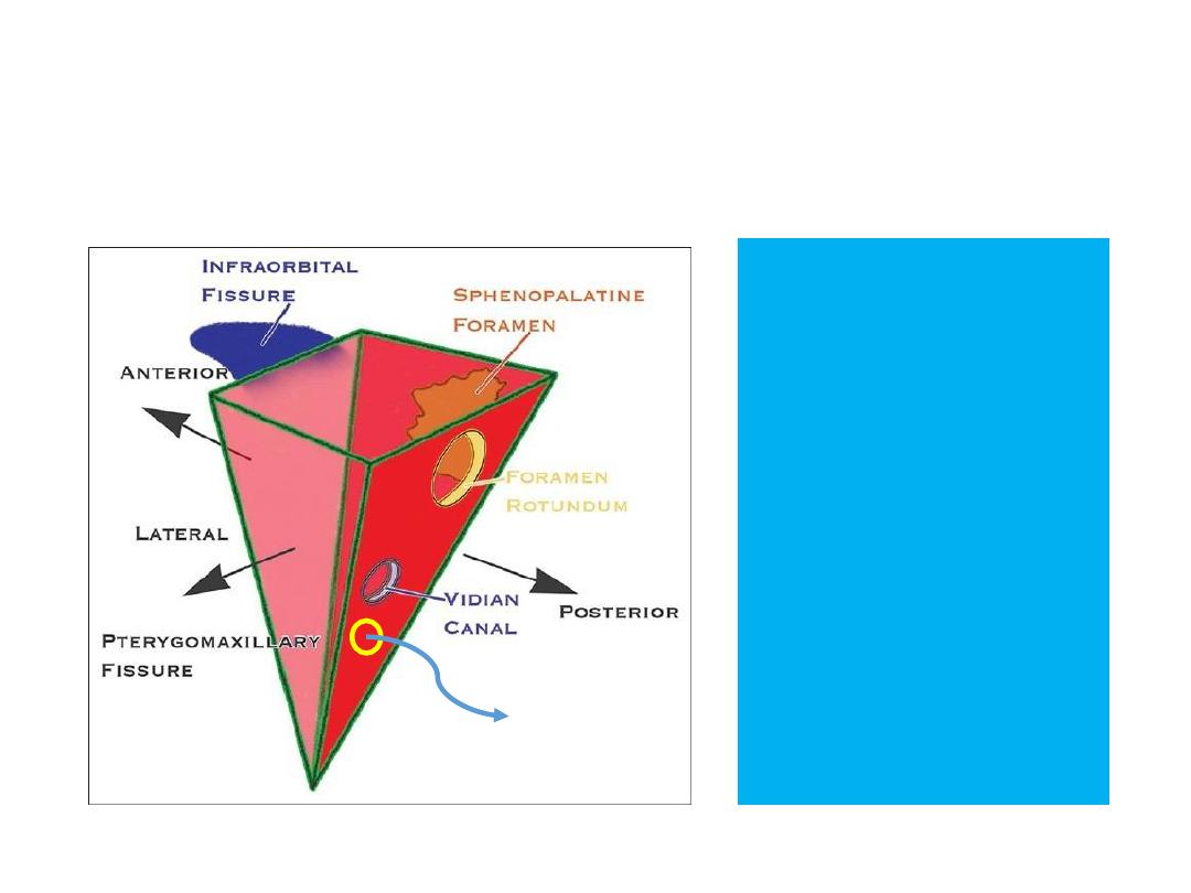
Pterygopalatine fossa - connections
1.
foramen rotundum
& pterygoid canal
middle cranial fossa
2.
palatovaginal canal
nasopharynx
3.
palatine canal leads
oral cavity (hard
palate)
4.
sphenopalatine
foramen
nasal cavity
5.
pterygomaxillary
fissure
infratemporal fossa
6.
inferior orbital
fissure
orbit
palatovaginal canal
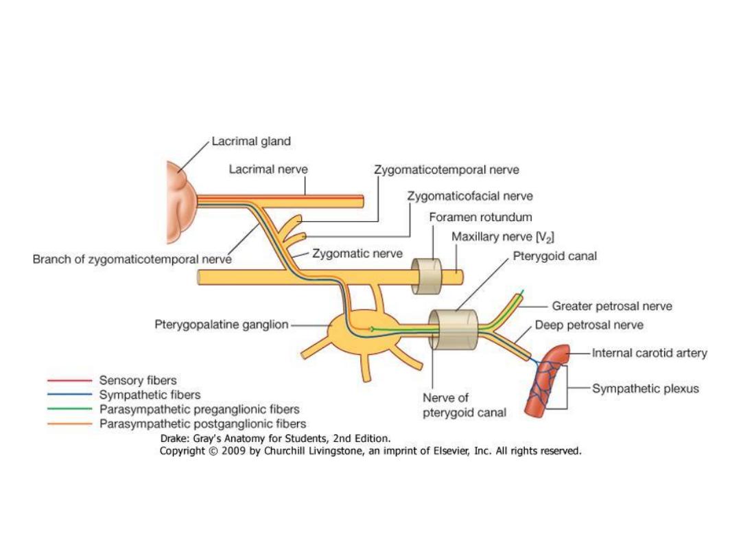
Pterygopalatine ganglion
Pterygopalatine Ganglion
(Ganglion pterygopalatinum, Meckel's ganglion, Nasal ganglion, Sphenopalatine ganglion)
Largest of the 4 PS ganglia
in the head
Formed by the cell bodies
of the postganglionic
neurons associated with
preganglionic
parasympathetic fibers of
the facial nerve [VII] carried
by the greater petrosal
nerve and the nerve of the
pterygoid canal.
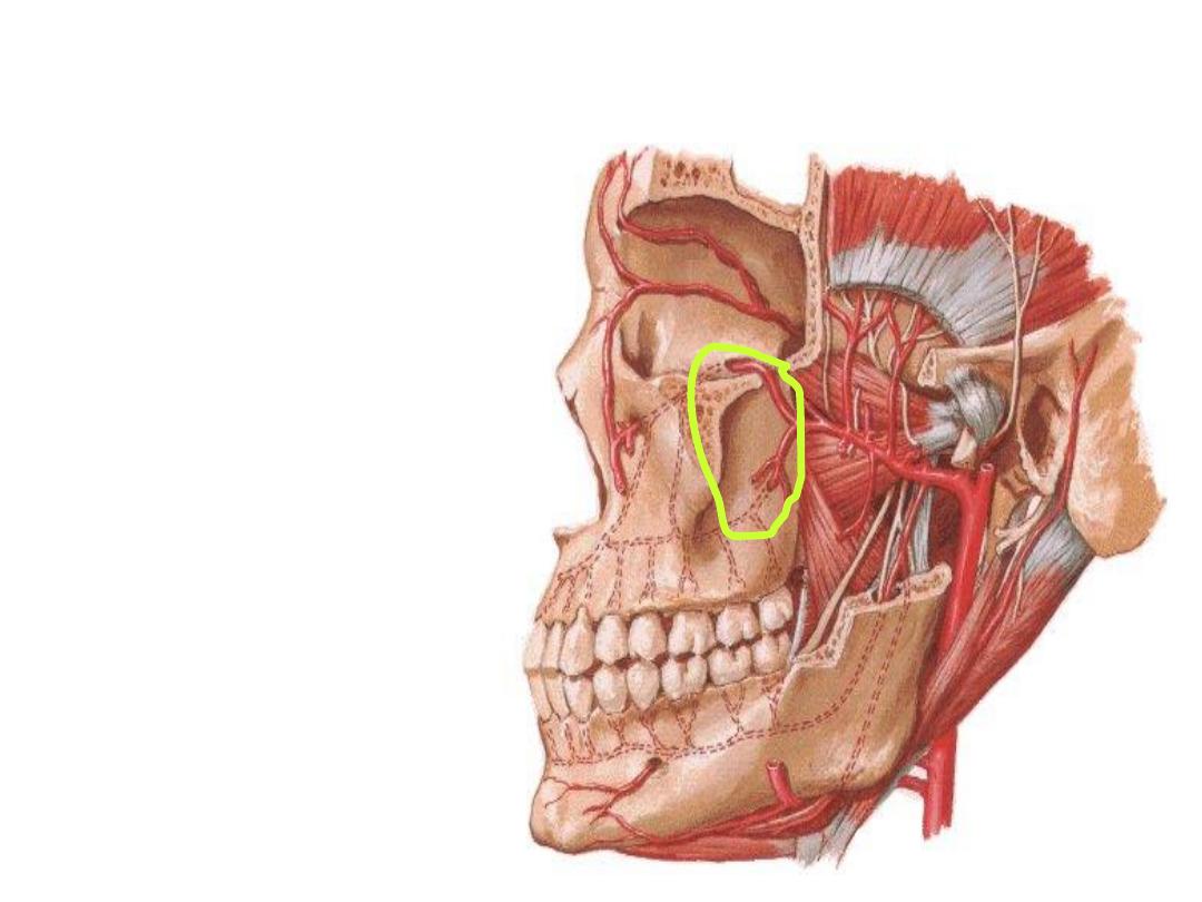
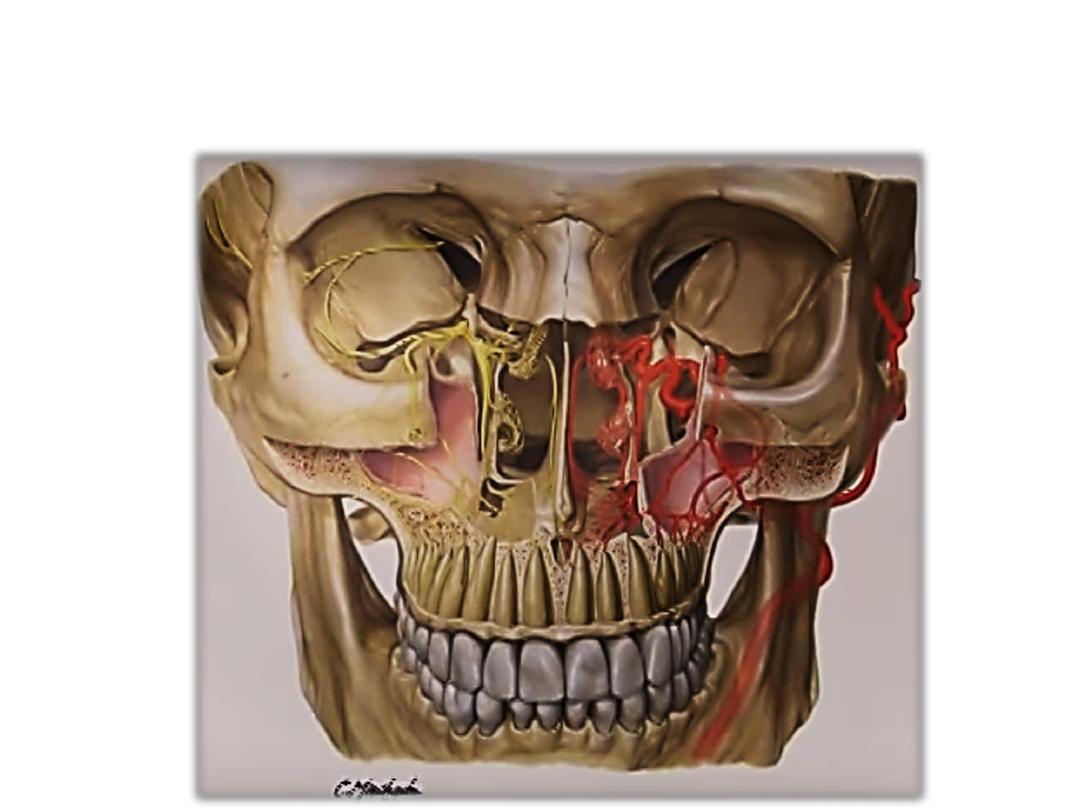
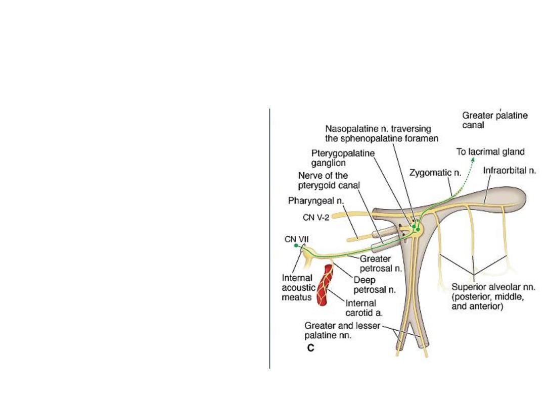
Maxillary artery and Nerve
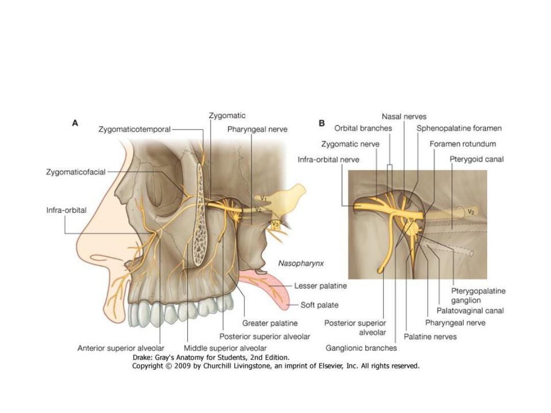
Maxillary Nerve and
Pterygopalatine ganglion
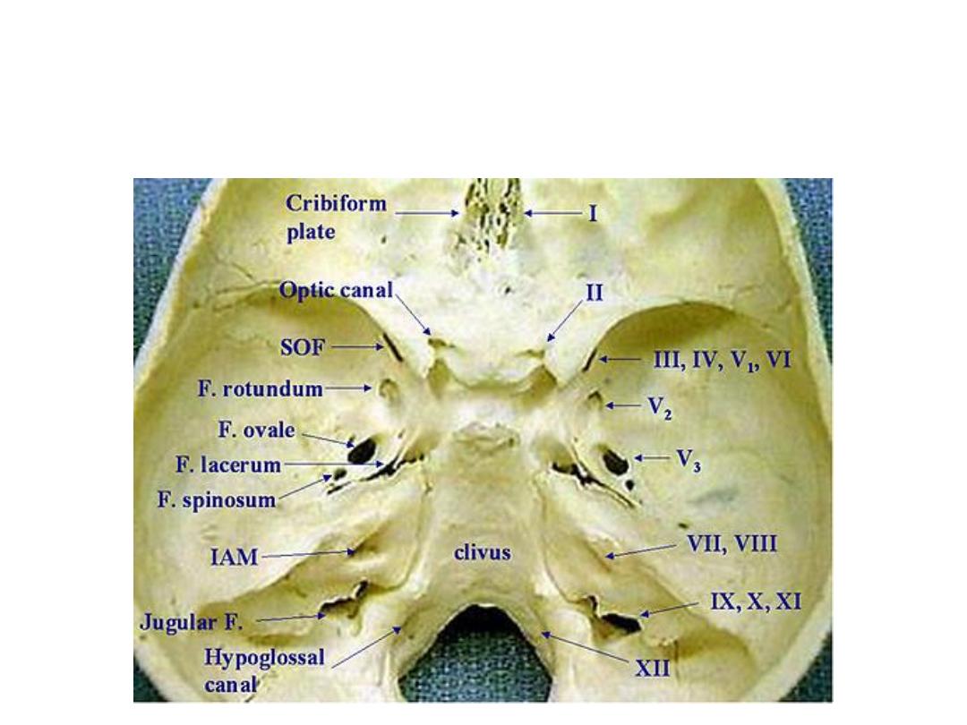
To those who want to study skull foramina!