
Lecture
Organs of the Immune
System
Dr. Nazik M. Hussein
The lymphoid organs and tissues are classified
into primary ( central ) and secondary( peripheral).
Lymphocytes are produced in the primary
lymphoid organs and function within the
secondary lymphoid organs and tissues.
Primary lymphoid organs: Immature lymphocytes
generating in hematopoiesis, mature and become
committed to a particular antigenic specificity
within the primary lymphoid organs. In mammals,
T cells mature in the thymus and B cells mature in
the bone marrow.
1.
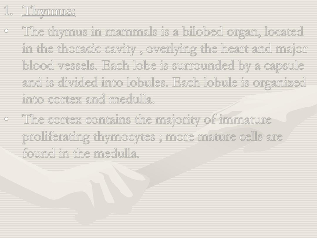
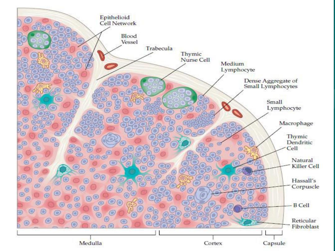
Thymus:
•
The thymus in mammals is a bilobed organ, located
in the thoracic cavity , overlying the heart and major
blood vessels. Each lobe is surrounded by a capsule
and is divided into lobules. Each lobule is organized
into cortex and medulla.
•
The cortex contains the majority of immature
proliferating thymocytes ; more mature cells are
found in the medulla.
•
At least three types of epithelial cells can be
distinguishes in the thymic lobules: the epithelial
nurse cells are in the outer cortex, the cortical
epithelial cells, and the medullary epithelial cells.
In addition, interdigitating dendritic cells(IDC)
and macrophages are found in thymic lobules,
particularly at the corticomedullary junction.
•
The mammalian thymus involutes with age. In
man, atrophy begins at puberty and continues
throughout life. Thymic involution begins
within the cortex and this region may disappear
completely, whereas medullary remnants
persist. Cortical atrophy is related to
corticosteroid sensitivity of the cortical
thymocytes. Thus, all conditions associated with
acute increase in steroids, for example
pregnancy and stress, promote thymic atrophy.
2.
Bone marrow: The soft vascular tissue that fills bone
cavities and cancellous bone spaces and consists
primarily of fat cells, hematopoietic cells, and
osteogenetic reticular cells.
Marrow types: There are two types of bone marrow: red
marrow and yellow marrow.
Red blood cells, platelets and most white blood cells
arise in red marrow; some white blood cells develop in
yellow marrow.
Red marrow is found mainly in the flat bones such as
hip bone, skull, ribs, vertebrae and shoulder blades,
and in the cancellous (spongy) material at the proximal
ends of the long bones (femur and humerus).
In mammals B cell maturation occurs in bone marrow.
In birds, a lymphoid organ called the bursa of
Fabricius is the primary site of B cell maturation.
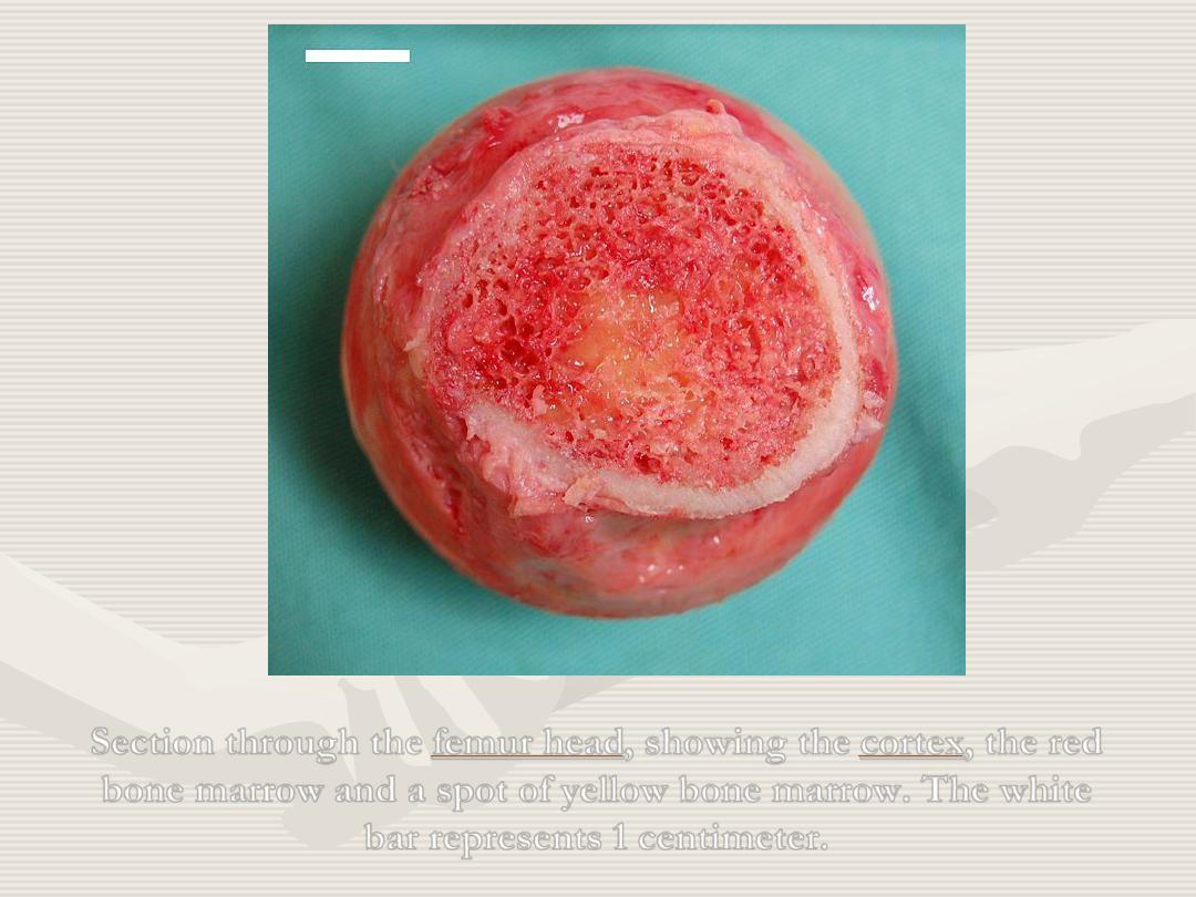
, the red
, showing the
Section through the
bone marrow and a spot of yellow bone marrow. The white
bar represents 1 centimeter.

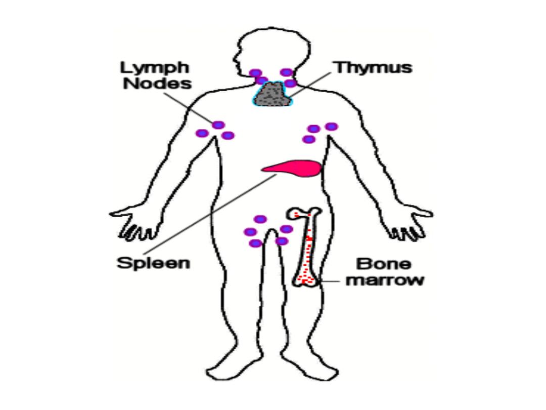
Secondary lymphoid organs and tissues:
•
The secondary lymphoid tissues comprise well
organized encapsulated organs, the spleen and lymph
nodes and non- encapsulated accumulations of
lymphoid tissue. Lymphoid tissue that is found in
association with mucosal surface is called the mucosa-
associated lymphoid tissue (MALT).
•
The spleen is responsive to blood- borne antigens
and patients who have had their spleens removed are
much more susceptible to pathogens that reach the
blood stream. The lymph nodes protect the body
from antigens that come from skin or from internal
surfaces and are transported via the lymphatic vessels.
1.
Spleen:
•
Two main types of splenic tissue: the white pulp and
the red pulp. The white pulp consists of lympoid
tissues, the bulk of which is arranged around a
central arteriole to form the periarteriolar lymphoid
sheaths (PALS).
•
PALS are composed of T and B cell areas. The T
cells are found around the central arteriole; the B
cells may be organized into either primary
(unstimulated) follicles , or secondary (stimulated)
follicles.
•
The red pulp consists of venous sinuses and cellular
cords containing resident macrophages, erythrocytes,
platelets, granulocytes, lymphocytes and numerous
plasma cells.
•
In addition to immunological functions, the spleen
serves as a reservoir for platelets, erythrocytes and
granulocytes. Aged platelets and erythrocytes are
destroyed in the spleen. This process is carried out in
the red pulp and is referred to as haemocatheresis.
Aged platelets and erythrocytes are recognized and
phagocytosed by macrophages.
2.
Lymph nodes:
•
The lymph nodes form part of a network which filters
antigens from the interstitial tissue fluid and lymph
during its passage from the periphery to the thoracic duct
and the other major collecting ducts.
•
Clusters of lymph nodes are strategically placed in areas
such as the neck, axillae, groin, mediastinum and the
abdominal cavity. Lymph nodes protect the skin
(superficial, subcutaneous nodes) and mucosal surfaces of
the respiratory, digestive and genitourinary tracts (visceral
or deep nodes).
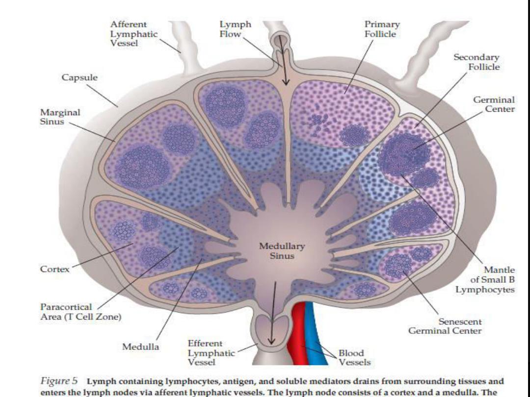
•
Human lymph nodes are round or kidney shaped, and
have an indentation called the hilus where blood
vessels enter and leave the node. Lymph arrives at the
lymph node via several afferent lymphatic vessels, and
leaves the node through one efferent lymphatic vessel
at the hilus.
•
A typical lymph node is surrounded by a collagenous
capsule.
•
The lymph node consists of a B cell area (cortex), a T
cell area (paracortex) and a central medulla consisting
of cellular cords containing T cells , B cells, abundant
plasma cells and macrophages.

3. Mucosa-associated lymphoid tissue (MALT):
• Aggregates of non-encapsulated lymphoid tissue
are found especially in the lamina propria and
submucosal areas of the gastrointestinal,
respiratory and genitourinary tracts.
• The tonsils contains a considerable amount of
lymphoid tissue, often with large secondary
follicles and intervening T cell zones with
endothelial venules.
• Similar aggregates of lymphoid tissue are seen
lining the bronchi and along the genitourinary tract.
•
Gut- associated lymphoid tissue includes Peyer
’s
patches and the appendix.
•
The intestinal epithelium overlying the Peyer's patches
is specialized to allow the transport of pathogens into
the lymphoid tissue. This particular function is carried
out by epithelial cells termed M cells. M cells contain
deep invaginations of the plasma membrane which
form pockets containing B and T lymphocytes,
dendritic cells and macrophages.