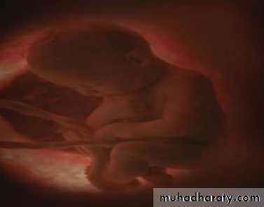
Embryology
What is embryology?
Embryo is a Greek word “Embryon”= unbornThe study of developmental events that occur during the prenatal stage .
The branch of biology and medicine concerned with the study of embryos and their development.INTRODUCTION
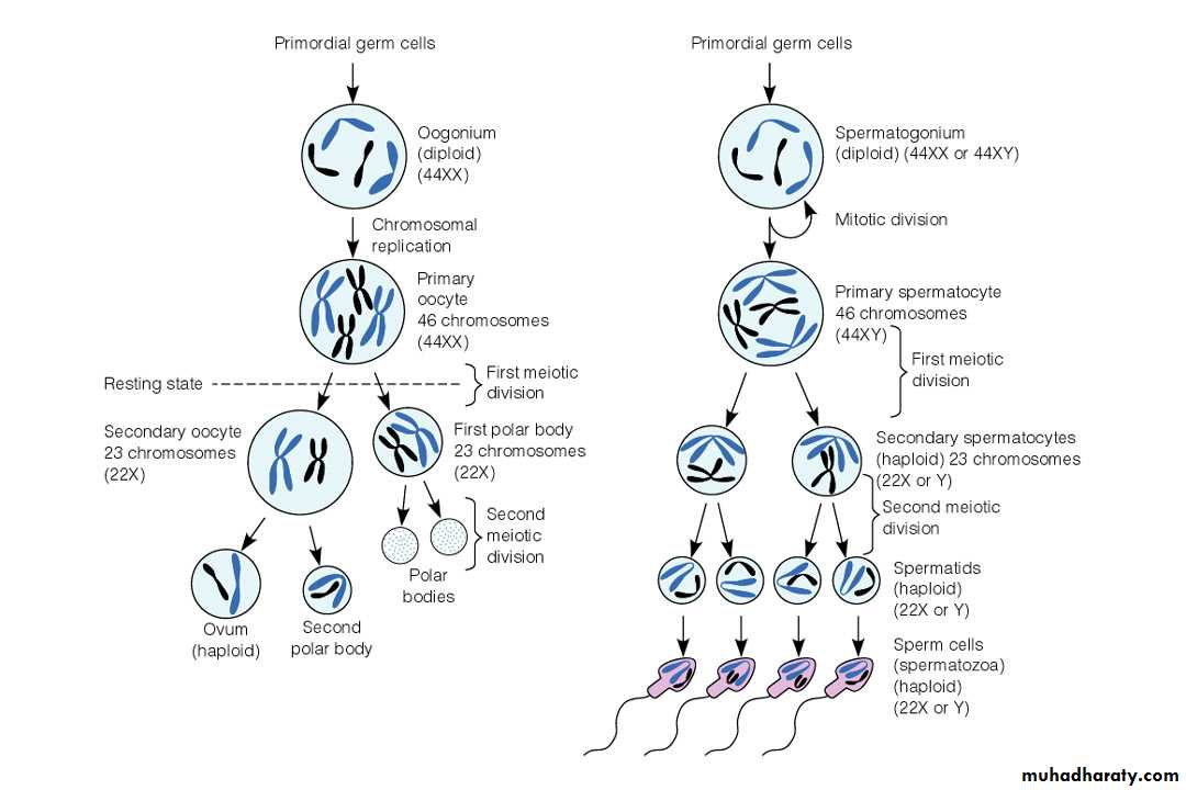
The human somatic cell contains 46 chromosomes, called as the diploid number. Out of which 44 are autosomes and the remaining 2 are sex chromosomes, designated as X and Y.The sex chromosomes in females are XX and in males are XY.
There are two series of division of somatic cells- MITOSIS and MEIOSIS.
MITOSIS produces the same number of chromosomes in the resulting daughter cell while MEIOSIS produces half the number i.e. 23 designated as haploid, with resultant formation of gametes .
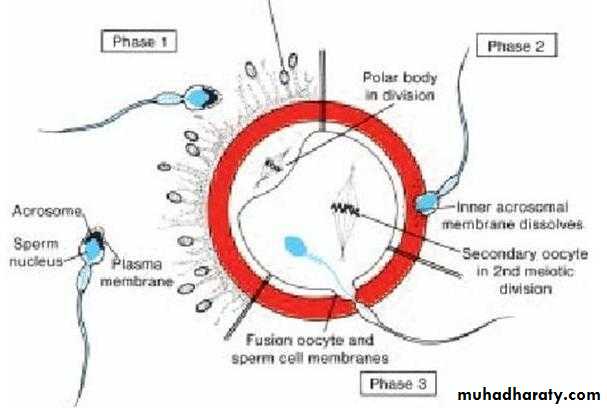
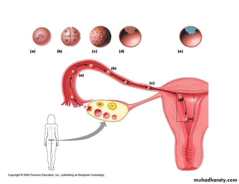
Development begins with FERTILIZATION, the process in which the male gamete- the sperm, and the female gamete- the oocyte, unite to form a ZYGOTE.
Embryonic period vs. Fetal period
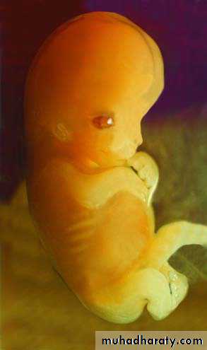
Embryonic – first 8 weeks. Development of the three primary germ layers give rise to all structures and Basic body plan takes shapeFetal period – remaining 30 weeks. Structures and organs continue to grow and develop.
Stages of Development
• 1. Fertilization• 2. Cleavage
• 3. Gastrulation Embryogenesis
• 4.Organogenesis
• 5.Maturation
1- Fertilization
The process of fusion or union of the spermatozoon with the mature ovum is known as conception / fertilizaiton/impregnantation.
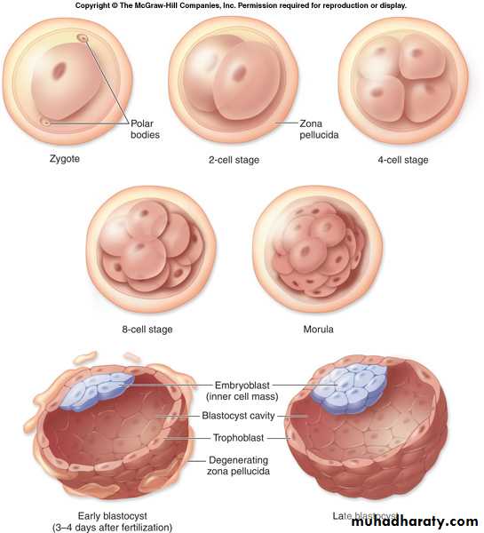
Which produced the fertilized single mono-nucleated cell called the zygote.
The sperm and ovum contribute half the complement of chromosomes to make a total of 46.The sperm and ovum is known as the male and female gametes and the fertilized ovum as the zygote
THE PROCESS OF FERTILIZATIONTHE MALE GAMETE (SPERM) FUSES WITH THE FEMALE GAMETE (OVUM)
2- Cleavage
Day 2 and 3: CleavageCleavage is a series of rapid mitotic divisions (without cell growth)
The original zygote divides about 30 hours after conception into two daughter cells called blastomeres.
Continued subdivisions of the original cell result in increasing numbers of blastomeres.
During cell division the dividing cells decrease in size. This type of cell division is called cleavage.
Morula
• By the time the zygote is ready to enter the uterus, it contains a solid ball of 16 blastomeres called the morula (from the latin word for mulberry).• The blastomeres continue to divide by binary division through 4, 8, and 16 cell stage until a cluster of cells is formed– Morula, resemblibg a mulberry.
Morula
Morula
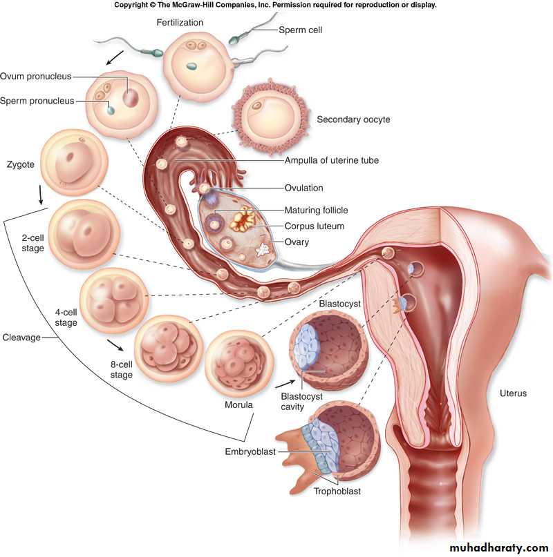
Day 4: Formation of the blastocyst
Fluid within the intercellular spaces of the morula gradually increases, and spaces on one side of the inner cell mass come together, forming a single cavity, the blastocele.The cavity of the blastocele fills with fluid, and the conceptus is now called the blastocyst.


Blastocele
cavity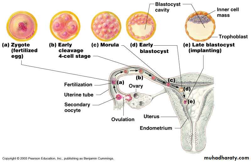
Blastocyst
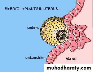
The cell of the outer cell mass forms the wall of the blastocyst and is known as trophoblast.The inner cell mass is concerned with the development of the embryo.
Two Distinct Cell Types
• 1. An outer cell layer, the trophoblast.• 2. An inner cell mass, the embryoblast.
•
Trophoblast
EmbryoblastBlastocyst
Trophoblast Inner cell mass (embryoblast)Placenta Chorion Fetus Amnion umbilical cord
IMPLANTATION : 6TH DAYThe trophoblast attaches to the sticky endometrial surface on the posterior wall of the body of the uterus.
2nd week
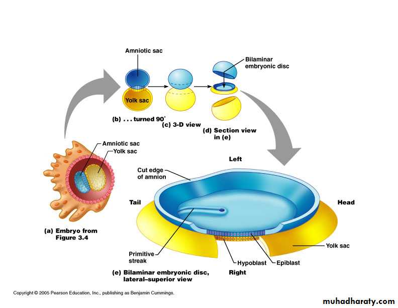
Embryoblast forms two layers:epiblast and hypoblast.
Together they make up the bilaminar embryonic disc.
Formation of 2 cavities:amniotic and yolk sac cavities.
21
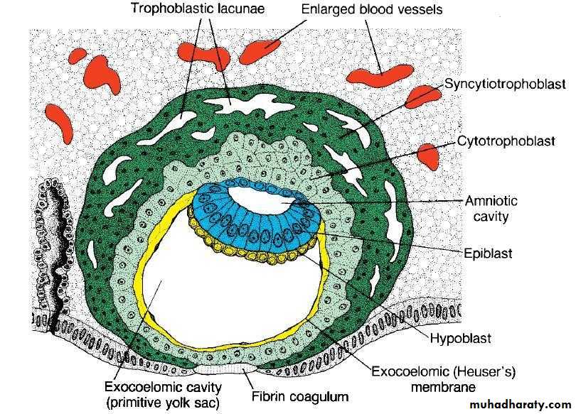
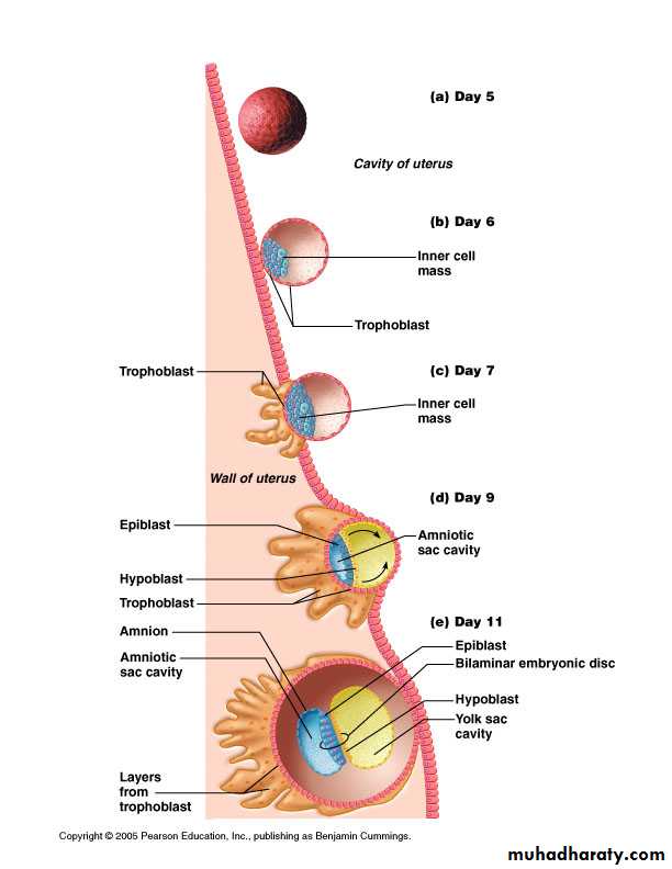
Week 2
2 fluid filled sacs
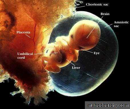
1. Amniotic sac from epiblast within which the embryo and later the fetus develop until birth.
2. Yolk sac from hypoblast which is one of the structures through which the mother supplies nutrients to the early embryo.
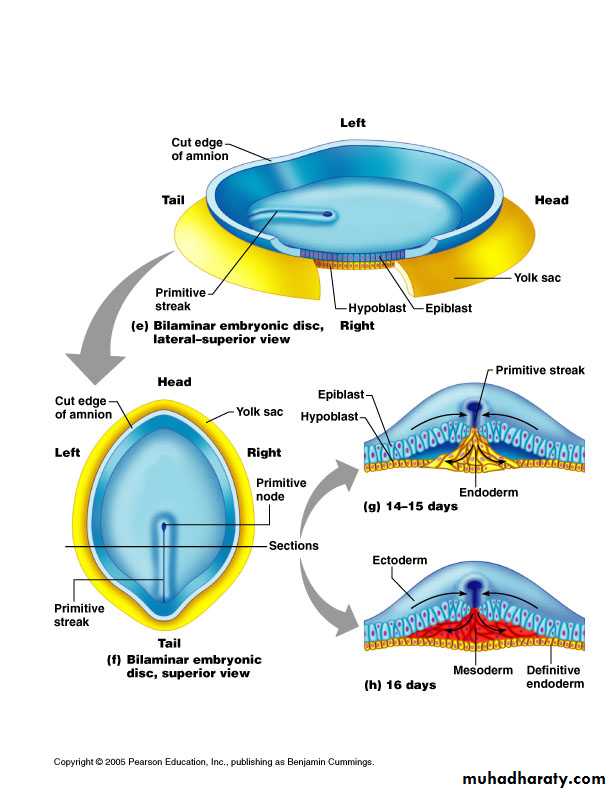
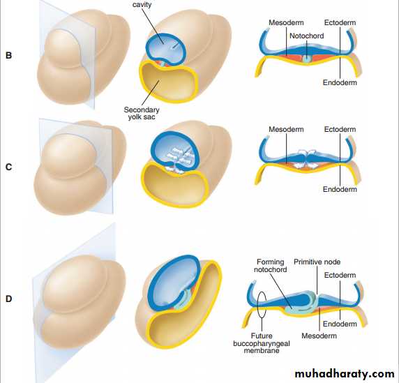
3. Gastrulation
The process by which the bilaminar disc is converted to a trilaminar disc (known as the ectoderm, mesoderm, and endoderm)Consist of the formation of primitive streak, three germ layer and the notochord.
The three germ layers*
1. Endoderm—formed from migrating cells that replace the hypoblast2. Mesoderm
3. Ectoderm—formed from epiblast cells that stay on dorsal surface
*All layers derive from epiblast cells
Bilaminar to trilaminar disc
Three primary “germ” layers: all body tissues develop from theseEctoderm
Endoderm
Mesoderm
Week 3
Each of the three germ layers gives rise to specific tissues and organs:Ectoderm forms the outer layer. Ectoderm forms skin, hair, sweat glands, epithelium, brain and nervous system.
Endoderm forms the inner layer. The endoderm forms digestive, respiratory systems, liver, pancreas, all bladder, and endocrine glands such as thyroid and parathyroid glands.
Mesoderm forms the middle layer. The mesoderm forms body muscles, cartilage, bone, blood, reproductive system organs and kidneys
Neurulation
The process of formation of the neural tube is known as neurulation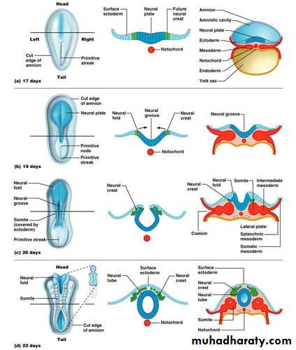
Neurulation
The nervous system develops as a thickening within the ectodermal layer at the rostral end of the embryo. This thickening constitutes the neural plate called neurectoderm, which rapidly forms raised margins (the neural folds).These folds in turn encompass and delineate a deepening midline depression, the neural groove.
The neural folds eventually fuse so that a neural tube separates from the ectoderm.
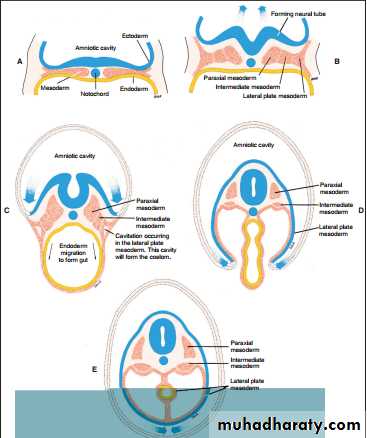
As the neural tube forms, changes occur in the mesoderm adjacent to the tube and the notochord. The mesoderm first thickens on each side of the midline to form:
1- Paraxial mesoderm: Along the trunk of the embryo, this paraxial mesoderm breaks into segmented blocks called somites.
Each somite has three components:
(a) the sclerotome, which eventually contributes to two adjacent vertebrae and their disks;(b) the myotome, which gives origin to a segmented mass of muscle;
(c) the dermatome, which gives rise to the connective tissue of the skin overlying the somite. In the head region, the mesoderm only partially segments to form a series of numbered somatomeres, which contribute in part to the head musculature.
2- Lateral plate mesoderm
Further laterally the mesoderm thickens again to form the lateral plate mesoderm, which gives rise to (1) the connective tissue associated with muscle and viscera; (2) the serous membranes of the pleura, pericardium, and peritoneum; (3) the blood and lymphatic cells; (4) the cardiovascular and lymphatic systems; and (5) the spleen and adrenal cortex.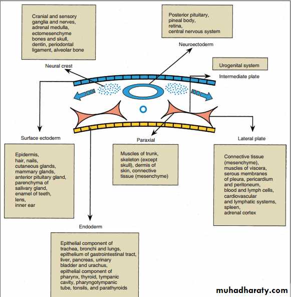
3- Intermediate mesoderm:
At the periphery of the paraxial mesoderm, the mesoderm remains as a thin layer, the intermediate mesoderm, which becomes the urogenital system.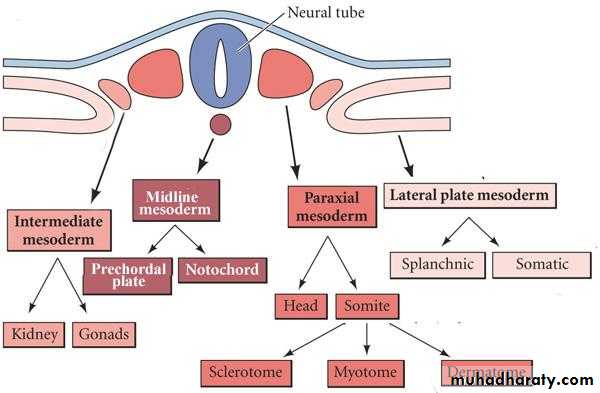
THE NEURAL CREST CELLS
As the neural tube forms, a group of cells separate from the neuroectoderm called neural crest cell. These cells have the capacity to migrate and differentiate extensively within the developing embryo.Neural crest cells in the head region have an important role. In addition to assisting in the formation of the cranial sensory ganglia, they also differentiate to form most of the connective tissue of the head.
Embryonic connective tissue elsewhere is derived from mesoderm and is known
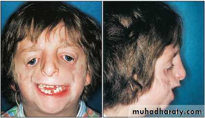
as mesenchyme, whereas in the head it is known as ectomesenchyme, reflecting its origin from neuroectoderm.In a dental context the proper migration of neural crest cells is essential for the development of the face and the teeth. In Treacher Collins syndrome , for example, full facial development does not occur because the neural crest cells fail to migrate properly to the facial region. All the tissues of the tooth (except enamel and perhaps some cementum) and its supporting apparatus are derived directly from neural crest cells, and their depletion prevents proper dental development.