1
Fifth stage
Medicine
Lec-
د.خالد نافع
8/11/2016
Lymphoma
Most present as tumor Involving lymph nodes or other lymphoid organs such as the spleen.
But extra nodal presentation may seen.
1. Hodgkin’s Lymphoma.
2. Non - Hodkin’s Lymphoma; Non-Hodgkin lymphomas are classified as low- or high-
grade tumours on the basis of their proliferation rate.
High-grade tumours divide rapidly, are typically present for a matter of weeks before
diagnosis, and may be life-threatening.
Low-grade tumours divide slowly, may be present for many months before diagnosis,
and typically behave in an indolent fashion.
Hodgkin lymphoma :
2
Clinical features :
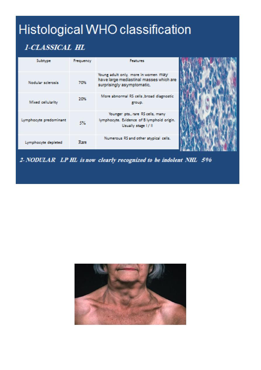
There is painless, rubbery lymphadenopathy, usually in the neck or supraclavicular fossae;
the lymph nodes may flucuate in size(wax an wan).
Hepatosplenomegaly may be present but does not always indicate disease in those organs.
Spread is contiguous from one node to the next and extranodal disease, such as bone, brain
or skin involvement, is rare.
3
Investigations :
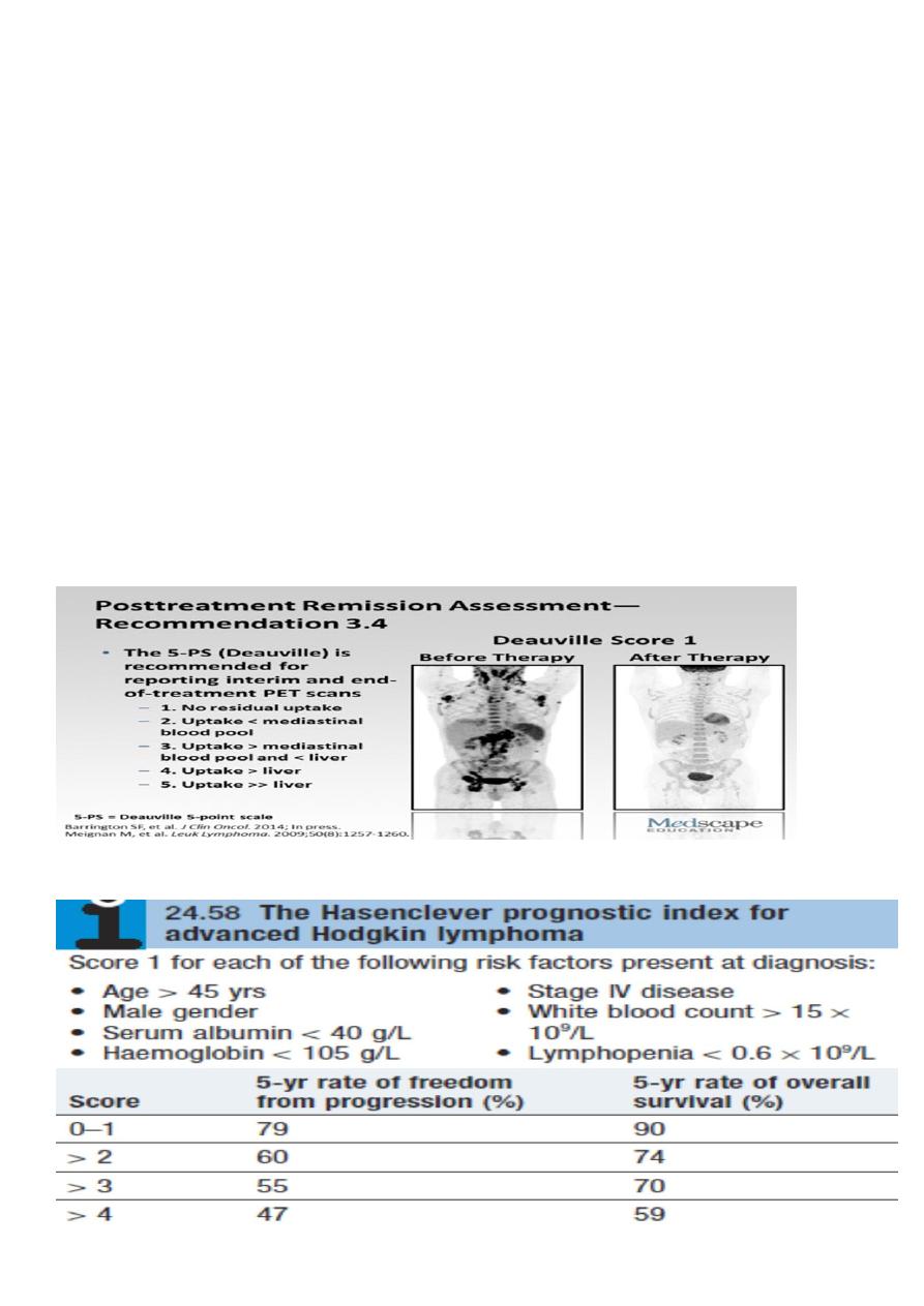
FBC may be normal. If a normochromic, normocytic anaemia or lymphopenia is present, this
is a poor prognostic factor. An eosinophilia or a neutrophilia may be present.
• ESR may be raised.
• Renal function tests are required to ensure function is normal prior to treatment.
• Liver function may be abnormal in the absence of disease or may reflect hepatic
infiltration. An obstructive pattern may be caused by nodes at the porta hepatis.
LDH measurements showing raised levels are an adverse prognostic factor.
• Chest X-ray may show a mediastinal mass.
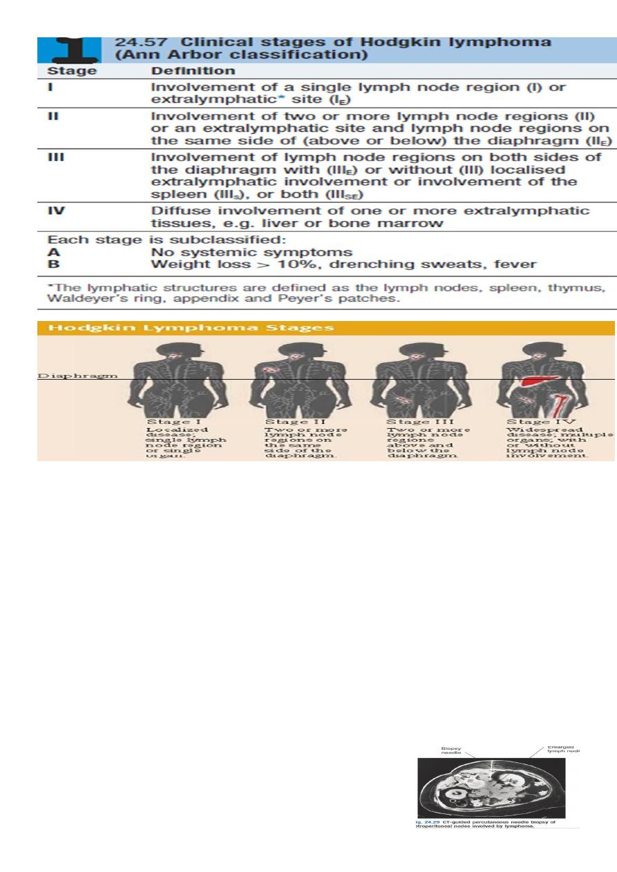
• CT scan of chest, abdomen and pelvis permits staging. Bulky disease (> 10 cm in a single
nodemass) is an adverse prognostic feature.
• Lymph node biopsy may be undertaken surgically or by percutaneous needle biopsy under
radiological guidance.
4
Management
:
1-chemotherapy;
The ABVD regimen (doxorubicin,vinblastine,bleomycin and dacarbazine)
* cardiac and pulmonary toxicity, due to doxorubicin and bleomycin respectively.
2-radiotherapy
* The majority of HL patients are now treated with chemotherapy and adjunctive
radiotherapy.
* Early-stage patients usually includes additional treatment with radiotherapy to the
involved lymph nodes after four courses of ABVD.
* Advanced-stage disease are most commonly managed with chemotherapy alone. 6–8
cycles of ABVD.
* Patients with disease which is resistant to therapy may be considered for autologous
HSCT.
* Treatment response is assessed clinically and by repeat CT and newer scanning
modalities such as positron emission tomography (PET)
Prognosis
:
5
:
Non-Hodgkin lymphoma
Non-Hodgkin lymphoma (NHL) represents a monoclonal proliferation of lymphoid cells of B
cell (70%) or T cell (30%) origin.
working formulation for lymphoma classification :
•
Low grade :
•
diffuse small lymphocytic.
•
follicular small cleaved cell.
•
follicular mixed small and large cell.
•
Intermediate grade :
•
diffuse small cleaved cell.
•
diffuse small and large cell.
•
diffuse large cell lymphoma.
•
follicular large cell lymphoma.
•
High grade :
•
immunoblastic lymphoma.
•
small non cleaved cell (Burkitt-type).
•
lymphoblastic lymphoma.

6
Lymphoma :
Non - Hodgkin’s disease
More random spread
Extranodal sites in unfavorable lesions
Systemic symptoms less common
More frequent involvement of
peripheral and mesenteric L.N.
waldeyer’s ring
Multiple diseases (at least two)
clinically, cure rare in low grade types.
:
Clinical presentation
1.
most pt present with one or several enlarge painless LN. cervical, inguinal and
axillary's nods are most frequently involved. Hepatosplenomegaly may be present
2. 1/5-1/3 of pt with intermediate and high grade lymphoma present in extra-
nodal site such as waldeyers ring, GIT, skin, salivary gland, bone.
3. compression syndromes may occur, gut obstruction, ascites, superior vena caval
obstruction and spinal cord compression.
4. BM involvement in high grade 10% and 50-60% in low grade.
5. T cell lymphoma are usually first diagnosed in the skin lead to cutaneous T cell
lymphoma (sezary syndrome), erthro-derma and circulating lymphoma cells with
hyper convoluted nuclei.
**The same staging system is used for both HL and NHL, but NHL is more likely to be stage
III or IV at presentation.
:
Investigations
Same as HD inaddition;
1-Bone marrow aspiration and trephine.
2-Immunophenotyping of surface antigens to distinguish T from B cell tumours.
3-Cytogenetic analysis to detect chromosomal translocations.
4-Immunoglobulin determination.
5- Measurement of uric acid levels. 6- HIV testing.
Hodgkin’s disease
Orederly spread by contiguity
Extranodal presentation rare
Systemic symptoms, prognostic
importance
Involve central and axial L.N. but rarely
mesenteric or lymphoid tissue of
waldeyer’s ring (oro-naso-pharynx)
Single disease in terms of classical
treatment: cure possible of all types of
stages.
7
Treatment :
Low grade NHL |:
• A symptomatic pts may not require treatment
• Radiotherapy : for stage I disease.
• Chemotherapy : oral therapy with chlorambucil.
• The anti-CD20 antibody rituximab.
* Rituximab (R) in combination with cyclophosphamide, vincristine and prednisolone
(R-CVP).
**Transplantation.
High-grade NHL *Prognosis
• Chemotherapy; R-CHOP
• Radiotherapy
• HSCT
Prognosisn:
8
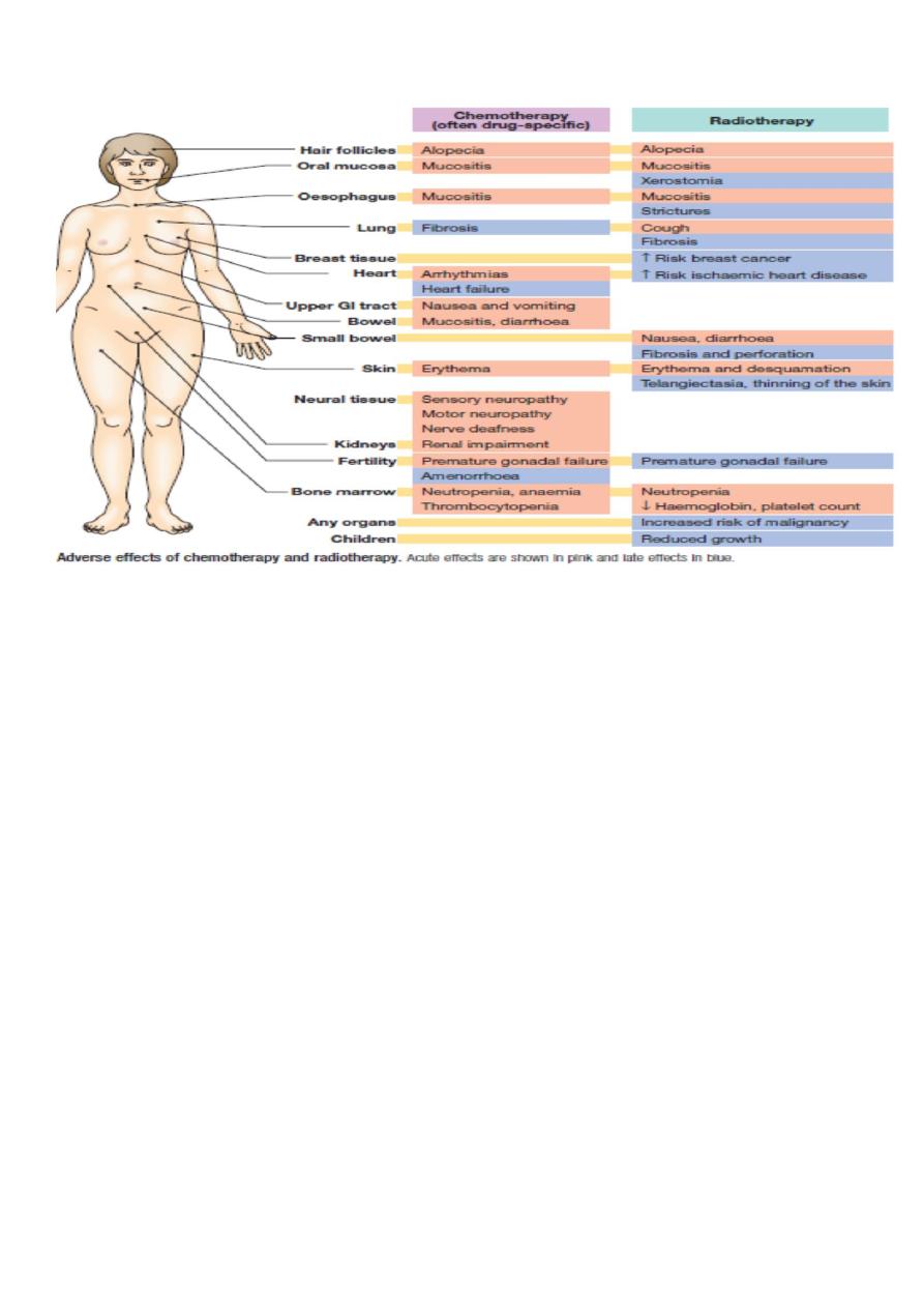
Adverse effect of chemotherapy and radiotherapy :