Interpretation ofPeriodontal Disease
Definition:Includes several disorders of the periodontium
- Gingivitis- Marginal Periodontitis
- Localized progressive Periodontitis
The term periodontium refers to the tissues that invest and support the teeth .
Periodontal disease refers to a group of disease that affects the tissues around the teeth.
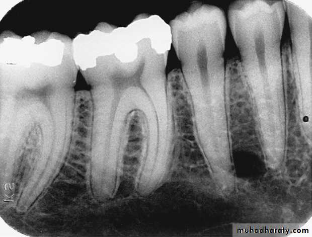
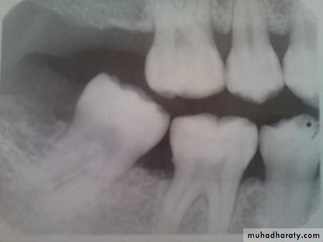
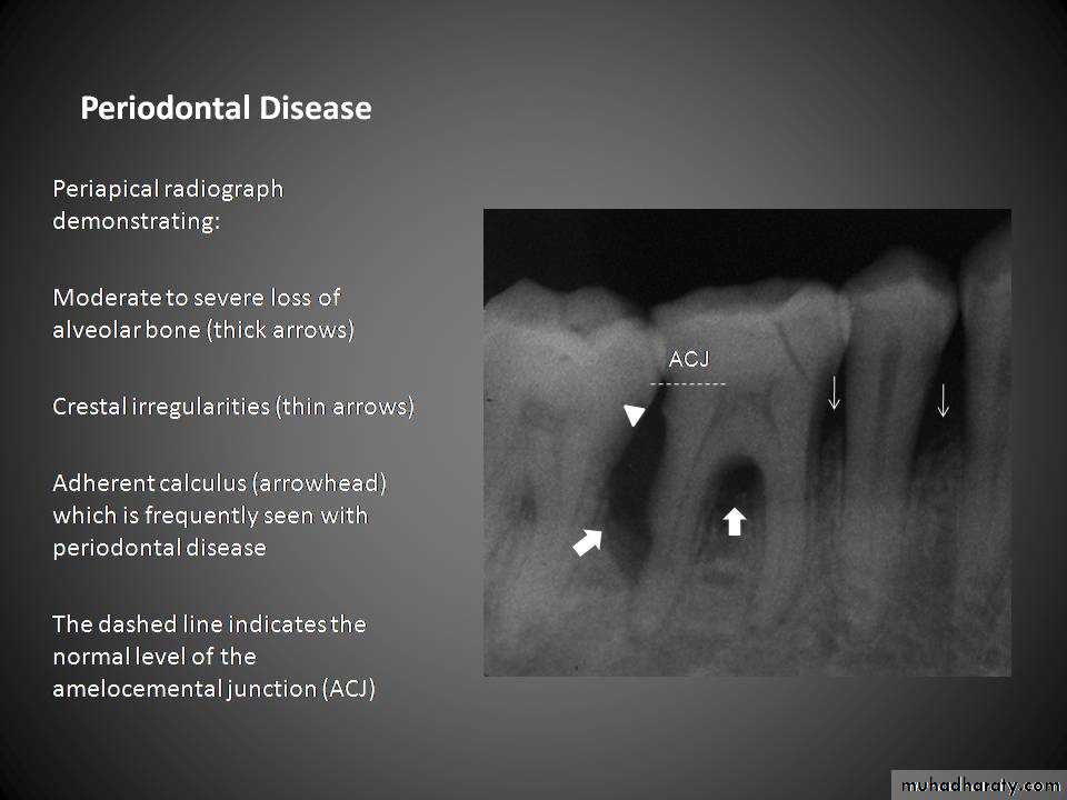
The normal anatomic landmarks of alveolar bone include the lamina dura, alveolar crest, and periodontal ligament space.
Lamina dura: In health, the lamina dura of teeth appears as a dense radiopaque line around the roots.
DESCRIPTION OF THE PERIODONTIUM
Alveolar crest: The normal healthy alveolar crest is located approximately 1.5 to 2.0 mm apical to the cementoenamel junctions (CEJs) of adjacent teeth.The shape and density of the alveolar crest vary between the anterior and posterior regions of the mouth. In the anterior regions, the alveolar crest appears pointed and sharp and is normally very radiopaque .In the posterior regions, the alveolar crest appears flat, smooth, and parallel to a line adjacent CEJs .The alveolar crest in the posterior regions appears slightly less radiopaque than that in the anterior regions
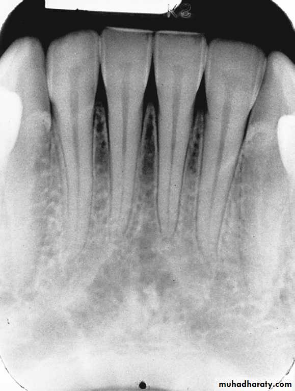
Healthy alveolar crest in the anterior region that appears pointed and highly radiopaque
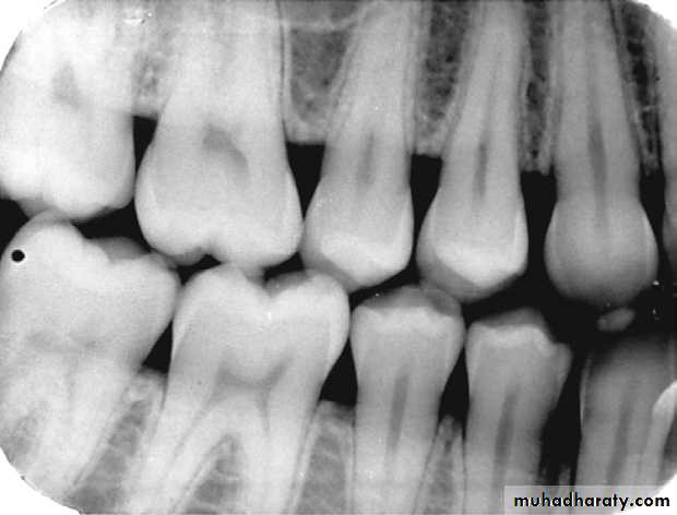
Healthy alveolar crest in the posterior region that appears flat, smooth, and radiopaque.
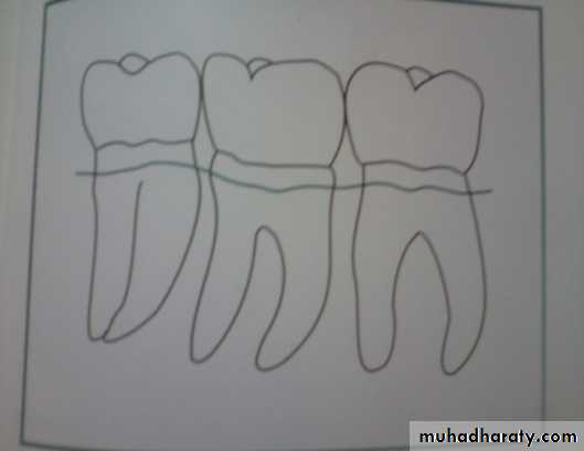
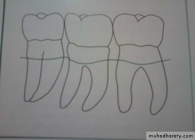
Periodontal ligament space: The normal periodontal ligament space appears as a thin radiolucent line between the root of the tooth and the lamina dura. In health, the periodontal ligament space is continuous around the root structure and is of uniform thicknessHealthy alveolar crest, normal lamina dura, and periodontal ligament space on a periapical image.
DESCRIPTION OF PERIODONTALDISEASE
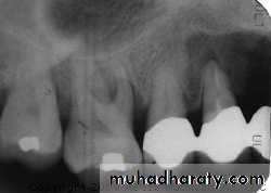
- Periodontal disease refers to a group of diseases that affect the tissuesaround teeth ,with periodontal disease, the alveolar crest is no longer located
1.5 to 2.0 mm apical to the CEJs and no longer appears radiopaque. Instead,
the alveolar crest appears indistinct, and bone loss is seen.
DETECTION OF PERIODONTALDISEASE
Clinical examination provides information about soft tissues, while dentalimages permit evaluation of bone.
1- Clinical Examination
include an evaluation of soft tissues (gingiva) for signs of inflammation (e.g.,
redness, bleeding, swelling, pus). A thorough clinical assessment must include
periodontal probing.
2- Dental Image Examination
Dental images provide an overview of the amount of bone present and
Indicate the pattern, distribution, and severity of bone loss resulting from
periodontal disease
Radiographs Techniques
The paralleling technique is the preferred periapical exposure method for the
demonstration of the anatomic features of periodontal disease. With the
paralleling technique, the height of crestal bone is accurately recorded in
relation to the tooth root. If the bisecting technique is used to expose
periapical images, a dimensional distortion of bone results due to vertical
angulation. Therefore, periapical images using the bisecting technique may
show more or less bone loss than actually present
A-Peri apical radiographic technique B-Bitewing radiographic techniqueC- Panoramic radiography
A -Peri apical radiographic technique:
The difference in bone level between two techniques
Bisecting technique distorting the level of bone present seen on an image because of the
vertical angulation used.Paralleling technique used to examine the same area
Radiographs Techniques
B- The vertical bite-wing :image can be used to examine bone levels and is best used for post-
treatment and follow-up purposes.
C- The panoramic image:
has little diagnostic value in the identification of periodontal disease and is
not recommended to demonstrate the anatomic features of this condition.
The Role of Radiology in Assessment of Periodontal Disease:
- Amount of bone present- Condition of the alveolar crests
- Bone loss in the furcation area- Width of the PDL space
Root length and morphology and crown to root ratio-1-Bony defects overlapped by existing bony walls.2-Buccal and lingual bone levels are not clearly seen on the radiograph.3-Radiographs show less severe destruction than that actually present.4-Radiograph do not demonstrate the soft tissue condition.5-Technique variation affect the appearance of the periodontium.
Limitations of Radiographs
Radiography is an indirect method for determining the amount of bone loss in periodontal disease. It shows the amount of remaining bone rather than the amount lost.
As a generally :
Radiographic examination of bone loss in periodontal
disease:
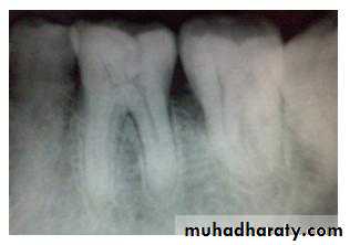
1- pattern: described as horizontal or vertical.
CEJs of adjacent teeth can be used as a plane of reference in determining the pattern of bone losspresent.
horizontal bone loss:
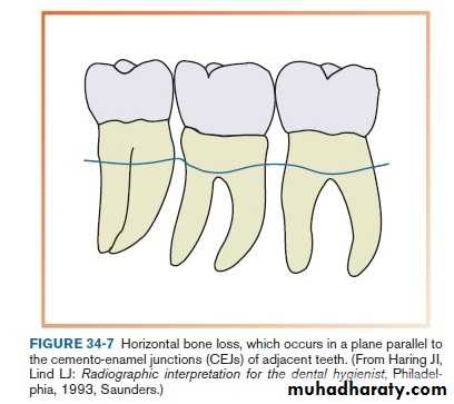
Bone loss occurs in a plane parallel to CEJs of adjacent teeth.
Horizontal bone lossis used to describe the radiographic appearance of the loss of bone height in the region of several adjacent teeth. Horizontal bone loss may be classified as localized or generalized, depending on the regions involved.
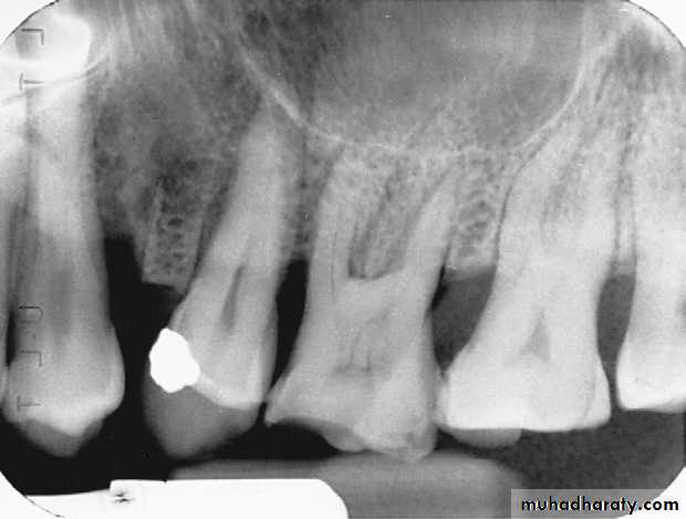
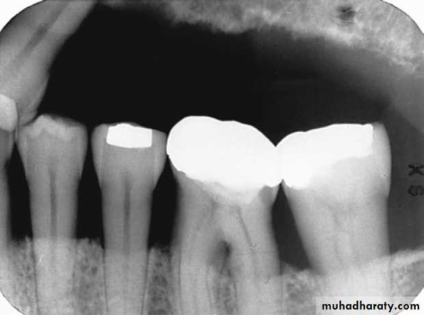
vertical (angular) bone loss:
Bone loss doesn’t occur in plane parallel to CEJs of adjacent teeth.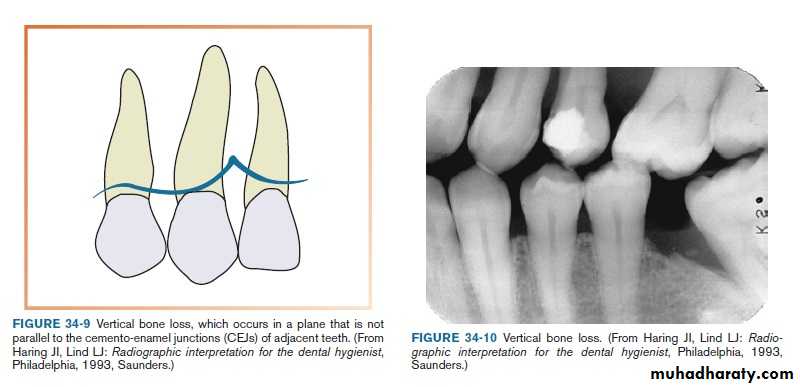
Vertical bone loss
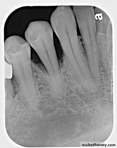
Vertical bone defects-are also called proximal intrabony defects. The defect extends apically2- Distribution
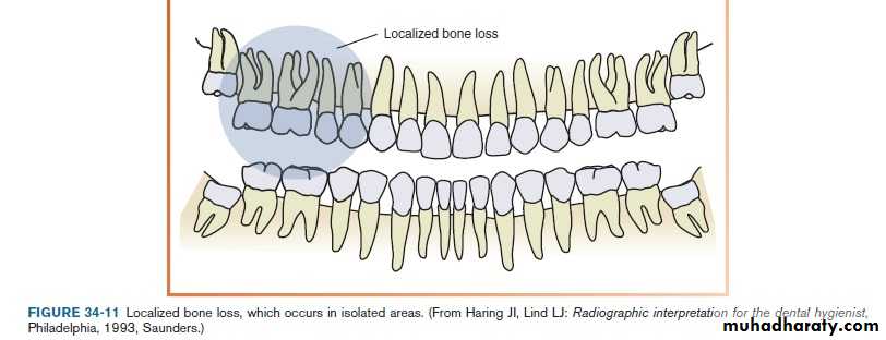
The distribution of bone loss seen on a dental image can be described as localized or generalizedLocalized: occurs in isolated area, with less than 30% of the sites involved.
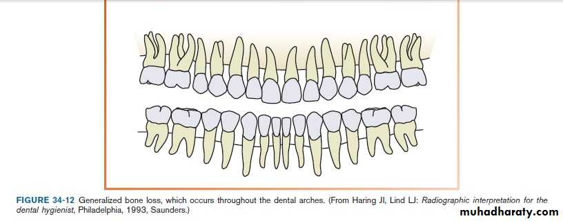
Generalized: occurs evenly throughout the dental arch, with more than 30% of the sites involved.
SEVERITY:
slightly, moderate ,or severe.Severity is measured by clinical attachment loss (CAL).
CAL is a measurement of the distance in mm from CEJ to the base of the sulcus or periodontal pocket: CAL is measured by calibrated periodontal probe.
- slightly bone loss: 1 to 2mm
- moderate bone loss: 3to 4mm- severe bone loss: 5mm or greater
slightly
moderate
severe

Furcation Bone Loss:
Furcation exposure comes from bone loss at the furcation of multirooted teeth. Furcation exposure may occur before advanced periodontal disease. It is sometimes difficult to determine radio graphically, whether the interradicular space is involved, unless there is a radiolucent area in the region of the furcation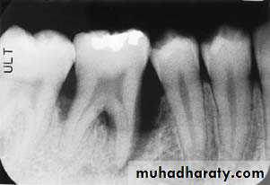
Classification of Periodontal Disease
Dental images can be used in the classification of periodontaldisease. On the basis of the amount of bone loss, periodontal
disease can be classified as follows: the American Dental
Association (ADA) Case Type I (gingivitis), ADA Case Type II (mild
periodontitis), ADA Case Type III (moderate periodontitis), or
ADA Case Type IV (advanced or severe periodontitis).
Predisposing Factors
The effects of certain medications, tobacco use, and variousmedical conditions are all considered risk factors for periodontal disease
Dental images play a major role in the detection of
local irritants such as calculus and defective restorations.
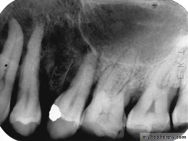
Calculus
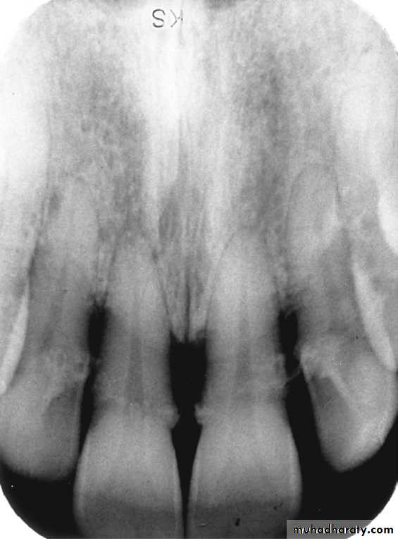
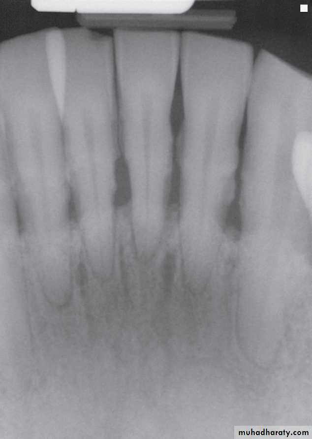
Subgingival calculus that appears as irregular radiopaque projections in the maxillary anterior region.
Calculus that appears as sharp, pointed radiopacities along the surfaces of mandibular anterior teeth.
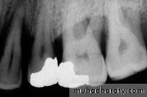
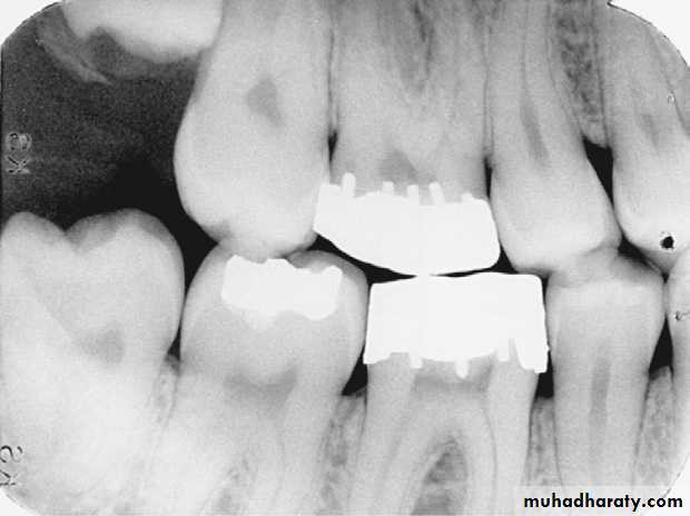
Defective Restorations
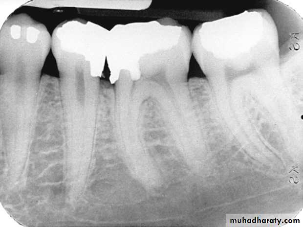
Amalgam overhang on the mesial surface of the mandibular first molar.
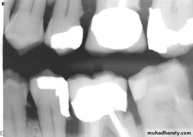
Ill-fitting restorations and open contacts between teeth contributing to the periodontal condition
Poorly contoured crowns on maxillary first molars.
Defective Restorations