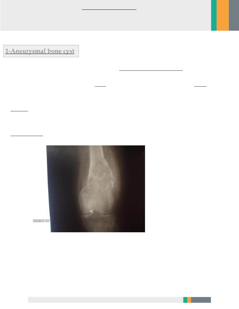
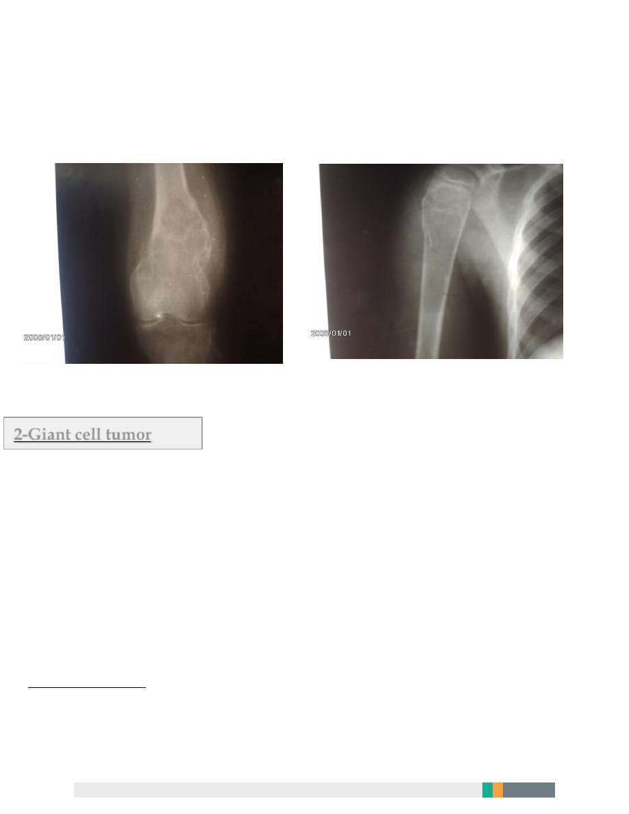
1-Aneurysmal bone cyst
It mainly affect young adult, and it locate in: The long bone metaphysis .
The patient mainly complain from pain and in large cyst visible or palpable mass may be
detected.
X-ray
: well defined radiolucent cyst , trabiculated and eccentrically located , usually in
the metaphysis , the lesion is
expandable (ballooning) .
Treatment :
by curettage and bone graft
Bone tumor
2
Surgery (orthopedic) Zakho hospital
Dr. Yakdhan Alsalem November 27, 2016
2
Comparison between simple bone cyst and aneurysmal bone cyst
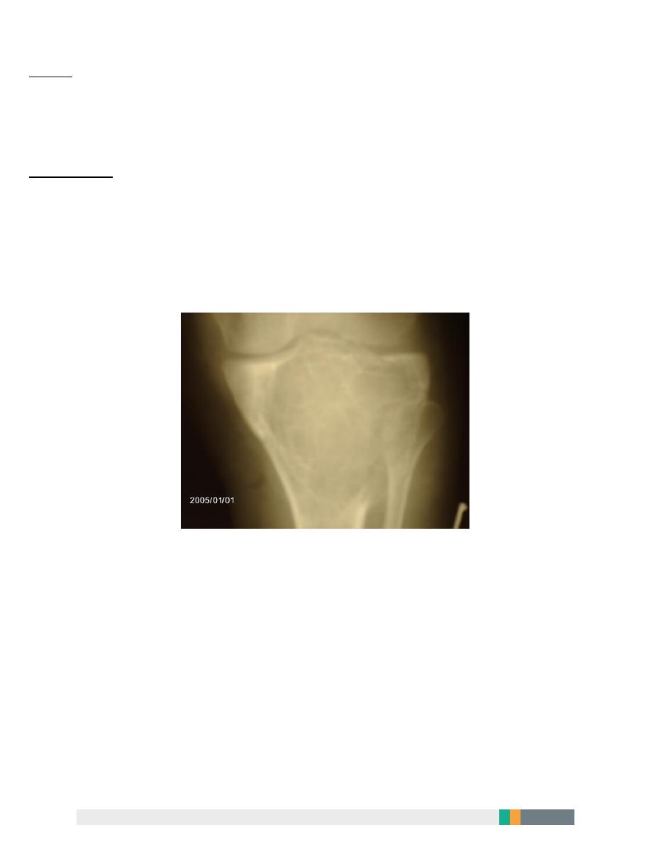
2-Giant cell tumor
It is a lesion of uncertain origin ; it appear in mature bone , most commonly in the
distal femur , proximal tibia , proximal humerus and distal radius .
It characteristically extends up to the subarticular bone plate .
The patient is usually young adult (20-40) years old who complains of pain at the
end of the long bone .
On examination
: there is palpable mass .
3
X-ray
: radiolucent area located eccentrically at the end of the long bone , the
cortex is thin and ballooned , the lesion is trabiculated and it is
characteristically extend to the subchondral bone .
Treatment
: well confined , slow growing lesion can be treated by curettage and
bone graft , bigger lesion treated by excision and bone graft some time with
prosthetic replacement .
If the tumor is rapidly enlarged and aggressive , amputation is indicated

4
3-Malignant bone tumors
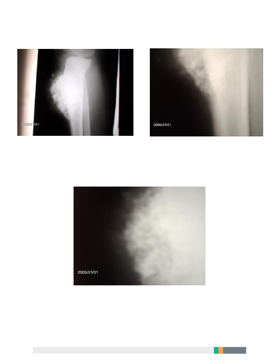
Osteogenic sarcoma
It is highly malignant bone forming tumor ,
It arise within the bone and spread rapidly out ward towards the periosteium and
the soft tissue .
It affect mainly the children , adolescent and old age group .
It affect any bone but mainly the metaphysis of the long bones especially around
the knee joint and proximal end of humerus .
Clinically :
1-
pain is the first symptom ,
2-
gradually increase in severity .
3- some time the patient presented with lump or pathological
fracture .
On examination :
in early case local tenderness , in late cases palpable mass can be felt and swelling
can be seen .

5
Investigation
Blood invest. : ESR increase serum alkaline phosphatase increase .
Radiological investigation :
1-
plain x-ray
:hazy osteolytic area which is non homogenous and alternate
with osteoblastic areas , the tumor destruct the cortex and extend to the
adjacent soft tissue ; when this happened streaks of new bone appear radiating
out ward from the cortex (sun burst) appearance
periosteal reaction is present in form called (Codman's triangle) ; these two
findings are typical for osteogenic sarcoma
2- Radioisotope scan .
3- C.T.
4- MRI.
5- Chest x-ray to detect metastasis .
6- Other invest. Incisional biopsy .
6
Osteogenic sarcoma (codman tiangle)
Osteogenic sarcoma (sun-burst) appearance

7
Treatment
Since this tumor is highly malignant and aggressive ;
radical surgery combined with chemotherapy and radiotherapy is indicated .
1- Radical surgery: it mean amputation through or above the joint proximal to
the lesion and proximal to the origin of any affected muscle .
2- Chemotherapy
:
methoterexate is used pre-operative and post-operative .
Prognosis : improved in the last few years due to improvement in the invest. ,
diagnosis and treatment .
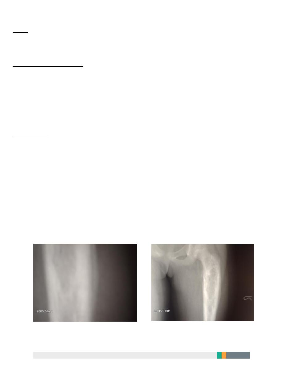
4-Ewing sarcoma
It arise from the bone marrow , the age affected are usually children and
adolescent (10-20)years old , it affect the tubular bone , it is highly malignant
tumor and carry bad prognosis .
Clinically :
pain
(which is usually throbbing in nature) , swelling (tender) , generalized illness
and pyrexia .
ESR is increase . C.T, MRI , Bone scan .All are helpful .
8
x-ray : destructive osteolytic diaphyseal lesion , new bone formation in fusiform
layering around the lesion so-called onion-peel appearance .
Differential diagnosis
:
1- bone infection .
2-osteoid osteioma
Treatment
: the prognosis is always poor , and surgery alone do little to improve
it .
The tumor is very sensitive to radiotherapy and chemotherapy but over all
survival is not much improved
Ewing sarcoma(periosteal reaction)

9
5-Metastatic bone tumor
Skeleton is one of the commonest site of the secondary cancer .
In patients over 50 years old , bone metastasis are seen more frequently than all
primary malignant bone tumors together .
The commonest source of secondary tumor in bone (from the commonest to less
common )are :
1- ca. breast .
2- ca. prostate .
3- ca. kidney .
4- ca. lung .
5- ca. thyroid .
The commonest site of secondary bone tumor are:
Vertebrae , pelvis ,proximal femur and humerus .

10
Metastasis is usually osteolytic but osteoblastic lesion seen in secondary tumor of
prostate .
Clinically
:the presenting symptoms of secondary bone tumor is usually , pain or
back ache and pathological fracture.
Investigation
:x-ray , bone scan , biopsy .
In children secondary bone tumor is usually from adrenal neuroblastoma .
Treatment
:by the time the patient develop sec. bone tumor the prognosis is
usually hopeless .
in most of cases the treatment is symptomatic i.e. to search for primary tumor is
value less .
It is important to get the patient live the remaining of his life comfortably by :
1- controlling the pain by analgesics or narcotics .
2- radiotherapy to decrease the pain and metastatic activity .
3- fixation of the pathological fracture .
4- sever pain some time need nerve or spinal tract ablation A.L.Y