Uroradiology & Slides 1مع الشرح على السلايدات
بسم الله الرحمن الرحيم


Rt side cysts
Description :-plain xray of abdomen,,showing longitudinal Radioapque shadow extends from upper part of lt urter to bladder used To divert the urine internally, to drain the upper UT,after surgeries of UT.Description :- IVU in pt with renal mass,compression of calyces of lt kidney , eating of calyces by the tumour , soft tissue mass +loss of lower calyces(eating of lower calyces).

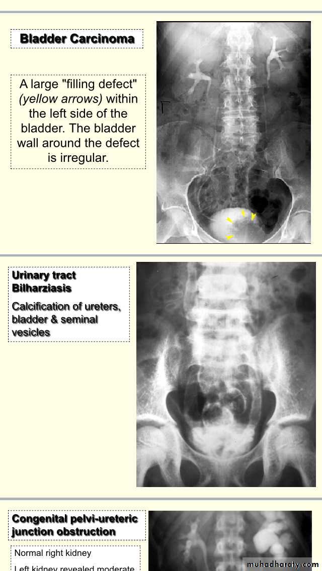
IVU Cystogram phase :- filling defect in the lt lateral side of the bladder DDX:- stone,tumour, blood clot,bladder mass, fungal ball,F.B”catheter part”.
Cystogram IVU ,time predicted <30min,irregular filling defect in the lt side of bladder.
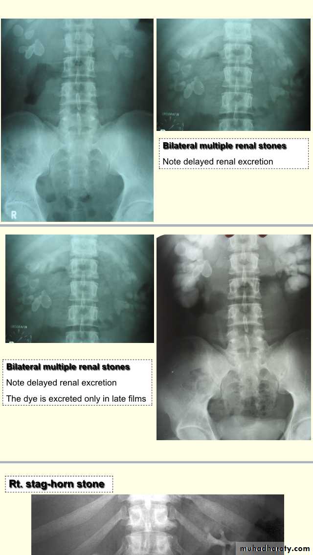
Advanced bilhariziasis,linear calcification, while in T.b it is dot like calcification .
Hydronephrosis ,dilated PCS,buldging calyces,with parenchymal thicknessPUJ obstruction

Hyperechoic with acoustic shadow **if no acoustic shadow infection,F.B,,calcification.
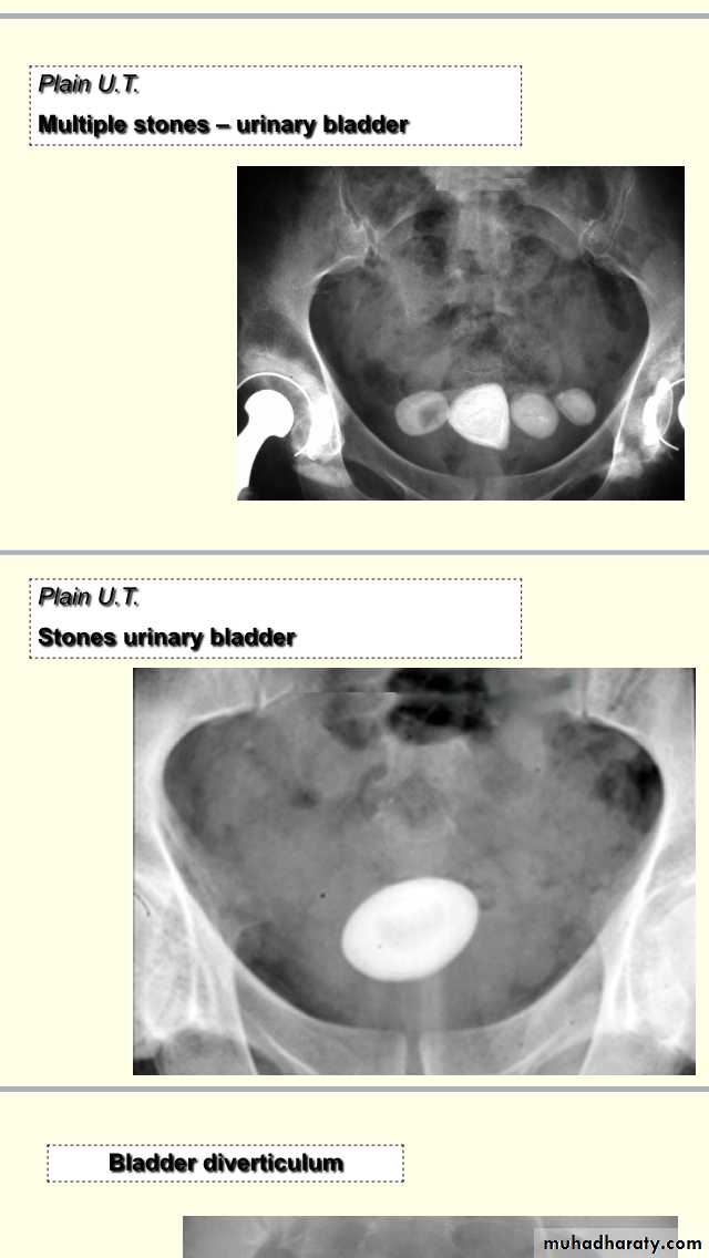
Description :-Xray of pelvis (NOT KUB),show multiple radiopaque shadow, rounded shape smooth surface,above pubis 2.5cm size.what you recognize from the picture ?Old age pt ,bedridden for long time,due to OA changes,,he may be taken VIT D supplements ,he may have BPH.
Total hip replacement,Austin moore .
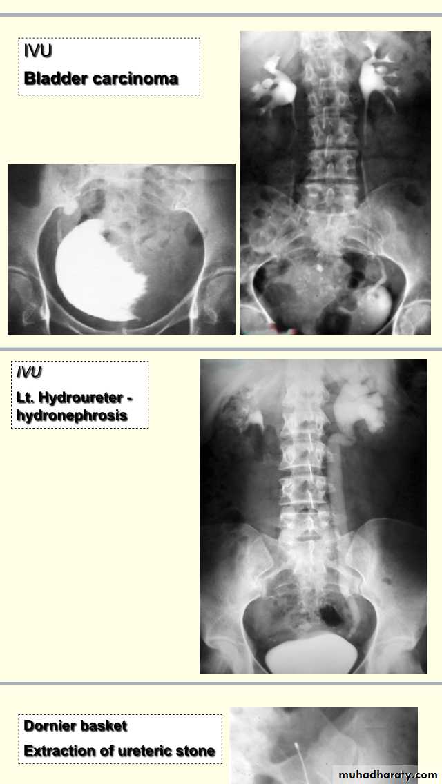
Radiopaque shadow ,smooth ,round surface,(2,5cm)size,solitary.Management of vesical stone:-if no underlying UT pathology ,Rx depend on the size,in pediatric age group by open surgery,in adult:endoscopic Mx,if >3cm cystolithotomy,if 2.5cm endoscopic crushing(cystolithopaxy) or equalIVU cystogram phase :-showing opacity with thickening and irregular of bladder wall(outline) due to neurogenic bladder (multiple saccule in the bladder commonly seen +trabeculation) with diverticulum (paraureteric buldging).

Description:- IVU showing filling defect in the base of bladder ,elevated base,buldging and curvature of bladder extending above symphysis pubis Dx:BPH(median lobe of prostate)
Description:-IVU study ,soft tissue

Description :-KUB :plain x-ray showing pubic symphysis with radiopaque shadow below it. .