
1
Fifth stage
GYNE
Part 1
د.أسماء
28/11/2016
Abnormal uterine bleeding
It is an descriptive term applied to any alteration in the normal pattern of
menstrual flow and it is the uterine bleeding that is abnormal in amount, duration
or timing
.
This term describes abnormalities of menstruation and do not describe the
underlying pathology
.
Early pregnancy and its complications should always be ruled out as the cause of
AUB in women of reproductive age.
pattern of abnormal uterine bleeding :-
menorrhagia : The average menstrual period ,
The normal duration of bleeding is 4.5 to 8 days, and the amount of normal
flow is less than 80 mL of blood.
Menorrhagia ('heavy mentrual bleeding HMB')
(hypermenorrhea): Excessive and/or pro-longed menses (>80 mL and >7
days) occurring at normal intervals ,this level represents the level at which a
fall in haemoglobin and haematocrit concentration commonly occurs
Polymenorrhoea:it is a frequent menstruation as menses occurring at < 21
days interval associated with a shortened follicular phase or inadequate luteal
phase.
Metrorrhagia – bleeding at irregular intervals with excessive flow and
duration
Menometrorrhagia: Heavy and irregular uterine bleeding
Dysfunctional uterine bleeding: Bleeding caused by ovulatory dysfunction
Intermenstrual bleeding: Uterine bleeding of variable amounts occurring
between regular menstrual periods.
Midcycle spotting: is scanty intermenstrual discharge occurring just before
ovulation that is associated with a decrease in estrogen at midcycle.
Postcoital bleeding: is non-menstrual bleeding that occurs immediately after
sexual intercourse.
-With drawl bleeding: bleeding occurred after stopping oestrogen and
progestrone use or progestrone use.

2
-Postmenopausal bleeding : Recurrence of bleeding in a menopausal woman
at least 6 months to 1 year after cessation of cycles.
Prevalence
The presentation of HMB is common because women are having fewer children
and Consequently more menstrual cycles.
Indeed, each year in the UK,5 per cent of women between the ages of 30 and 49
years consult Their general practitioner with this complaint, and is the reason
for up to 20% of outpatient clinic visits by women,with a substantial number
being referred on to secondary care.
Heavy menstrual bleeding can cause severe anemia, but less significant blood
loss can diminish a woman’s quality of life and even her income when workplace
activity is adversely affected
Classification
Menorrhagia can be classified as:
1. idiopathic: where no organic pathology can be found: idiopathic
menorrhagia is otherwise known as dysfunctional uterine bleeding-
(DUB).now (bleeding of endometrial origin) The majority of women who
present with menorrhagia will have DUB,
2. secondary to an organic underlying cause, such as fibroids.
Aetiology:-
A. Organic causes:
1. Local disorders:
• Uterine fibroids.
• Endometrial/ Endocervical polyp.
• Adenomyosis.
• Pelvic endometriosis.
• Intrauterine device (IUD).
• Cervicitis
• PCOS
• Pelvic inflammatory disease (PID).
• Oestrogen-secreting ovarian tumour.(granulosa or theca cell tumour).

3
• Cervical carcinoma.
• Uterine body carcinoma.
• Trauma of lower genital tract
• Urethral caruncle.
• Arteriovenous malformation, is a congenital or acquired localized
collection of abnormally connected arteries and veins. When they occur in
the uterus, they have been associated with episodes of acute excessive
Bleeding. Colour Doppler imaging is a useful diagnostic.
2. Systemic disorders :
Menorrhagia is a feature of a number of systemic Conditions,which should be
considered in the Differential diagnosis. These include :
1- Endocrine disorders may interfere with normal feedback mechanisms that
regulate secretion of gonadotrophin- releasing hormone (GnRH),
gonadotrophin, sex steroid.
Thyroid disorder (Hypothyroidism or hyperthyroidism).
Diabetes mellitus.
E. Prolactin disorders
3. Haemostasis disorder:
Von Willebrand's disease.
Idiopathic Thrombocytopenic purpura (ITP).
4. Liver disorder
5. Renal disease
6. Medications as steroid hormones, anticoagulants and cytotoxic agents,
contraceptive method
7.Psychological and emotional cause: Excessive exercise, stress, and weight
changes. All these can cause hypothalamic suppression leading to abnormal
uterine bleeding due to disruption along the hypothalamus-pituitary-ovarian
pathway
8. Pregnancy : Should be considered in women of reproductive life in any
patient presenting with abnormal uterine bleeding

4
B. Non –organic cause (Dysfunctional uterine bleeding (DUB)
It is defined as abnormal uterine bleeding in the absence of organic disease.
It is the most common cause of abnormal vaginal bleeding during a woman's
reproductive years especially at the extreme ages of a woman's reproductive
years.
It is a diagnosed by the exclusion of other causes.
Aetiology of DUB
Despite extensive research, the aetiology of DUB remains unclear. Disordered
endometrial prostaglandin production has been implicated in the aetiology of this
condition, as have abnormalities of endometrial vascular Development
There are clearer reasons why many more women complain of menorrhagia
now than they did a century Ago,:
because of decreasing family size, women now Experience many more
menstrual cycles,
changing role of women in society and women are now much less likely
to tolerate menstrual loss that they consider to be excessive.
clinical assessment:
History:
Age, parity, marital status and reproductive wishes
description of the pattern of abnormal menstrual bleeding and it's severity
and it's duration and amount of blood loss.
Patients will have different ideas as to what constitutes a ‘heavy period’ . Useful
questions include:
How often does soaked sanitary wear need to be changed?
Is there presence of clots?
Is the bleeding so heavy (flooding) that it spills over your towel/tampon
and on to your pants,clothes or bedding?
Have you had to take any time off work due to this bleeding?
Do you ever find you are confined to your house when the bleeding is at
its worst?
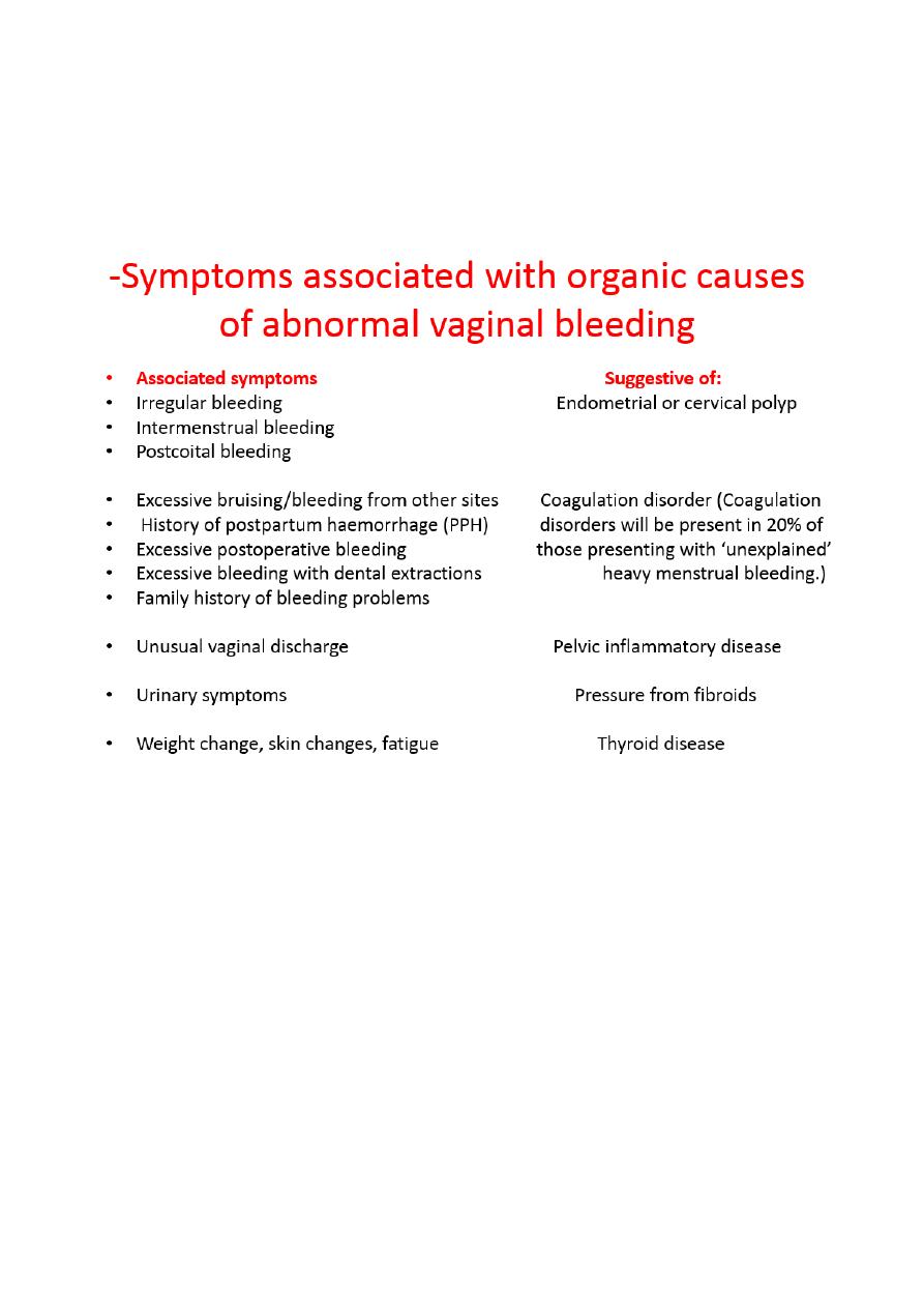
5
It is also important to determine the onset and Duration of the current
problem.
determine the presence of other cyclical symptoms as dysmenorrhoea,
breast tenderness, Psychological disturbance, fatigue, dizziness, and
syncope.
Ask about : Recent illness, psychological stress, excessive exercise, or weight
change
*Past medical history:Diabetes mellitus, Thyroid disease, Endocrine problems,
pituitary tumors, Liver disease.
*Past surgical history.
*Drug history:Medication usage, including exogenous hormones, anticoagulants,
aspirin, anticonvulsants, and antibiotics

6
Clinical examination
height and weight and body mass index (BMI).
signs of anemia or hypovolemia, vital signs.
evidence of systemic coagulopathy (bruising, petechiae)
thyroid disease (goitre).
An abdominal examination should be performed to reveal a pelvic mass
(fibroid);
a speculum examination should be performed to assess the vulva, vagina
and cervix (this may reveal sources of bleeding, such as a tumour, or a
discharge suggesting infection);
bimanual examination should be performed to elicit uterine enlargement.
General looking for stigmata of underlying systemic disease is important.
investigations
Initial investigations
1. Full blood count : A full blood count (FBC) should be carried out in all
women with HMB to ascertain the need for iron therapy (and in certain
cases, blood transfusion). Blood count with reticulocyte count and
differential Serum iron and iron-binding capacity Serum ferritin .
2. Coagulation screen : Referral for a haematological opinion should be
considered in women with a history consistent with a coagulation
disorder ( PT, PTT, and INR).
3. B-hCG if any possibility of pregnancy exists.
4. thyroid function tests: Only from women with other symptoms of
thyroid disease
5. Liver function tests
6. Renal function test.
7. Pelvic ultrasound scan
Pelvic ultrasonography
Saline infusion sonography
A pelvic ultrasound scan (USS) should be performed
when a pelvic mass is palpated on examination (suggestive of fibroids)
when symptoms suggest an endometrial polyp, e.g.irregular or
intermenstrual bleeding;

7
when drug therapy for HMB is unsuccessful .
By US we can diagnose :fibroid, endometrial thickening ,poly cystic ovary,
adenomyosis
8. High vaginal and endocervical swabs : High vaginal and endocervical
swabs should be taken:
• when unusual vaginal discharge is reported or observed on examination;
• where there are risk factors for PID.
9. Biopsies: as necessary
• Cervical biopsy
• Endocervical biopsy
• Endometrial biopsy :
Biopsy should be performed
• those aged >45 years;
• in younger women
- if irregular or intermenstrual bleeding;
- drug therapy has failed.
• all women Prior to surgical therapy .
UK study data demonstrate categorically that the age cut-off of 45 Years has the
highest sensitivity in detecting the maximum proportion of all types of
endometrial hyperplasia and carcinoma, while having a reasonably high
specificity
so as to avoid false negatives. Reassuringly,these data support the guidelines
produced in the UK by the National Institute of Clinical Excellence
( NICE.)
1- A Pipelle™ endometrial biopsy can be performed in the outpatient
setting. It is performed as follows:
- a speculum examination is carried out and the cervix is completely
visualized;
- a vulsellum instrument may be required to grasp the cervix and
provide gentle traction, thereby straightening the endocervical canal

8
- the Pipelle sampler is carefully inserted through the cervical os until it
reaches the fundus of the uterus. The length of the uterus is noted;
- the inner part of the Pipelle is withdrawn to create a vacuum and the
device is gently moved in and out to obtain a sample of endometrial
tissue;
- the Pipelle is removed and the tissue is expelled into a histopathology
container of formalin.
2.An outpatient hysteroscopy with endometrial biopsy may be indicated if:
Pipelle biopsy attempt fails.
Pipelle biopsy sample is insufficient for histopathology assessment;
there is an abnormality on USS, e.g. suggested endometrial polyp or
submucosal fibroid;
patient is known to poorly tolerate speculum examinations (more
comfortable vaginoscopic approach can be used).
If the patient fails to tolerate an outpatient procedure or the cervix
needs to be dilated to enter the cavity,
Endometrial biopsy can be done by.
then a hysteroscopy and endometrial biopsy under general anaesthetic (may be
required)
Hysteroscopically directed biopsy is the gold standered procedure as it provides
direct visualization of uterine cavity and allows to take biopsy from specific
lesion. It is ideally done in proliferative phase of menstrual cycle when the
endometrium is at it's thinnest.
3.Curettage. SH.J
ღ

9