
Conjunctiva, cornea and sclera
INTRODUCTION
Disorders of the conjunctiva and cornea are a common cause of symptoms.The ocular
surface is regularly exposed to the external environment and subject to trauma, infection and
allergic reactions which account for the majority of diseases in these tissues. Degenerative
and structural abnormalities account for a minority of problems.
Symptoms :
Patients may complain of the following:
1 Pain and irritation. Conjunctivitis is seldom associated with anything more than mild
discomfort. Pain signifies something more serious such as corneal injury or infection.
This symptom helps differentiate conjunctivitis from corneal disease.
2 Redness. In conjunctivitis the entire conjunctival surface including that covering the
tarsal plates is involved. If the redness is localized to the limbus ciliary flush the
following should be considered:
(a) keratitis (an inflammation of the cornea);
(b) uveitis;
(c) acute glaucoma.
3 Discharge. Purulent discharge suggests a bacterial conjunctivitis. Viral conjunctivitis is
associated mainly with a watery discharge.
4 Visual loss. This occurs only when the central cornea is affected. Loss of vision is thus
an important symptom requiring urgent action.
5 Patients with corneal disease may also complain of photophobia.
Signs
The following features may be seen in conjunctival disease:
• Papillae.These are raised lesions on the upper tarsal conjunctiva, about 1 mm in
diameter with a central vascular core. They are non-specific signs of chronic
inflammation. Giant papillae, found in allergic eye disease, are formed by the
coalescence of papillae (see Fig. 7.4).
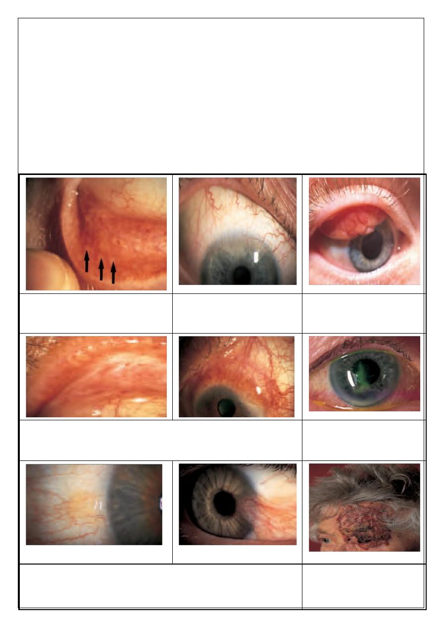
• Follicles (Fig. 7.1).These are raised, gelatinous, oval lesions about 1 mm in diameter
found usually in the lower tarsal conjunctiva and upper tarsal border, and occasionally
at the limbus. Each follicle represents a lymphoid collection with its own germinal
centre. Unlike papillae, the causes of follicles are more specific (e.g. viral and
chlamydial infections).
• Dilation of the conjunctival vasculature (termed ‘injection’).
• Subconjunctival haemorrhage, often bright red in colour because it is fully
oxygenated by the ambient air, through the conjunctiva.
Fifth stage Lec-
Dr.Nazar
Ophthalomology
28/11/2016

The features of corneal disease are different and include the following:
• Epithelial and stromal oedema may develop causing clouding of the cornea.
• Cellular infiltrate in the stroma causing focal granular white spots.
• Deposits of cells on the corneal endothelium (termed keratic precipitates or KPs,
usually lymphocytes or macrophages, see p. 92).
• Chronic keratitis may stimulate new blood vessels superficially, under the epithelium
(pannus; Fig. 7.2) or deeper in the stroma.
• Punctate Epithelial Erosions or more extensive patches of epithelial loss which are
best detected using fluorescein dye and viewed with a blue light.
CONJUNCTIVA
Inflammatory diseases of the conjunctiva
BACTERIAL CONJUNCTIVITIS :
Patients present with:
1. redness of the eye;
2. discharge;
3. ocular irritation.
The commonest causative organisms are Staphylococcus, Streptococcus, Pneumococcus and
Haemophilus. The condition is usually self-limiting although a broad spectrum antibiotic
eye drop will hasten resolution. Conjunctival swabs for culture are indicated if the condition
fails to resolve.
ANTIBIOTICS Chloramphenicol Ciprofloxacin Fusidic acid Gentamicin Neomycin
Ofloxacin Tetracycline
Ophthalmia neonatorum, which refers to any conjunctivitis that occurs in the first 28 days
of neonatal life, is a notifiable disease. Swabs for culture are mandatory. It is also important
that the cornea is examined to exclude any ulceration.
The commonest organisms are:
1. Bacterial conjunctivitis (usually Gram positive).
2. Neisseria gonorrhoea. In severe cases this can cause corneal perforation.
Penicillin given topically and systemically is used to treat the local and systemic
disease respectively.
3. Herpes simplex, which can cause corneal scarring.Topical antivirals are used to
treat the condition.
4. Chlamydia. This may be responsible for a chronic conjunctivitis and cause sight-
threatening corneal scarring.Topical tetracycline ointment and systemic
erythromycin is used is used to treat the local and systemic disease respectively.

VIRAL CONJUNCTIVITIS
This is distinguished from bacterial conjunctivitis by:
1. a watery and limited purulent discharge;
2. the presence of conjunctival follicles and enlarged pre-auricular lymph nodes;
3. there may also be lid oedema and excessive lacrimation.
The conjunctivitis is self-limiting but highly contagious. The common- est causative agent is
adenovirus and to a lesser extent Coxsackie and picornavirus. Adenoviruses can also cause a
conjunctivitis associated with the formation of a pseudomembrane across the conjunctiva.
Certain adenovirus serotypes also cause a troublesome punctate keratitis. Treatment for the
conjunctivitis is unnecessary unless there is a secondary bacterial infection. Patients must be
given hygiene instruction to minimize the spread of infection (e.g. using separate towels).
Treatment of keratitis is controversial. The use of topical steroids damps down symptoms
and causes corneal opacities to resolve but rebound inflammation is common when the
steroid is stopped.
CHLAMYDIAL INFECTIONS
Different serotypes of the obligate intracellular organism Chlamydia trachomatis are
responsible for two forms of ocular infections.
Inclusion keratoconjunctivitis
This is a sexually transmitted disease and may take a chronic course (up to 18 months)
unless adequately treated. Patients present with a muco- purulent follicular conjunctivitis
and develop a micropannus (superficial
peripheral corneal vascularization and scarring) associated with sub- epithelial scarring.
Urethritis or cervicitis is common. Diagnosis is confirmed by detection of chlamydial
antigens, using immunofluorescence, or by identification of typical inclusion bodies by
Giemsa staining in conjunctival swab or scrape specimens.
Inclusion conjunctivitis is treated with topical and systemic tetracycline. The patient should
be referred to a sexually transmitted diseases clinic.
Trachoma (Fig. 7.3)
This is the commonest infective cause of blindness in the world although it is uncommon
in developed countries.The housefly acts as a vector and the disease is encouraged by poor
hygiene and overcrowding in a dry, hot climate. The hallmark of the disease is
subconjunctival fibrosis caused by frequent re-infections associated with the unhygienic
conditions. Blindness may occur due to corneal scarring from recurrent keratitis and
trichiasis.
Trachoma is treated with oral or topical tetracycline or erythromycin. Azithromycin, an
alternative, requires only one application. Entropion and trichiasis require surgical
correction.

ALLERGIC CONJUNCTIVITIS
This may be divided into acute and chronic forms:
2-Acute (hayfever conjunctivitis). This is an acute IgE-mediated reaction to airborne
allergens (usually pollens). Symptoms and signs include:
(a) itchiness;
(b) conjunctival injection and swelling (chemosis);
(c) lacrimation.
2- Vernal conjunctivitis (spring catarrh) is also mediated by IgE. It oftenb affects male
children with a history of atopy. It may be present all year long. Symptoms and signs
include:
(a) itchiness;
(b) photophobia;
(c) lacrimation;
(d) papillary conjunctivitis on the upper tarsal plate (papillae may coalesce to form giant
cobblestones; Fig. 7.4);
(e) limbal follicles and white spots;
(f) ) punctate lesions on the corneal epithelium;
(g) an opaque, oval plaque which in severe disease replaces an upper zone of the corneal
epithelium.
Initial therapy is with antihistamines and mast cell stabilizers (e.g. sodium
cromoglycate; nedocromil; lodoxamide). Topical steroids are required in severe cases but
long-term use is avoided if possible because of the possibility of steroid induced
glaucoma or cataract.
Contact lens wearers may develop an allergic reaction to their lenses or to lens cleaning
materials leading to a giant papillary conjunctivitis (GPC) with a mucoid discharge.
Whilst this may respond to topical treatment with mast cell stabilizers it is often
necessary to stop lens wear for a period or even permanently. Some patients are unable to
continue contact lens wear due to recurrence of the symptoms.
Conjunctival degenerations
Pingueculae and pterygia are found on the interpalpebral bulbar
conjunctiva. They are thought to result from excessive exposure to the reflected or direct
ultraviolet component of sunlight. Histologically the collagen structure is altered.
Pingueculae are yellowish lesions that never impinge on the cornea. Pterygia are wing
shaped and located nasally, with the apex towards the cornea onto which they progressively
extend (Fig. 7.5). They may cause irritation and, if extensive, may encroach onto the visual
axis. They can be excised but may recur.
CONJUNCTIVAL TUMOURS
These are rare. They include:
• Squamous cell carcinoma. An irregular raised area of conjunctiva which may invade
the deeper tissues.

• Malignant melanoma. The differential diagnosis from benign pigmented lesions (for
example a naevus) may be difficult. Review is necessary to assess whether the lesion
is increasing in size. Biopsy, to achieve a definitive diagnosis, may be required.
CORNEA
Infective corneal lesions
HERPES SIMPLEX KERATITIS :
Type 1 herpes simplex (HSV) is a common and important cause of ocular disease. Type 2
which causes genital disease may occasionally cause keratitis and infantile chorioretinitis.
Primary infection by HSV1 is usually acquired early in life by close contact such as kissing.
It is accompanied by:
• fever;
• vesicular lid lesions;
• follicular conjunctivitis;
• pre-auricular lymphadenopathy;
• most are asymptomatic.
The cornea may not be involved although punctate epithelial damage may be seen.
Recurrent infection results from activation of the virus lying latent in the trigeminal
ganglion of the fifth cranial nerve. There may be no past clinical history. The virus travels in
the nerve to the eye. This often occurs if the patient is debilitated (e.g.. Emotional stress,
systemic illness, immunosuppression,fever, menstruation, topical or systemic steroid). It is
characterized by the appearance of dendritic ulcers on the cornea (Fig. 7.6). These usually
heal without a scar. If the stroma is also involved oedema develops causing a loss of
corneal transparency. Involvement of the stroma may lead to permanent scarring. If
corneal scarring is severe a corneal graft may be required to restore vision. Uveitis and
glaucoma may accompany the disease.
Disciform keratitis is an immunogenic reaction to herpes antigen in the stroma and presents
as stromal clouding without ulceration, often associated with iritis.
Dendritic lesions are treated with topical antivirals which typically heal within 2 weeks.
Topical steroids must not be given to patients with a dendritic ulcer since they may cause
extensive corneal ulceration (Ameboid or geographic ulcer). In patients with stromal
involvement (keratitis) steroids are used under ophthalmic supervision and with antiviral
cover.
ANTIVIRAL AGENTS Vidarabine Trifluorothymidine Aciclovir Ganciclovir
HERPES ZOSTER OPHTHALMICUS (OPHTHALMIC SHINGLES) (Fig. 7.7)
This is caused by the varicella-zoster virus which is responsible for chickenpox. The
ophthalmic division of the trigeminal nerve is affected. Unlike herpes simplex infection
there is usually a prodromal period with the patient systemically unwell. Ocular

manifestations are usually preceded by the appearance of vesicles in the distribution of the
ophthalmic division of the trigeminal nerve. Ocular problems are more likely if the naso-
ciliary branch of the nerve is involved (vesicles at the root of the nose).
Signs include:
• lid swelling (which may be bilateral);
• keratitis;
• iritis;
• secondary glaucoma.
Reactivation of the disease is often linked to unrelated systemic illness. Oral antiviral
treatment (e.g. aciclovir and famciclovir) is effective in reducing post-infective neuralgia
(a severe chronic pain in the area of the rash) if given within 3 days of the skin vesicles
erupting. Ocular disease may require treatment with topical antivirals and steroids.
The prognosis of herpetic eye disease has improved since antiviral treatment became
available. Both simplex and zoster cause anaesthesia of the cornea. Non-healing indolent
ulcers may be seen following simplex infection and are difficult to treat.
BACTERIAL KERATITIS
Pathogenesis
A host of bacteria may infect the cornea.
• Staphylococcus epidermidis
• Staphylococcus aureus
• Streptococcus pneumoniae
• Coliforms
• Pseudomonas
• Haemophilus
Some are found on the lid margin as part of the normal flora. The conjunctiva and cornea
are protected against infection by:
• blinking;
• washing away of debris by the flow of tears;
• entrapment of foreign particles by mucus;
• the antibacterial properties of the tears;
• the barrier function of the corneal epithelium (Neisseria gonnorrhoea
is the only organism that can penetrate the intact epithelium).
Predisposing causes of bacterial keratitis include:
• keratoconjunctivitis sicca (dry eye);
• a breach in the corneal epithelium (e.g. following trauma);
• contact lens wear;
• prolonged use of topical steroids.

Symptoms and signs
These include:
• pain, usually severe unless the cornea is anaesthetic;
• purulent discharge;
• ciliary injection;
• visual impairment (severe if the visual axis is involved);
• hypopyon sometimes (a mass of white cells collected in the anterior chamber; see pp.
91–92);
• a white corneal opacity which can often be seen with the naked eye (Fig. 7.8).
Treatment
Scrapes are taken from the base of the ulcer for Gram staining and culture. The patient is
then treated with intensive topical antibiotics often with dual therapy (e.g. cefuroxime
against Gram +ve bacteria and gentamicin for Gram —ve bacteria) to cover most
organisms. The use of fluoro- quinolones (e.g. Ciprofloxacin, Ofloxacin) as a
monotherapy is gaining popularity. The drops are given hourly day and night for the first
couple of days and reduced in frequency as clinical improvement occurs. In severe or
unresponsive disease the cornea may perforate. This can be treated initially with tissue
adhesives (cyano-acrylate glue) and a subsequent corneal graft. A persistent scar may
also require a corneal graft to restore vision.
ACANTHAMOEBA KERATITIS (Fig. 7.9)
This freshwater amoeba is responsible for infective keratitis. The infection is becoming
more common due to the increasing use of soft contact lenses. A painful keratitis
with prominence of the corneal nerves results. The amoeba can be isolated from the
cornea (and from the contact lens case) with a scrape and cultured on special plates
impregnated with Escherichia coli. Topical chlorhexidine, polyhexamethylene
biguanide (PHMB) and propamidine are used to treat the condition.
FUNGAL KERATITIS
more common in warmer climates. It should be considered in:
• lack of response to antibacterial therapy in corneal ulceration;
• cases of trauma with vegetable matter;
• cases associated with the prolonged use of steroids.
The corneal opacity appears fluffy and satellite lesions may be present. Liquid and
solid Sabaroud’s media are used to grow the fungi. Incubation may need to be prolonged.
Treatment requires topical antifungal drops such as pimaricin 5%.

INTERSTITIAL KERATITIS
This term is used for any keratitis that affects the corneal stroma without epithelial
involvement. Classically the most common cause was syphillis, leaving a mid stromal scar
with the outline (‘ghost’) of blood vessels seen. Corneal grafting may be required when the
opacity is marked and visual acuity reduced.
Corneal dystrophies (Fig. 7.10)
These are rare inherited disorders. They affect different layers of the cornea and often
affect corneal transparency. They may be divided into:
• Anterior dystrophies involving the epithelium. These may present with recurrent
corneal erosion.
• Stromal dystrophies presenting with visual loss. If very anterior they may cause
corneal erosion and pain.
• Posterior dystrophies which affect the endothelium and cause gradual loss of vision
due to oedema. They may also cause pain due to epithelial erosion.
Disorders of shape
KERATOCONUS
This is usually a sporadic disorder but may occasionally be inherited. Thinning of the
centre of the cornea leads to a conical corneal distortion. Vision is affected but there is no
pain. Initially the associated astigmatism can be corrected with glasses or contact lenses. In
severe cases a corneal graft may be required.
Central corneal degenerations
BAND KERATOPATHY (Fig. 7.11)
the subepithelial deposition of calcium phosphate in the exposed part of the cornea. It
is seen in eyes with chronic uveitis or glaucoma. It can also be a sign of systemic
hypercalcaemia as in hyperparathyroidism or renal failure. and may cause visual loss or
discomfort if epithelial erosions form over the band. If symptomatic it can be scraped off
aided by a chelating agent such as sodium edetate.The excimer laser can also be effective in
Degenerative Changes of Cornea
Keratoconus:
القرنية المخروطية
Progressive coning of corneal due to thinning of inf. Paracentral stroma.
Usually bilateral but may be asymmetrical
Start in teens age (puberty), more in females.
Associations:
1. Vernal catarrh
2. Atopic dermatitis
3. Down syndrome

4. Turner syndrome
5. Marfan syndrome
6. Ehler Danlos Syndrome
Present: Progressive blurring of vision (irregular myopic astigmatism & corneal opacities).
O/E:
1- Thinning & forward bowing of inf. Paracentral cornea stroma (slit lamp ex.)
2- Brownish ring (Fleischer) around the base of cone due to hemosiderin deposit.
3- Distortion of the corneal light reflection (placido disc).
4- Altered ophthalmoscopic & retinoscopic light reflexes.
5- Munson’s sign (indentation of the lower lid by the conical corneal when patient
looks downward).
6- Acute hydrops=stromal oedema due to rupture descemet membrane.
Nowadays ,it is mostly diagnosed by corneal topography examination during the pre-
oprative assessment of refractive surgery
Treatment:
1- Corneal Collagen Cross-linking, in early cases to arrest the disease
2- Glasses
3- Contact lenses (Rigid)
4- Intra-stromal corneal rings
5- Penetrating keratoplasty or Lamellar keratoplasty..in advanced cases or corneal
scarring
Peripheral corneal degenerations
CORNEAL THINNING
A rare cause of painful peripheral corneal thinning is Mooren’s ulcer, a condition
with an immune basis. Corneal thinning or melting can also be seen in collagen diseases
such as rheumatoid arthritis and Wegener’s granulomatosis. Treatment can be difficult and
both sets of disorder require systemic and topical immunosuppression. Where there is an
associated dry eye it is important to ensure adequate corneal wetting and corneal protection
(see pp. 59–60).
LIPID ARCUS
This is a peripheral white ring-shaped lipid deposit, separated from the limbus by a
clear interval. It is most often seen in normal elderly people (arcus senilis) but in young
patients it may be a sign of hyperlipidaemia. No treatment is required.

Corneal grafting (Fig. 7.12)
Donor corneal tissue can be grafted into a host cornea to restore corneal clarity or
repair a perforation. Donor corneae can be stored and are banked so that corneal grafts can
be performed on routine operating lists. The avascular host cornea provides an immune
privileged site for grafting,
with a high success rate. Tissue can be HLA-typed for grafting of vascular- ized corneae at
high risk of immune rejection although the value of this is still uncertain. The patient uses
steroid eye drops for some time after the operation to prevent graft rejection. Complications
such as astigmatism can be dealt with surgically or by suture adjustment.
GRAFT REJECTION
Any patient who has had a corneal graft and who complains of redness, pain or visual
loss must be seen urgently by an eye specialist, as this may indicate graft rejection.
Examination shows graft oedema, iritis and a line of activated T-cells attacking the graft
endothelium. Intensive topical steroid application in the early stages can restore graft clarity.
SCLERA
EPISCLERITIS
This inflammation of the superficial layer of the sclera causes mild discomfort. It is
rarely associated with systemic disease. It is usually self-limiting but as symptoms are
tiresome, topical anti-inflammatory treatment can be given. In rare, severe disease, systemic
non-steroidal anti-inflammatory treatment may be helpful.
SCLERITIS (Fig. 7.13)
This is a more severe condition than episcleritis and may be associated with the
collagen-vascular diseases, most commonly rheumatoid arthritis. It is a cause of intense
ocular pain. Both inflammatory areas and ischaemic areas of the sclera may occur.
Characteristically the affected sclera is swollen. The following may complicate the
condition:
• scleral thinning (scleromalacia), sometimes with perforation;
• keratitis;
• uveitis;
• cataract formation;
• glaucoma.
Treatment may require high doses of systemic steroids or in severe cases cytotoxic
therapy and investigation to find any associated systemic disease.
Scleritis affecting the posterior part of the globe may cause choroidal effusions or
simulate a tumour.
• Avoid the unsupervised use of topical steroids in treating ophthalmic conditions since
complications may be serious.
• In contact lens wearers a painful red eye is serious; it may imply an infective keratitis.
• Redness, pain and reduced vision in a patient with corneal graft suggests rejection and is
an ophthalmic emergency.
Box 7.4 Key points in corneal disease.
Fig.
7.1
The clinical appearance of
follicles.
Fig.
7.2
Pannus.
Fig.
7.4
The appearance of
giant (cobblestone) papillae in
vernal conjunctivitis
Fig.
7.3
Scarring of (
a
) the upper lid (everted) and (
b
) the cornea in
trachoma.
Fig.
7.5
The clinical appearance of: (
a
) a pingueculum; (
b
) a pterygium.
Fig.
7.6
A dendritic ulcer seen
in herpes simplex infection.
Fig.
7.7
The clinical appearance
of herpes zoster ophthalmicus
.
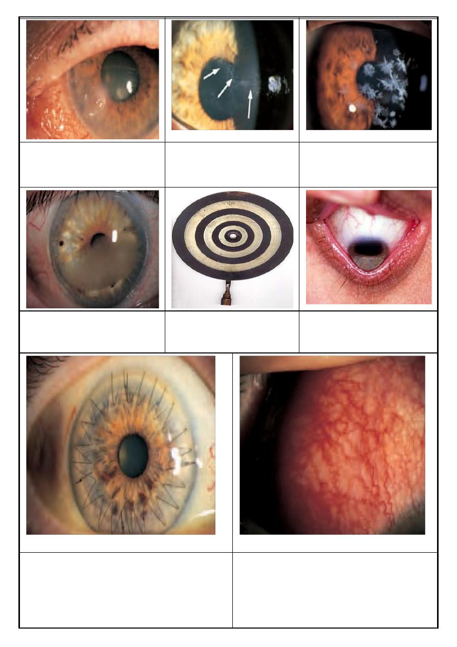
Fig.
7.8
Clinical appearance of a
corneal ulcer.
Fig.
7.9
The clinical appearance
of acanthamoeba keratitis. Arrows
indicate neurokeratitis.
Fig.
7.10
Example of a corneal
dystrophy (granular dystrophy).
Fig.
7.11
Placido disc
Munson’s sign
.
7.12
A corneal graft, nFigote the interrupted and the
continuous sutures at the interface between graft and
host.
Fig.
7.13
The appearance of scleritis.