
Dr.Hameda abdulmahdi College of Medicine /Dep. of anatomy & histology 2nd
stage Date:21/11/2016
1
Lecture Objectives
By the end of this lecture, students are expected to:
1. List and describe the layers of the hart.
2. Outline the histological features of the pericardium.
3. Outline the different types of epithelium simple squamous cells found in heart.
4. nSummarize the functional and histological structure of Purkinje fibres.
The circulatory system: introduction
The circulatory system pumps and directs blood cells and substances carried in blood to
all tissues of the body. It includes both the blood and lymphatic vascular systems, and in an
adult the total length of its vessels is estimated at between 100,000 and 150,000 kilometers.
The blood vascular system , or cardiovascular system, consists of the following structures:
■
The heart propels blood through the system.
■
Arteries , a series of vessels efferent from the heart that become smaller as they branch
into the various organs, carry blood to the tissues.
■
Capillaries , the smallest vessels, are the sites of O 2 , CO 2 ,n nutrient, and waste product
exchange between blood and tissues. Together with the smallest arterial and venous
branches carrying blood to and from them, capillaries in almost every organ form a complex
network of thin, anastomosing tubules called the microvasculature or microvascular bed.
■
Veins result from the convergence of venules into a system of larger channels that
continue enlarging as they approach the heart, toward which they carry the blood to be
pumped again. two major divisions of arteries, microvasculature, and veins make up the
pulmonary circulation , where blood is oxygenated in the lungs, and the systemic circulation
, where blood brings nutrients and removes wastes in tissues throughout the body.
The lymphatic vascular system , begins with the lymphatic capillaries , which are thin-
walled, closed-ended tubules carrying lymph, that merge to form vessels of steadily
increasing size. The largest lymph vessels connect with the blood vascular system and empty
into the large veins near the heart. This returns fluid from tissue spaces all over the body to
the blood.
The internal surface of all components of the blood and lymphatic systems is lined by a
single layer of a squamous epithelium, called endothelium . As the interface between blood
and the organs, cardiovascular endothelial cells have crucial physiologic and medical
importance. Not only must endothelial cells maintain a selectively permeable,
antithrombogenic (inhibitory to clot formation) barrier, they also determine when and
where white blood cells leave the circulation for the interstitial space of tissues and secrete a
variety of paracrine factors for vessel dilation, constriction, and growth of adjacent cells.
Dr.Hameda abdulmahdi College of Medicine /Dep. of anatomy & histology 2nd
stage Date:21/11/2016
2
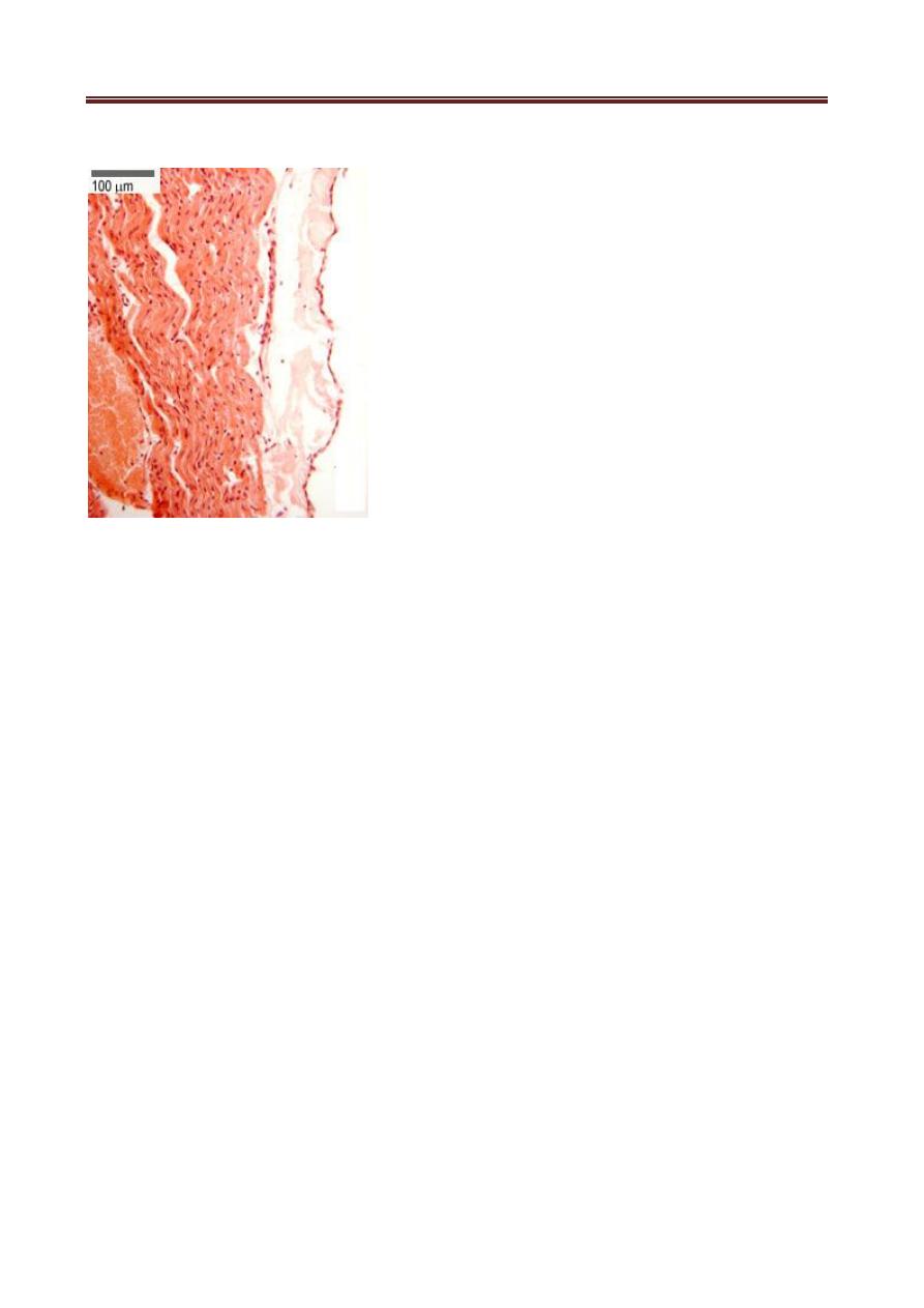
Heart
Cardiac muscle in the four chambers of the heart wall contracts rhythmically, pumping
the blood through the circulatory system . The right and left ventricles propel blood to the
pulmonary and systemic circulation, respectively; right and left atria receive blood from the
body and the pulmonary veins, respectively. The walls of all four heart chambers consist of
three major layers: the internal endocardium; the middle myocardium; and the external
epicardium.
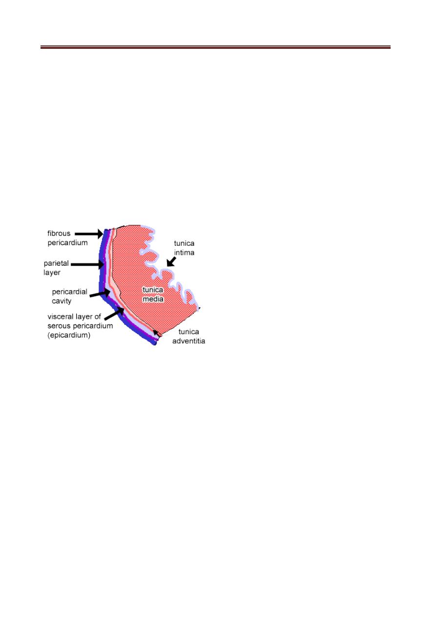
The simple squamous epithelium of the tunica adventitia layer of the heart
(mesothelium) is also the visceral layer of the serous pericardium, the pericardium is a two-
layered connective tissue sac that encloses the heart. The fibrous pericardium is
the outer layer, and the serous pericardium is the inner layer. The space between the two
layers is the pericardial cavity, that contains serous fluid. This facilitates the pumping action
of the heart.
The layers of haert
The endocardium consists of a very thin inner layer of endothelium and supporting
connective tissue, a middle myoelastic layer of smooth muscle fibers and connective tissue,
and a deep layer of connective tissue called the subendocardial layer that merges with the
myocardium Branches of the heart’s impulse-conducting system, consisting of modified
cardiac muscle fibers, are also located in the subendocardial layer. The thickest layer, the
myocardium, consists mainly of cardiac muscle with its fibers arranged spirally around each
heart chamber. Because strong force is required to pump blood through the systemic and
pulmonary circulations, the myocardium is much thicker in the walls of the ventricles,
particularly the left, than in the atrial walls .
The epicardium is a simple squamous mesothelium supported by a layer of loose
connective tissue containing blood vessels and nerves . The epicardium corresponds to the
visceral layer of the pericardium, the membrane surrounding the heart. Where the large
vessels enter and leave the heart, the epicardium is reflected back as the parietal layer lining
the pericardium.
Dr.Hameda abdulmahdi College of Medicine /Dep. of anatomy & histology 2nd
stage Date:21/11/2016
3
Tunica Adventitia (Epicardium)
The thickest layer, the myocardium, consists mainly of cardiac muscle with its fibers
arranged spirally around each heart chamber. Because strong force is required to pump
blood through the systemic and pulmonary circulations, the myocardium is much thicker in
the walls of the ventricles, particularly the left, than in the atrial walls
During heart movements, underlying structures are cushioned by deposits of adipose
tissue in the epicardium and friction within the pericardium is prevented by lubricant fluid
produced by both layers of serous mesothelial cells within these major layers the heart
contains other structures important for its overall function of moving blood. Within the
subendocardial layer and adjacent myocardium, modified cardiac muscle cells make up the
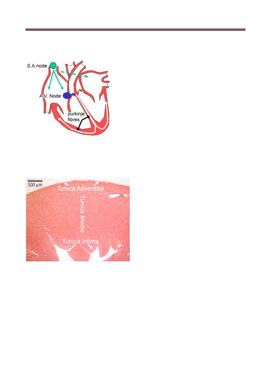
impulse conducting system of the heart, which generates and propagates waves of
depolarization that spread through the myocardium to stimulate rhythmic contractions.
First, impulses are generated by the sinoatrial node (SA), which is found in the wall of the
superior vena cava. It is a small mass of specialized cardiac muscle fibers and associated
connective tissue, and is supplied by nerve fibers from the autonomic nervous system,
excitation of the SA node sets of a wave of depolarization around the atria via gap
junctions between the muscle fibers, next the atrioventricular node (AV) starts impulse
generation around the ventricles , the AV node lies in the interatrial septum, impulses are
sent from the AV node into the AV bundle, or bundle of his, which branches to form Purkinje
fibers, the AV node is also supplied by nerve fibers from the autonomic nervous system that
speed up and slow down the heart rate.
Purkinje fibers lie in the deepest layer of the endocardium and supply the papillary
muscles. Hence the apex of the heart contracts first, followed by the papillary muscles, and
Dr.Hameda abdulmahdi College of Medicine /Dep. of anatomy & histology 2nd
stage Date:21/11/2016
4
then the wave of depolarization spreads up the walls of the ventricles from the base
upwards, as shown in the diagram.
The layers of heart: tunica intima, media and adventitia