
Dr. Hameda abdulmahdi College of Medicine
Dep. of anatomy& histology 2nd stage
1
Lecture Objectives
By the end of this lecture, students are expected to:
1. Summarize the structures of muscular arteries and veins differ.
2. identify the three layers of the vein.
3. identify the different types of capillaries, and how their structure is
related to their function.
4. identify the three layers of the artery.
5. Compare the histological features of conducting and distributing
arteries.
6. Compare the histological features of large arteries and large veins
in tunica adventitia .
7. Summarize the functional and histological structure of capillary bed.
8. Outline the histological structure of arteriole and venules.
Elastic Arteries
Elastic arteries are the aorta, the pulmonary artery, and their largest
branches; these large vessels are also called conducting arteries because their
major role is to carry blood to smaller arteries, the most prominent feature of
elastic arteries is the thick media in which elastic lamellae, each about 10 μm
thick, alternate with layers of smooth muscle fibers. The adult aorta has about 50
elastic lamellae (more if the individual is hypertensive).
Tunica intima is made up of an epithelium, which is a single layer of flattened
epithelial cells, together with a supporting layer of elastin rich collagen. It is well
developed, with many smooth muscle cells in the subendothelial connective
tissue, and often shows folds in cross section as a result of the loss of blood
pressure and contraction of the vessel at death .The internal elastic lamina is not
easily discerned because it is similar to the elastic laminae of the next layer.
Tunica media is broad and elastic with concentric fenestrated sheets of elastin,
and collagen and only relatively few smooth muscle fibers.
Tunica adventitia has small 'vasa vasorum' as the large arteries need their own
blood supply.
Dr. Hameda abdulmahdi College of Medicine
Dep. of anatomy& histology 2nd stage
2
Muscular Arteries
The muscular arteries distribute blood to the organs and help regulate blood
pressure by contracting or relaxing, these arteries distribute blood to various
parts of the body, These included arteries such as the femoral and coronary
arteries, the walls of these arteries have lots of smooth muscle, which means
that they are able to contract or relax (dilate) to change the amount of blood
delivered, as needed.
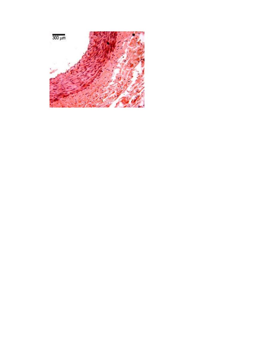
Comparing these arteries to the elastic arteries, the sheet of elastin is now
much reduced, and found at the border between the tunica intima and tunica
media in a layer called the internal elastic layer (IEL) which can be seen very
clearly. Less well defined is the external elastic layer (EEL), between the tunica
media and tunica adventitia. There is a well defined circular layer of smooth
muscle in the tunica media.
The tunica intima has an endothelium of flattened endothelial
cells. The tunica media is primarily a layer of smooth muscle, with some elastin
an collagen. muscle layer, and is sandwiched between the IEL and EEL.
The Tunica Adventitia is very broad, and mostly contains collagen and elastin.
Arteriole.
Arterioles almost always branch to form anastomosing networks or beds of
capillaries that surround the parenchymal cells of the organ. Smooth muscle
fibers act as sphincters closing arterioles and producing periodic blood flow into
capillaries . Acting as “resistance vessels,” muscle tone usually keeps arterioles

Dr. Hameda abdulmahdi College of Medicine
Dep. of anatomy& histology 2nd stage
3
partially closed In certain tissues and organs arterioles deviate from this simple
path to accommodate various specialized functions, for example,
thermoregulation by the skin involves arterioles that can bypass capillary
networks and connect directly to venules.
The media and adventitia are thicker in these arteriovenous shunts (or
arteriovenous anastomoses) and richly innervated by sympathetic and
parasympathetic nerve fibers. The autonomic fibers control the degree of
vasoconstriction at the shunts, regulating blood flow through the capillary beds.
High capillary blood flow in the skin allows more heat dissipation from the body,
while reduced capillary blood flow conserves heat— important functions when
the environmental temperature is hot or cold, respectively
The internal elastic lamina layer is still present. The T.M. (tunica media layer)
has no more than six concentric rings of smooth muscle, and the tunica adventia
(T.A.) layer is approximately the same size as the T.M.
Larger arterioles have a lumen less than 100 to 300 µm in diameter. Arterioles
are small arteries that deliver blood to capillaries. Arterioles control blood flow
through capillary beds by contracting or dilating the size of the lumen, and
therefore the tunica media layer contains concentric rings of smooth muscle to
do this. This compartment is important in determining your blood pressure as
the narrow diameter of these blood vessels resists blood flow, and the back
pressure helps to stretch the walls of the arteries during heart contractions.
The tunica intima is very thin, and mostly consists of a single layer of squamous
epithelium.
The tunica media consists almost entirely of a single layer up to six layers of
smooth muscle cells, and there is no EEL.
The Tunica adventitia is about the same size as the tunica media layer,
merges in with surrounding tissue.
Dr. Hameda abdulmahdi College of Medicine
Dep. of anatomy& histology 2nd stage
4
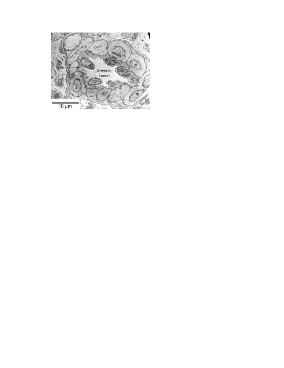
This is an EM of a a very small arteriole. There is only one layer of smooth muscle
(M), but there is still an internal elastic layer (IEL).