Dr. Maryam Mohammed 2
nd
stage College of Medicine /Dep. of anatomy &
histology
1
Lecture Objectives
By the end of this lecture, students are expected to:
1. Summarize the structures of muscular arteries and veins differ
2. identify the three layers of the vein:
3. identify the different types of capillaries, and how their structure is
related to their function.
4. identify the three layers of the artery
5. Compare the histological features of conducting and distributing
arteries
6. Summarize the functional and histological structure of capillary bed
Capillary Beds
Capillaries permit and regulate metabolic exchange between blood and
surrounding tissues, these smallest blood vessels always function in groups
called capillary beds, whose size and overall shape conforms to that of the
structure , the richness of the capillary network is related to the metabolic
activity of the tissues, tissues with high metabolic rates, such as the kidney,
liver, and cardiac and skeletal muscle, have an abundant capillary network; the
opposite is true of tissues with low metabolic rates, such as smooth muscle and
dense connective tissue.

Dr. Maryam Mohammed 2
nd
stage College of Medicine /Dep. of anatomy &
histology
2
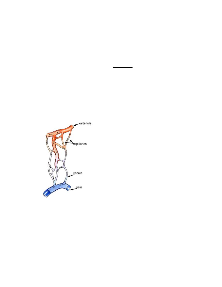
Capillary beds are supplied preferentially by one or more terminal arteriole
branches called metarterioles, which are continuous with thoroughfare
channels connected with the postcapillary venules .true capillaries branch from
the metarterioles, which are encircled by scattered smooth muscle cells, and
converge into the thoroughfare channels, which lack muscle, at the beginning of
each true capillary, muscle fibers act as precapillary sphincters that contract or
relax to control the entry of blood, these sphincters contract and relax cyclically,
with 5 to 10 cycles per minute, causing blood to pass through capillaries in a
pulsatile manner, when the sphincters are closed, blood flows directly from the
metarterioles and thoroughfare channels into postcapillary venules.
Capillaries are small, normally around 3‐4µm, but some capillaries can be 30‐
40 µm in diameter. The largest capillaries are found in the liver.
Capillaries connect arterioles to venules.
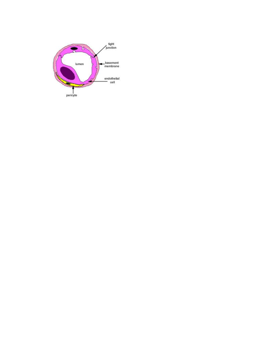
Capillaries have a single layer of flattened endothelial cells, as shown here
in the diagram, there are no muscular or adventitial layers, the thinness of the
capillaries helps efficient exchange between the lumen of the capillary and the
surrounding tissue.
There are three types of capillary:
o
continuous
o
fenestrated
o
discontinuous
Continuous capillaries often have pericytes associated with them. lie just
underneath the endothelium of blood capillaries, and are a source of new
fibroblasts. Continuous capillaries lack fenestrae and have a continuous basal
lamina. They are located in nervous tissue, muscle, connective tissue, exocrine
glands, and the lungs.
Sinusoids, possess many large fenestrae that lack diaphragms. Sinusoidal
capillaries are 30 to 40 _m in diameter, much larger than continuous and
fenestrated capillaries. Sinusoidal capillaries have a discontinuous basal lamina
and lack pinocytotic vesicles. Gaps may be present at the cell junctions,
permitting leakage between endothelial cells ,they are located in the liver,
spleen, bone marrow, lymph nodes, and adrenal cortex.
Dr. Maryam Mohammed 2
nd
stage College of Medicine /Dep. of anatomy &
histology
3
fenestrated capillaries found
in a kidney glomerulus, these are found in some tissues where there is
extensive molecular exchange with the blood such as the small intestine,
endocrine glands and the kidney. The 'fenestrations' are pores that will
allow larger molecules though, these capillaries are more permeable
than continuous capillaries.
Venules have a diameter of 0.2 to 1 mm and are involved in exchange of
metabolites with tissues and in diapedesis (exiting of blood cells through
vessel walls).
Veins
conduct blood away from the organs and tissues and return it to the heart, veins
contain about 70% of the body’s total blood volume at any given time. Their
walls are composed of three layers: the tunica intima (inner), tunica media
(middle), and tunica adventitia (outer), the thickest and most prominent. Vasa
vasorum are more numerous in veins than arteries. A distinct internal elastic
lamina is also absent in veins.
Comparison with arteries. Veins have thinner walls and larger, more irregular
Lumina than the companion arteries. They may have valves in their Lumina that
prevent retrograde flow of the blood .

Dr. Maryam Mohammed 2
nd
stage College of Medicine /Dep. of anatomy &
histology
4
Types of veins
Large veins include the vena cava and pulmonary veins. These veins
possess cardiac muscle in the tunica adventitia for a short distance as
they enter the heart. This layer also contains vasa vasorum and nerves.
Small and medium‐sized veins include the external jugular vein. These
veins have a diameter of 1 to 9 mm.
Venules have a diameter of 0.2 to 1 mm and are involved in exchange of
metabolites with tissues and in diapedesis (exiting of blood cells through vessel
walls).