1
Rickets
Definition:
Rickets signifies a failure in mineralisation of the growing bone or osteoid tissue.
Failure of mature bone to mineralise is osteomalacia.
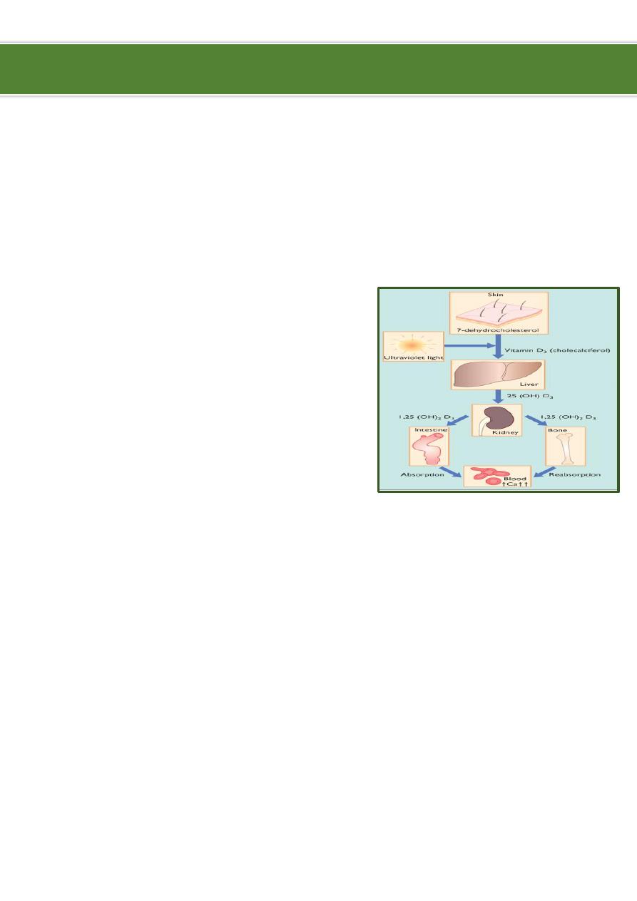
Pathophysiology:
Bone consists of a protein matrix called osteoid
and a mineral phase, principally composed of
calcium and phosphate.
Rickets is a disease of growing bone that is
caused by unmineralized matrix at the growth
plates and occurs in children only before fusion
of the epiphyses.
Because growth plate cartilage and osteoid
continue to expand but mineralization is
inadequate, the growth plate thickens.
There is also an increase in the circumference of the growth plate and the metaphysis,
increasing bone width at the location of the growth plates and causing some of the
classic clinical manifestations, such as widening of the wrists and ankles.
There is a general softening of the bones that causes them to bend easily when subject
to forces such as weight bearing or muscle pull. This softening leads to a variety of
bone deformities.
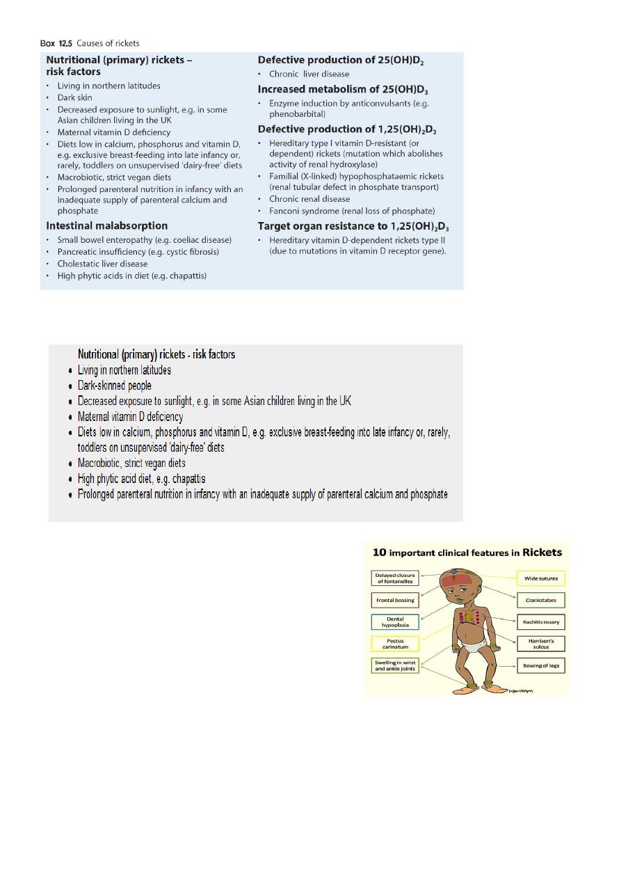
Etiology:
Vitamin D disorders nutritional, congenital, secondary, chronic renal failure,
malabsorption.
Calcium deficiency low intake, dietary inhibitors of calcium absorption,
malabsorption, premature infants (rickets of prematurity)
Phosphorous deficiency low intake, premature infants (rickets of prematurity),
aluminum-containing antacids.
Renal losses X-linked hypophosphatemic rickets, RTA.
Ibnlatef
Notes
Pediatrics
2
Nutritional Vitamin D Deficiency:
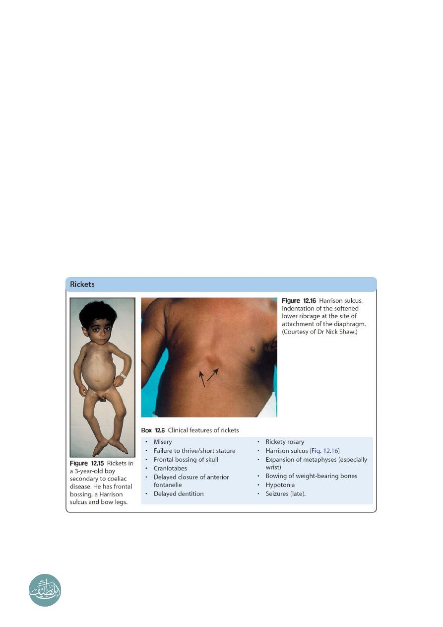
Clinical fractures of rickets:
General:
o
Failure to thrive.
o
Listlessness.
o
Protruding abdomen.
o
Muscle weakness (especially proximal).
o
Fractures.
Head:
o
Craniotabes (softening of the cranial bones and can be detected by applying
pressure at the occiput or over the parietal bones. The sensation is similar to the
feel of pressing into a Ping-Pong ball and then releasing).
o
Frontal bossing.
o
Delayed fontanel closure.
o
Delayed dentition; caries.
o
Craniosynostosis.
3
Chest
:
o
Rachitic rosary.
o
Harrison groove.
o
Respiratory infections and atelectasis.
Back
:
o
Scoliosis.
o
Kyphosis.
o
Lordosis.
Extremities:
o
Enlargement of wrists and ankles.
o
Valgus or varus deformities.
o
Windswept deformity (combination of valgus deformity of 1 leg with varus
deformity of the other leg).
o
Anterior bowing of the tibia and femur.
o
Coxa vara.
o
Leg pain.
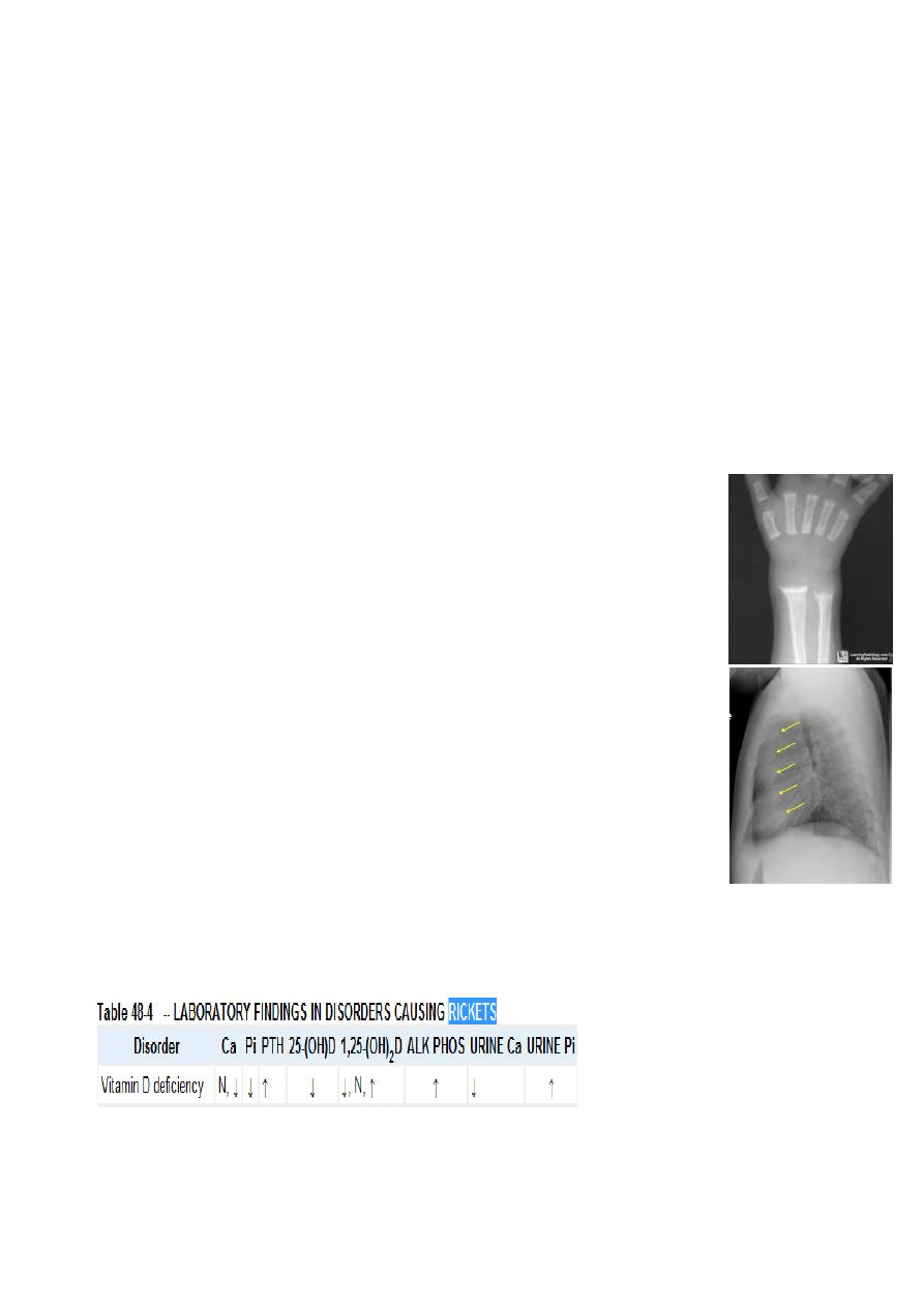
Radiological findings in rickets:
The findings are best seen at the knee, wrist, ankles.
Frayed, cupped, splaying of the metaphysis.
Increased distance between the growing epiphysis & metaphysis.
Generalized decrease in bone density.
Bossing and bowing deformity (due to softening).
Greenstick fractures.
Rackety rosary of the ribs.
Laboratory tests:
The initial laboratory tests in a child with rickets should include serum calcium,
phosphorus, alkaline phosphatase, parathyroid hormone (PTH), 25-hydroxyvitamin D,
1,25-dihydroxyvitamin D3, creatinine, and electrolytes.
Note: Most cases of rickets are diagnosed based on the presence of classic radiographic
abnormalities. The diagnosis is supported by physical examination findings and a history
and laboratory test results that are consistent with a specific etiology
4
Treatment:
Children with nutritional vitamin D deficiency should receive vitamin D and adequate
nutritional intake of calcium and phosphorus (usually provided by milk, formula, and
other dairy products).
There are 2 strategies for administration of vitamin D:
o
Stoss therapy, 300,000-600,000 IU of vitamin D are administered orally or
intramuscularly as 2-4 doses over 1 day. Stoss therapy is ideal in situations where
adherence to therapy is questionable.
o
The alternative is daily, high-dose vitamin D, with doses ranging from 2,000-5,000
IU/day over 4-6 wk.
Either strategy should be followed by daily vitamin D intake of:
o
400 IU/day if <1 yr old.
o
600 IU/day if >1 yr old.
Children who have symptomatic hypocalcemia: might need intravenous calcium
acutely, followed by oral calcium supplements, which typically can be tapered over 2-6
wk in children who receive adequate dietary calcium.
----------------------------------------------------------------------------------------------
www.facebook.com/ibnlatef
https://goo.gl/RpvNsl