Cell Biology
Lec. 9 Dr: Buthaina Al- Sabawi
Date:10/12/2016
Eukaryotic Cytoskeleton
Structure and Function:
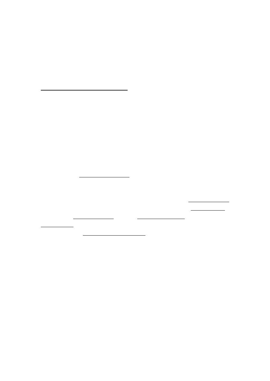
The cytoskeleton is a network of fibers throughout the cell's
cytoplasm.
The structure, function and dynamic behavior of the
cytoskeleton can be very different, depending on organism and
cell type. Even within one cell the cytoskeleton can change
through association with other proteins.
There is a multitude of functions that the cytoskeleton can
perform. Primarily, it gives the cell shape and mechanical
resistance to deformation, and through association with
extracellular connective tissue and other cells it stabilizes entire
tissues. The cytoskeleton can also actively contract, thereby
deforming the cell and the cell's environment and allowing cells
to migrate. Moreover, it is involved in many cell signaling
pathways, in the uptake of extracellular material (endocytosis),
segregates chromosomes during cellular division, is involved in
cytokinesis (the division of a mother cell into two daughter
cells), and for intracellular transport
typically divided into three categories (microfilaments,
intermediate filaments, and microtubules) based on the diameter
and composition of the filaments.
1- Actin filaments (also called microfilaments)
2- Intermediate filaments
3- Microtubules
1
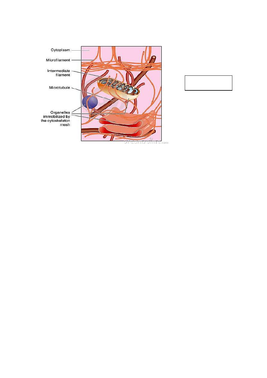
Microfilaments:
Microfilaments are solid rods made of contractile protein
known as actin. When it is first produced by the cell, actin
appears in a globular form (G-actin). In microfilaments,
however, which are also often referred to as actin filaments,
Microfilaments are 5- 7 nm in diameter, represent the active or
motile part of the cytoskeleton. Microfilaments are long
polymerized chains of the molecules are intertwined in a helix,
creating a filamentous form of protein (F-actin).
Microfilaments considered part of the cell cortex, which
regulates the shape and movement of the cell’s surface.
Functions of microfilaments:
1- Provides mechanical strength to the cell
2- Link transmembrane proteins (e.g., cell surface receptor)
to cytoplasmic proteins.
3-Anchors the centrosomes at opposite poles of the cell
during mitosis.
4-Pinches the dividing animal cells during cytokinesis.
5-Supports the plasma membrane.
2
Cytoskeleton
7-Microfilaments association with the protein myosin is
responsible for muscle contraction.
8- They function in the maintenance of cell-shape.
9- These filaments are associated with membrane activities
such as endocytosis and exocytosis.
Microfilaments
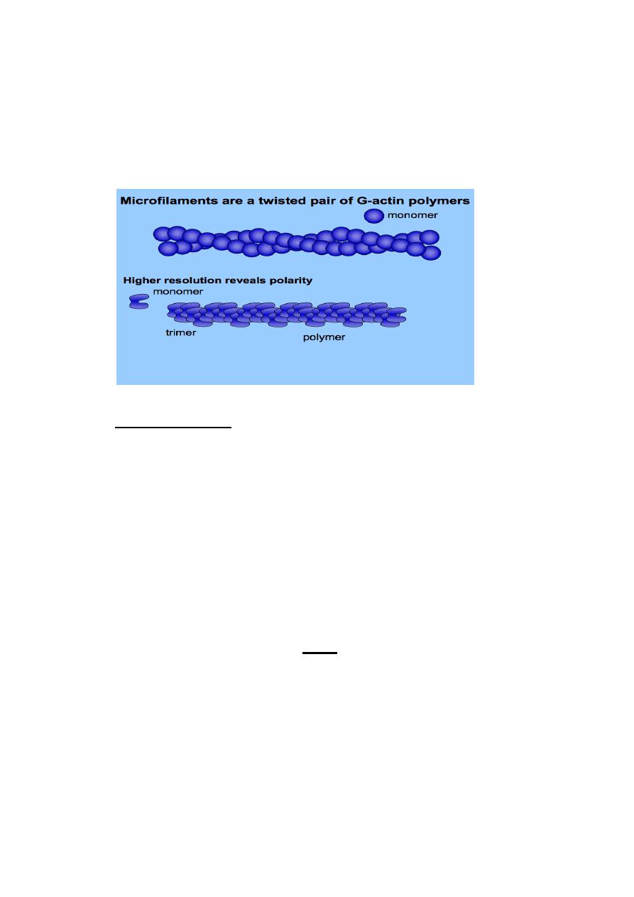
Microtubules:
Microtubules are filamentous intracellular structures that are
responsible for various kinds of movements in all eukaryotic
cells. Microtubules are involved in nucleic and cell division,
organization of intracellular structure, and intracellular
transport.
Microtubules are hollow cylindrical tubes, 20- 25 nm in
diameter (lumen = approximately 15nm in diameter), most
commonly comprised of 13 protofilaments which, in turn, are
polymers of alpha and beta tubulin .They have a very
dynamic behavior, binding GTP for polymerization.
In animal cells, microtubules arise from a region of the cell
called the microtubule organizing center (MTOC) located
near the nucleus.
3
In nine triplet sets (star-shaped), they form the centrioles ,
and in nine doublets oriented about two additional
microtubules (wheel-shaped) they form cilia and flagella
The latter formation is commonly referred to as “9+2”
arrangement, where in each doublet is connected to another
by protein dynein.
They play key roles in:
Intracellular transport (associated with dyneins and
kinesis) they transport organelles like mitochondria or
vesicles) and chromosomes during mitosis.
The axoneme of cilia and flagella .
The mitotic spindle .
Intermediate Filaments
Intermediate filaments have a diameter of about 10nm, which
is intermediate between the diameter of two other principle
4
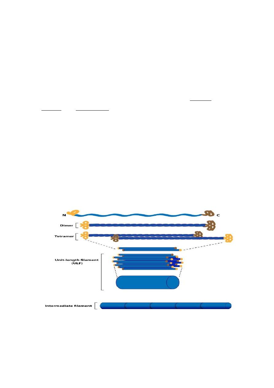
elements of the cytoskeleton , actin filaments and microtubules.
the intermediate filaments are not directly involved in cell
movements. Instead, they appear to play basically a structural
role by providing mechanical strength to cells and tissues.
Like actin filaments, they function in the maintenance of
cell-shape, its form a 'basket' around the nucleus.
Filaments serving as structural components of the nuclear
lamina and sarcomeres. Different intermediate filaments are:
The IFs can be divided into five major classes:-
Class: Name Tissue
i : Acidic Keratins Epithelia
ii : Basic Keratins Epithelia
iii: Desmin : Muscle
Glial: Glial cells and astrocytes
Peripherin : Peripheral neurones
Vimentin: Mesenchyme
iv : Neurofilaments Neuron
v : Lamins Nuclear envelopes
5