NECROSIS
“Necrosis is the morphological changes that follow cell death in a living tissue or organ,
Resulting from the progressive degenerative action of enzymes on the lethally injured cell.”
So,
Necrosis begins with an impairment of the cell’s ability to maintain homeostasis, leading
to an influx of water and extracellular ions
Apoptosis vs Necrosis
The word apoptosis mean falling off.“Apoptosis is a process of programmed and targeted
cause of cellular death”Necrosis is differ from apoptosis:
Apoptosis
Necrosis
Physiological or pathological
Always pathological
Cell shrinkage
Cell swelling
Apoptotic bodies form
Do not form
Dna cleavageNo DNA cleavage
Beneficial
Detrimental
Characteristic nuclear changes
Nuclei lost
No leak of lysosomal enzymes
Leak of lysosomal enzymes
Causes of necrosis
AnoxiaIschemia
Physical agents
Chemical agents
Biological agents
Immunological
Pathogenesis
Necrotic changes in tissues are caused By
Digestion of cell by enzymes
Denaturation of proteins
Digestion of cell by enzymes
This digestion is of two typesAutolysis: Digestion of cell by enzymes derived from their own lyosomes
Heterolysis: Digestion of cell by enzymes derived from lysosmes of leukocytes.
By Denaturation of proteins
Denaturation of proteins caused by intracellular acidosisand due to this result is that:
Injury to the cell membrane
Severe impairment of phosphorylation of cellIncrease permeability of the cell
Influx of Na+ and Ca+ in the cell
Decreased intracellular activity of the cell
Changes in Necrosis
Changes inside the cell
Changes in mitochondriaChanges in Nucleus
Changes in cytoplasmChanges inside the cell
Endoplasmic reticulum is disorganizedThere is rupture of membrane
Ribosomes are shed off
Disorganization of polysomes & their structures
Changes in mitochondria
Mitochondria become swallonLoss of interamitochondrial granules
Loss of cristae & change their shape
Rupture of outer membrane of Mitochondria
Changes in Nucleus
Nucleus becomes smaller
Chromatin loses & become clumped
Nucleus shows following changes
Pyknosis
Karyorrhexis
Karyolysis
PYKNOSIS
“When the DNA is broken down by endonucleasesfragments are formed & the nucleus becomes acid
and stains basophillic”
KARYORRHEXIS
“The pyknotic nucleus may break up into fragmentsand disappear. This process is called karyorrhexis”
KARYOLYSIS
“The pyknotic nucleus may undergo lysis by the
enzyme DNAse”
Changes in cytoplasm
Cytoplasm becomes more eosinophilic:
Due to loss of RNA & denaturation of cytoplasmic proteins
Cytoplasm becomes opaque.TYPES OF NECROSIS
Basic typesCoagulative necrosis
Liquefactive necrosis
Caseous necrosis
In special sites
Fat necrosis
Fibrinoid necrosis
Gangrenous necrosis
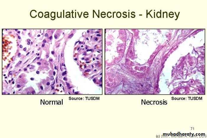
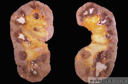
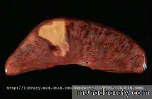
COAGULATIVE NECROSIS
“In this type of necrosis, the necrotic cell retains itscellular outline for several days”
Coagulative necrosis typically occurs in solid organs such as kidney, heart and adrenal gland usually as a result of deficient blood supply and anoxia.
Examples
Myocardial infarction
Mechanism
Denaturation of protein is the basic mechanism of coagulative necrosisThe injury and the subsequent increasing acidosis
denatures not only the structural proteins but also the enzymic proteins, thus blocking the cellular proteolysis.Morphology
Preservation of basic structural outline of the coagulated cells
Appears as a mass of coagulated, pink staining homogenous cytoplasm
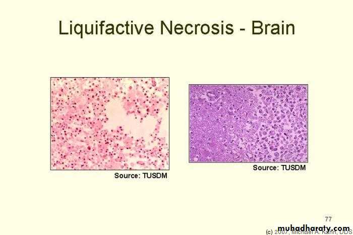
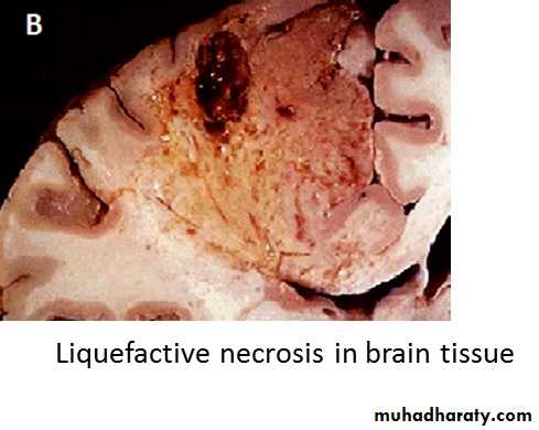
LIQUEFACTIVE NECROSIS
It is the type of necrosis that occurs due to autolytic and
heterolytic actions of enzymes that convert the proteins
of cells into liquid.
It is characterized by softening and liquifaction of tissue.
ExamplesIschemic necrosis of brain
Suppurative inflammation.
Mechanism
Enzymatic degradation of proteins is the basic mechanism of liquefactive necrosisMorphology
Complete loss of cellular detailCellular outline is also destroyed
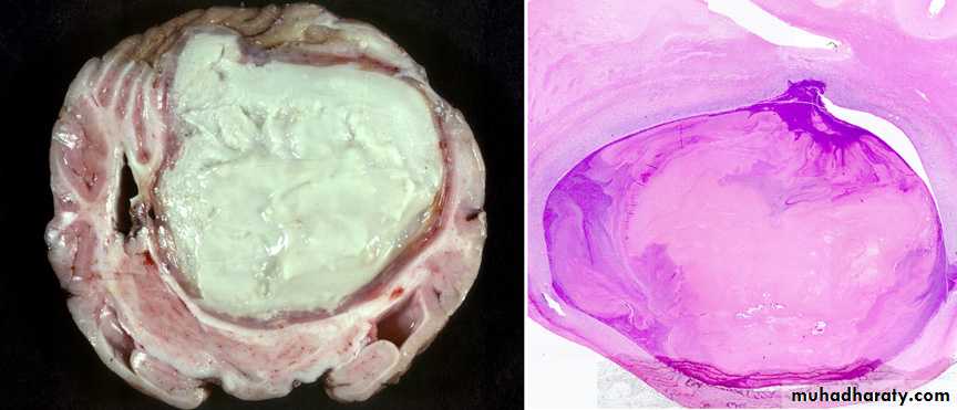
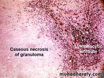
CASEOUS NECROSIS
Combination of coagulative and liquefactive necrosisCharacterized by the presence of soft, dry, cheesy homogenous necrotic material.
It is not liquefied
Examples
Principaly in the center of tuberculous granulomaMorphology
Microscopically the necrotic focus is composed of structureless amorphous granular debris enclosed within a ring of granulomatous inflammation.
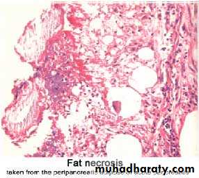
Necrosis in special sites Fat necrosis
It occurs in two forms:
Enzymatic fat necrosisTraumatic fat necrosis
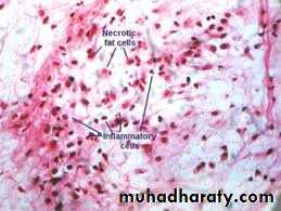
Enzymatic fat necrosis
Most commenly seen in acute pancreatitis.“Refers to the necrosis in adipose tissue, induced by the
action of pancreatic enzymes which are lead due to
trauma to the pancreas”
Morphology
Chalky white opaque spots surrounded by inflammatory margins are seenNecrotic area shows acute inflammatory changes with dissolved fat cells
Traumatic fat necrosis
It occur following severe injury to the tissues with high fat content such as the breast, subcutaneous tissue and abdomen.Morphology
Foam cells and gaint cells are seen.
necrotic foci contain a lot of phagocytes containing fat known as foam cells
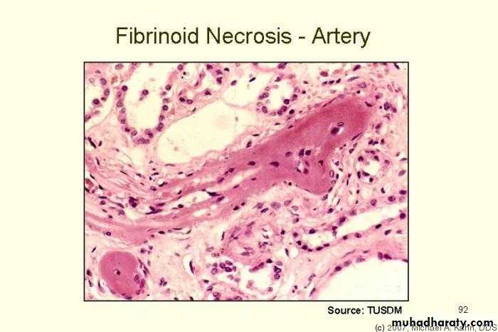
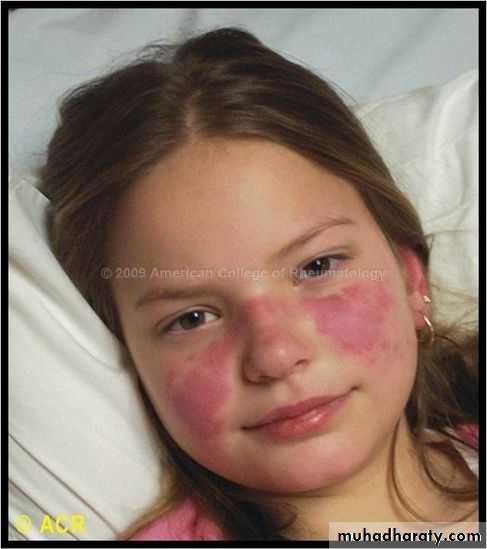
FIBRINOID NECROSIS
Type of connective tissue necrosis especially affecting arterial walls.Mostly seen in two conditions
Auto immune diseases e.gRheumatic fever
SLE
Malignant hypertension
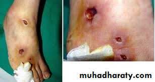
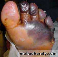
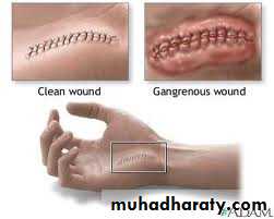
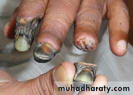
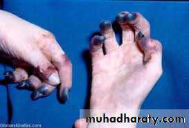
GANGRENOUS NECROSIS
Gangrene is the necrosis of tissue with superadded putrefaction (enzymatic decomposition).It is the clinical condition in which extensive tissue necrosis is complicated to a variable degree by secondary bacterial infection.
Gangree= Necrosis + infection + putrefaction
Causes of gangrene
Arterial obstructon due to:Thrombosis of atherosclerotic artery
Embolus
Diabetes:- atherosclerotic artery , loss of sensation
results reapeted trauma & increase chances of infection
Infection
Gas gangrene
Gangrene of scrotum
Trauma
Crush injuries
Physical agents
Burns
Chemicals
Types of gangrene
Dry gangrene
Wet gangreneGas gangrene
Dry gangrene of foot