
1
Lecture 07 Pathology D. Rasha
CARDIOVASCULAR SYSTEM
Congenital heart diseases:-
It's most common types of congenital malformations and it's most common cause of
heart disease in children.
Its causes in 90%
idiopathic
, while 10% of it reveal either
genetic factors
or
environmental factors
such as congenital rubella infection.
The types of congenital heart diseases include:
1- Malformations causing a left-to-right shunt.
2- Malformations causing a right-to-left shunt (cyanotic congenital heart diseases).
3- Malformations causing obstruction.
(I) Left-to-right shunts:-
is most common types of cardiac malformations, this is usually a cyanotic in early stage
but in later stages can cause cyanosis when produced significant pulmonary hypertension
and reversal blood flow through the shunt occurs, that include:-
1- Atrial septal defects.
2- Ventricular septal defects.
3- Patent ductus arteriosus.
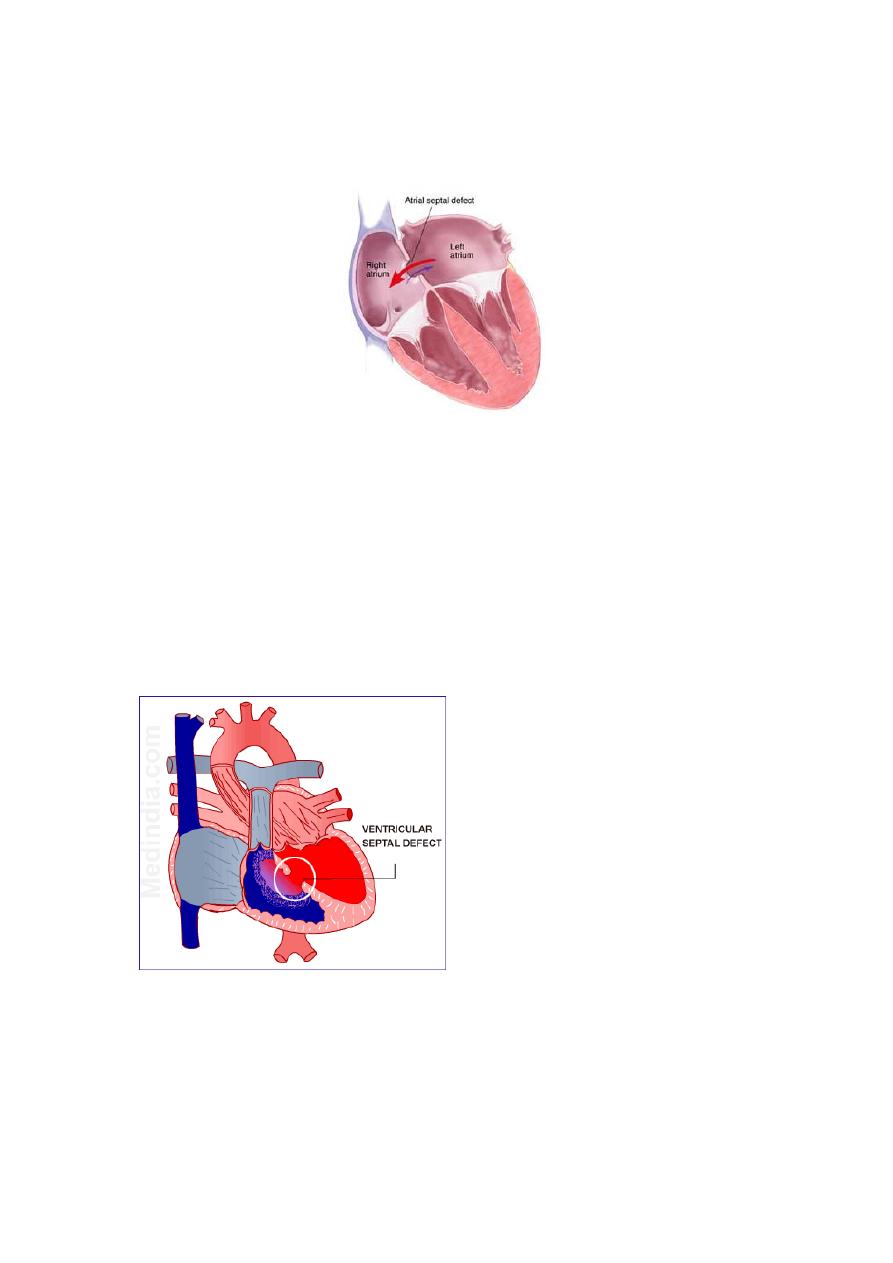
1- Atrial septal defects:-
Is caused most commonly by failure to close of foramen ovale after birth, the foramen
ovale is a flap of tissue in septum between 2 atrium act as a one-way valve allowing blood
to keep flowing from right to left during intrauterine life at the time of birth as pulmonary
vascular resistance fall and systemic arterial pressure increases, so pressure in the left
atrium rises above that in right atrium and must cause functional closure of the foramen
ovale.
Patency persist in about 25% of general population and this will cause shunt of blood from
left to right atrium, this defect usually well tolerated if it's less than 1 cm in diameter but
even larger lesions do not produce any symptom in childhood because the flow of blood
is from left to right, but with time when pulmonary vascular resistance increase and
pulmonary hypertension developed so reversal of shunt so the shunt become right to left
and developed cyanosis.
2
Morphology:-
Manifested as right atrial and ventricular dilation, right ventricular hypertrophy and
dilation of pulmonary artery, pulmonary hypertension developed.
Ventricular septal defect:-
VSDs are the most common congenital heart defects and this like ASDs occur in isolation
or in association with other cardiac malformations, the size and location of defect is
variable ranging from minute to large defect and this may close spontaneously during
infancy or childhood.
Morphology:-
In large defect associated with significant left to right shunt so right
ventricle is hypertrophied and dilated, with pulmonary hypertension developed if this
occur so reverse of shunt and cyanosis occur.
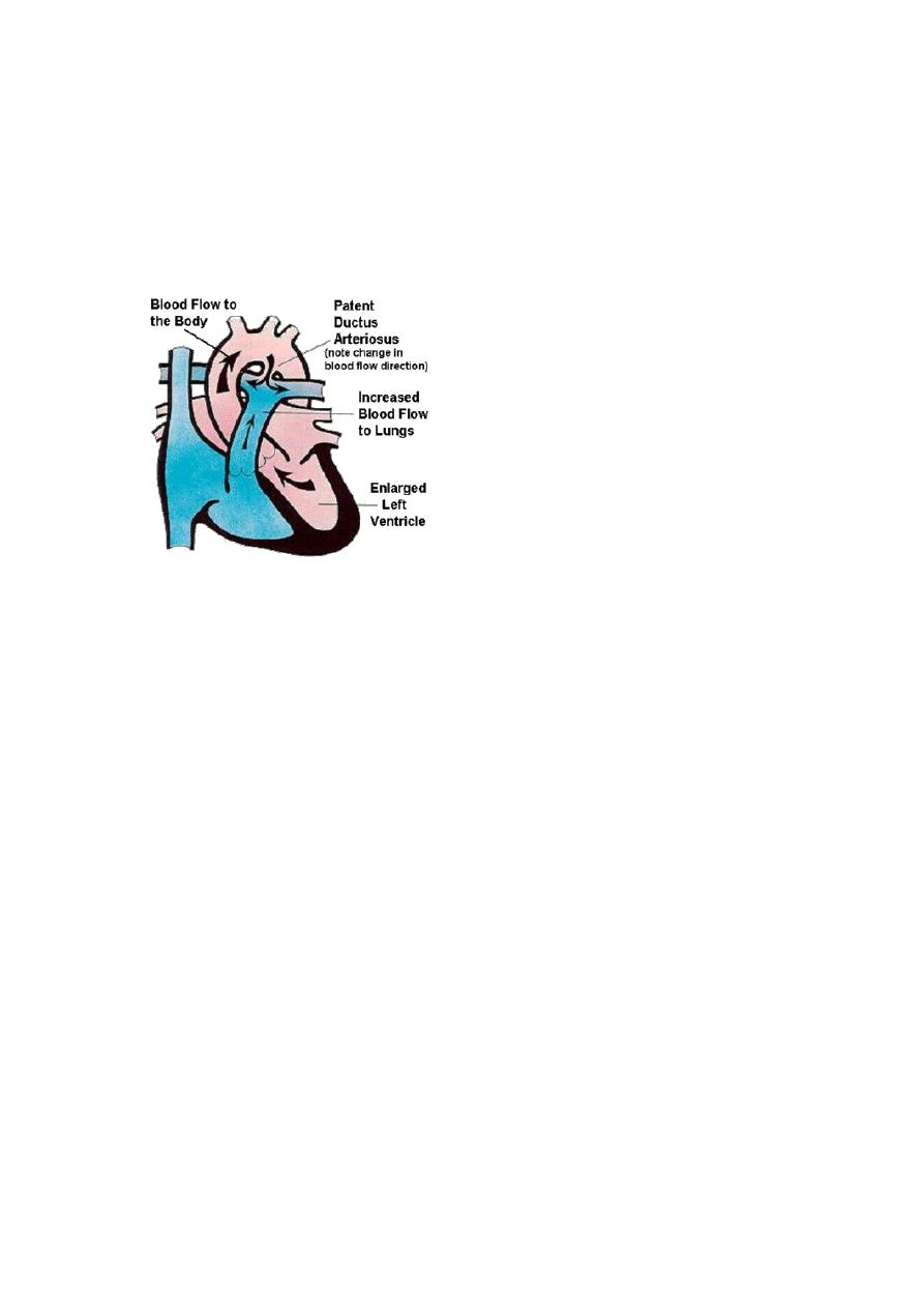
Patent ductus arteriosus:-
Ductus arteriosus:
is an arterial channel that courses between the pulmonary artery and
aorta. During intrauterine life, the DA permits blood to flow freely from the pulmonary
artery to the aorta. Complete irreversible closure occurs within the 1
st
few months after
birth, this closure may be delayed or failed to occur to give PDA condition.
3
Morphology:-
The oxygenated blood flows from the left ventricle to the lungs and is returned to the left
atrium, so form volume overload to cause dilation and hypertrophy of left atrium and
ventricle.
The proximal pulmonary arteries are also dilated with development of pulmonary
hypertension and cause right ventricular hypertrophy and dilation and right atrial dilation.
(II) Right-to-left shunts:-
Cardiac malformations associated with right-to-left shunts are distinguished by cyanosis
at or near the time of birth, this occur because poorly oxygenated blood from the right
side of the heart is introduced directly into the arterial circulation.
2 important conditions cause cyanotic congenital heart disease:
1- Tetralogy of Fallot
2- Transposition of great vessels.
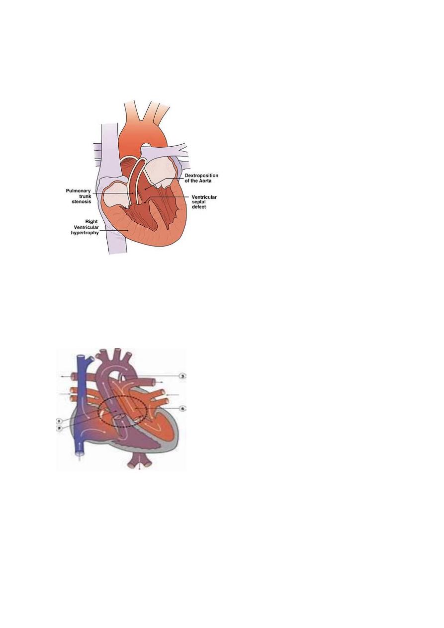
Tetralogy of Fallot:-
TOF is the most common cause of cyanotic congenital heart disease, the four components
of TOF:
1- VSD.
2- Dextraposed aortic root that overrides the VSD.
3- Right outflow obstruction.
4- Right ventricular hypertrophy.
Patient with TOF is at risk to develop infective endocarditis, systemic emboli and brain
abscesses.
4
Morphology:-
The heart is enlarged externally by right ventricular hypertrophy, the
proximal aorta is larger than pulmonary trunk, so that because of stenotic pulmonary
artery, this will lead to shunt unoxygenated blood from right ventricle through VSD to go
to left ventricle and aorta.
Transposition of great arteries:-
There's abnormal truncal septation, the aorta arises from the right ventricle and the
pulmonary artery from the left ventricle, so all unoxygenated blood with pump from right
ventricle to systemic circulation by aorta and cause cyanosis but with presence of another
defect as
ASD, VSD
and
PDA
, this allows oxygenated blood to reach aorta.
Congenital obstructive lesions:-
As :
1- valvular aortic stenosis.
2- Pulmonic stenosis.
3- Coarctation of aorta.

5
Pericardial diseases:-
The most diseases of pericardium are:
1- Inflammatory condition (pericarditis).
2- Pericardial effusion.
Pericarditis:-
It's divided into:
1- Primary
pericarditis: is uncommon and any organism may cause the infection but
viruses are the most causative agents.
2- Secondary
pericarditis: is most common and the most common
systemic
cause of
it is uremia.
Other causes are: rheumatic fever, SLE and metastatic carcinoma in this case the
effusion is bloody.
Local
causes of secondary pericarditis are acute M.I, cardiac surgery and radiation
to medaistinum.
The pericarditis is either resolve without significant sequelae or progress to chronic
fibrosing process.
Pericardial effusion:-
It's accumulation of fluid in pericardial space is often asymptomatic if it accumulate
slowly, but rapid developing effusions may cause tamponade the most cause of effusions
are congestive heart failure, hypoalbuminemia, malignancy, and mediastinal lymphatic
obstruction.
Cardiac tumors:-
Metastatic carcinoma:-
It's more common than primary one, the most common sites of metastasis to the heart
are: lungs, breast and malignant melanoma, the metastatic cells usually goes to
pericardium to cause pericarditis and hemorrhagic pericardial effusions.
Primary neoplasms:-
Less common and the common primary neoplasm are:
1- Myxoma: is a benign tumor,
2- Cardiac rhabdomyoma: is a benign tumor.
3- Lipoma.