Page
1
L3/PHYSIOLOGY OF
FEMALE REPRODUCTION.
Dr. HANAN AL-TAEE.
IF THE OVA IS FERTILIZED:
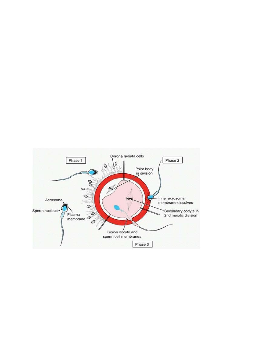
Fertilization:
Is the fusion of male and female gametes and formation of the zygote.
First step is capacitation of the sperm which is the process by which the sperm
acquire the ability to fertilize the ovum in female genital tract.
Figure demonstrates the steps of fertilization.
Second step is the Acrosome Reaction:
1. Hyaluronidase : dissolves the matrix of cumulus‐oophorous. 2. A trypsin‐ like
enzyme known as the acrosomal proteinase (acrosin) acts on the zona pellucida.
The oocyte will complete meiosis II and second pollar body will be extruded.

Page
2
Third step is the fusion of the sperm with the ovum and formation of the
pronuclei. Thereafter a discharge of cortical granules leading to deposition of the
granules contents in the perivitelline‐space of the ovum. The secreted materials
diffuse rapidly into the zona pellucida and provoke a change in its physiological
and biochemical properties .The setting of a reduction in the membrane potential
of the ovum may be responsible for prevention of polyspermy & polyploidy.
Implantation:
It takes 6–7 days for the embryo (after subsequent divisions) to travel down to
the uterus. By then it is a blastocyst. Implantation involves an interactive process
between the uterus and the embryo, with both responding mutually to one
another. The process of implantation, usually on the dorsal wall of the uterus.
Implantation may be separated into a series of developmental phases, starts with
apposition, adhesion, penetration and invasion. Early embryo is capable of
producing enzymes that degrade the basement membrane and, with
prostaglandin‐ induced local edema; trophoblastic cells enter the endometrial
stroma. The trophoblast and the decidua produce a large variety of growth
factors, cytokines and paracrine factors. Of them, are platelet‐ derived growth
factor (PDGF), fibroblast growth factor (FGF), insulin‐like growth factor (IGF) and
transforming growth factor (TGF). The trophoblast and decidua also synthesize
receptors for these and other implantation‐ promoting factors. The trophoblast
actually digests its way into the endometrium, which then covers it up.
Gestation:
Gestation period refers to the pregnancy period. The average gestation period is
about 280 days or 40 weeks from the date of last menstrual period (LMP). If the
menstrual cycle is normal 28 day cycle, the fertilization of ovum by the sperm
occurs on 14th day after LMP. Thus the actual duration of human pregnancy is
280 – 14 = 266 days. During gestation, the pregnancy is maintained by:
a) First by the corpus luteum.
b) Then by the blastocyst, which maintains the corpus luteum (hCG)

Page
3
c) Then by the placenta.
Placentation:
The chorion of the trophoblast and the surrounding endometrium react to each
other to form a placenta.
Placenta is a temporary membranous vascular organ that develops in females
during pregnancy. It is expelled after childbirth. Placenta forms a link between the
fetus and mother. It is the physical attachment and physiological connection
between fetus and mother. Placenta is implanted in the wall of the uterus. It
consists of two parts namely the fetal part and the mother’s part. It is connected
to the fetus by umbilical cord, which contains blood vessels and connective tissue.
The delivery of fetus is followed by the expulsion of placenta. After expulsion of
the placenta, the umbilical cord is cut. The site of attachment of placenta in the
center of anterior abdomen of fetus is called navel or umbilicus.
Functions of placenta:
1. Nutritive function:
Nutritive substances, electrolytes and hormones necessary for the development
of fetus diffuse from mother’s blood into fetal blood through placenta.
2. Excretory function:
Metabolic end products and other waste products from the fetal body are
excreted into the mother’s blood through placenta.
3. Respiratory function:
Fetal lungs are nonfunctioning and placenta forms the respiratory organ for fetus.
Oxygen necessary for fetus is received by diffusion from the maternal blood and
carbon dioxide from fetal blood diffuses into the mother’s blood through
placenta. Exchange of respiratory gases between fetal blood and maternal blood
occurs mainly because of pressure gradient. Partial pressure of oxygen in the

Page
4
maternal blood is 50 mm Hg. In fetal blood, the partial pressure of oxygen is 30
mm Hg. This pressure gradient of 20 mm Hg causes the diffusion of oxygen into
the fetal blood. This pressure gradient is very low, compared to the gradient
existing between partial pressure of oxygen in arterial blood and alveoli in adults.
Still, an adequate quantity of oxygen is available for fetus. This is because of two
reasons:
1. The hemoglobin in fetal blood (Hb‐F) has 20 times more affinity for oxygen than
the adult hemoglobin. 2. The concentration of hemoglobin is about 50%more in
fetal blood than in adult blood.
4. Endocrine function:
Hormones secreted by placenta are:
a.Human chorionic gonadotropin.
b. Estrogen.
c. Progesterone.
d. Human chorionic somatomammotropin.
e. Relaxin.
a. Human Chorionic Gonadotropin: (hCG):
IS a glycoprotein with chemical structure similar to that of LH. It comprised of
two non‐identical polypeptide chains (alpha and β), with almost exactly the same
properties as LH. hCG is secreted by growing chorion of the developing embryo
and later by the placenta, it forms the basis of pregnancy tests, urine tests usually
detect hCG 14 days after fertilization.
Actions of hCG:
i. On corpus luteum: hCG is responsible for the preservation and the secretory
activity of corpus luteum. Progesterone and estrogen secreted by corpus
luteum are essential for the maintenance of pregnancy. Deficiency or absence

Page
5
of hCG during the first 2 months of pregnancy leads to termination of
pregnancy (abortion), because of involution of corpus luteum.
ii.Immune tolerance for the embryo.
iii.On fetal testes: Action of hCG on fetal testes is similar to that of LH in adults.
It stimulates the interstitial cells of Leydig and causes secretion of
testosterone. The testosterone is necessary for the development of sex organs
in male fetus.
iv. it helps Invasion of the trophoblast into the endometrium.
v. it has an association to the severity of morning sickness of pregnancy.
b. Estrogen:
Placental estrogen is similar to ovarian estrogen in structure and function.
Actions of placental estrogen:
i. On uterus: Causes enlargement of the uterus so that, the growing fetus can be
accommodated.
ii. On breasts: Responsible for the enlargement of the breasts and growth of the
duct system in the breasts.
Estrogen suppresses prolactin and thus inhibits milk
production during pregnancy.
iii. On external genitalia: Causes enlargement of the female external genitalia.
iv. On pelvis: Relaxes pelvic ligaments. It facilitates the passage of the fetus
through the birth canal at the time of labor.
d. Progesterone:
Placental progesterone is similar to ovarian progesterone in structure and
function.
Actions of placental progesterone

Page
6
i. On endometrium of uterus: Accelerates the proliferation and development of
decidual cells in the endometrium of uterus. The decidual cells are responsible for
the supply of nutrition to the embryo in the early stage.
ii. On the uterus: Inhibits the contraction of muscles in the pregnant uterus. It is
an important function of progesterone as it prevents expulsion of fetus during
pregnancy.
iii. On breasts: Causes enlargement of breasts and growth of duct system of the
breasts.
Progesterone is responsible for further development and preparation of
mammary glands for lactation.
d. Human Chorionic Somatomammotropin: human placental lactogen.
Human chorionic somatomammotropin (HCS) is a protein hormone secreted from
placenta. It is often called placental lactogen. It acts like prolactin and growth
hormone secreted from pituitary. Actions of HCS:
i. On breasts: In experimental animals, administration of HCS causes enlargement
of mammary glands and induces lactation.
ii. On protein metabolism:
HCS acts like GH on protein metabolism. It causes anabolism of proteins and
accumulation of proteins in the fetal tissues. Thus, the growth of fetus is
enhanced.
iii. On carbohydrate metabolism: It reduces the peripheral utilization of glucose in
the mother leading to availability of large quantity of glucose to the growing
fetus.
iv. On lipid metabolism: It mobilizes fat from the adipose tissue of the mother. A
large amount of free fatty acid is made available as the source of energy in the
mother’s body. It compensates the loss of glucose from the mother’s blood to
fetus.
Page
7
e. Relaxin:
Relaxin is a polypeptide, which is secreted by corpus luteum. It is also secreted in
large quantity by placenta at the time of labor.
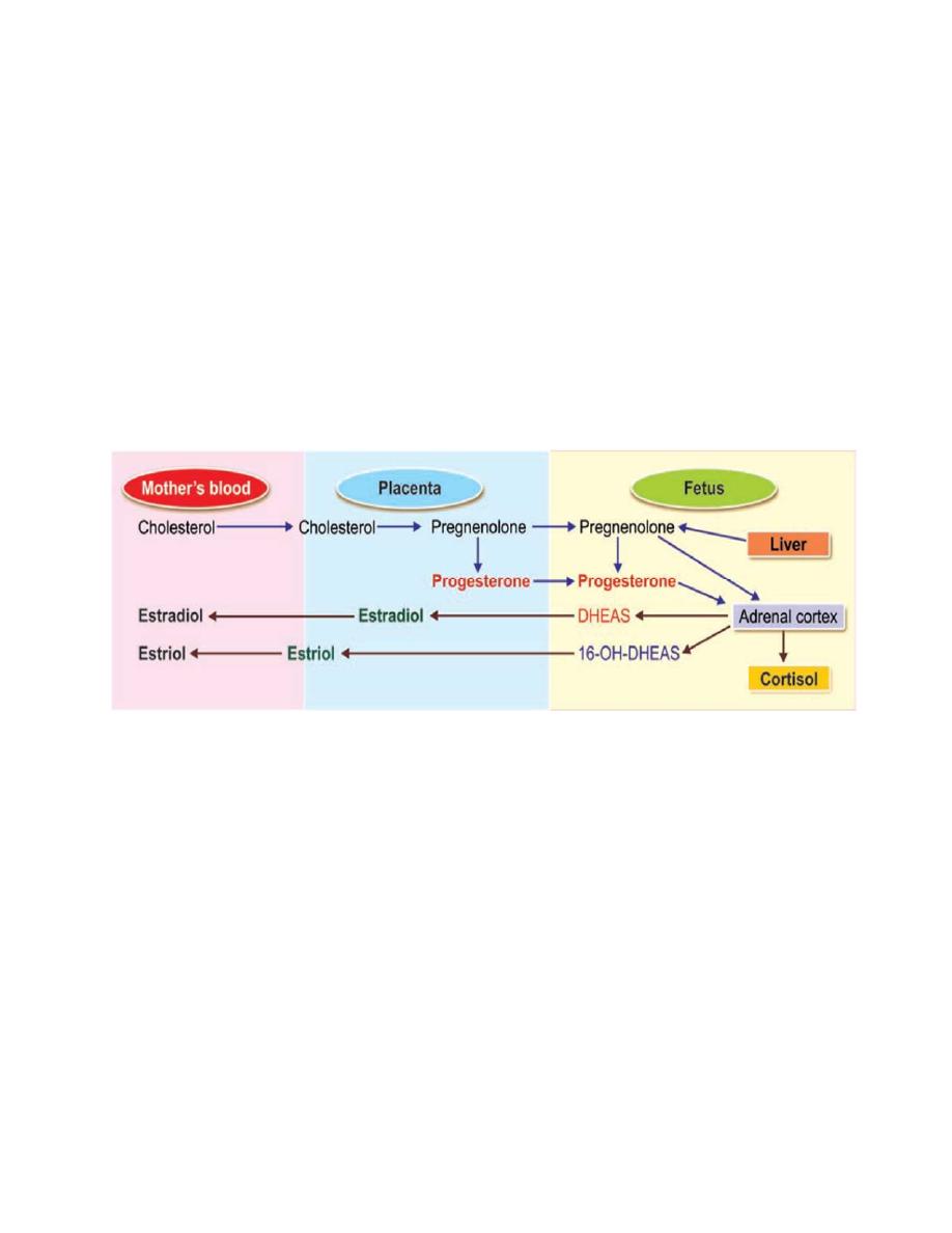
Fetoplacental unit:
Fetoplacental unit refers to the interaction between fetus and placenta in the
formation of steroid hormones. The interaction between fetus and placenta
occurs because some of the enzymes involved in steroid synthesis present in fetus
are absent in placenta and those enzymes, which are absent in fetus are present
in placenta.
Figure demonstrates the fetoplacental unit of steroid synthesis.
Fetoplacental unit. DHEAS = Dehydroepiandrosterone sulfate, 16 OHDHEAS =
16hydoxydehydroepiandrosteronesulfate.