Brachial plexus

OBJECTIVES
…
To define the parts of brachial plexus
To list the branches of each
To apply some clinical conditions on
important injuries affecting the branches
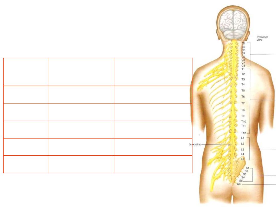
The spinal nerves:
-31 pairs of nerves spring from the spinal cord, and are
transmitted through the intervertebral foramina
Number of vertebrae
Number of nerves
Region
7
8
Cervical
12
12
Thoracic
5
5
Lumbar
5
5
Sacral
1
1
Coccygeal
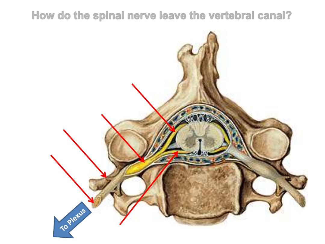
How do the spinal nerve leave the vertebral canal?
Dorsal root
Sensory
Ventral root
Motor
Spinal nerve
Mixed
Dorsal ramus
Mixed
Ventral ramus
Mixed
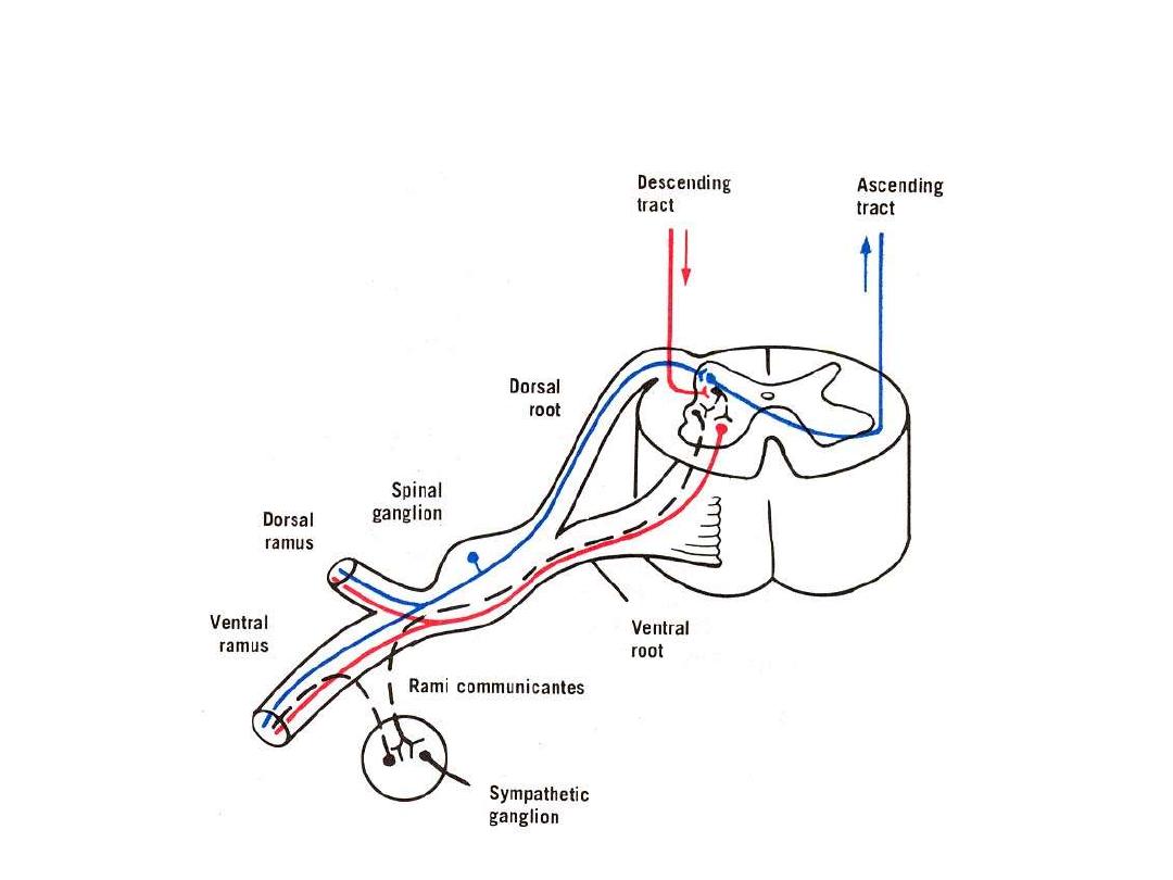
All plexuses arising from the ventral rami of spinal nerves contain
sensory, motor, and autonomic fibers
Brachial plexus:
A major nerve network supplying the upper limb
The plexus is formed by the union of the anterior rami of lower 4 cervical
(C5-C8) & most of the first thoracic (T1) nerves
Originates in the neck, passes laterally and inferiorly over the first rib to
enter the axilla
Most of its branches are given in the axilla
While descending from the neck to UL, these nerves exhibit different
branching & union events so it is divided into four major parts:
ROOTS
TRUNKS
DIVISIONS
CORDS
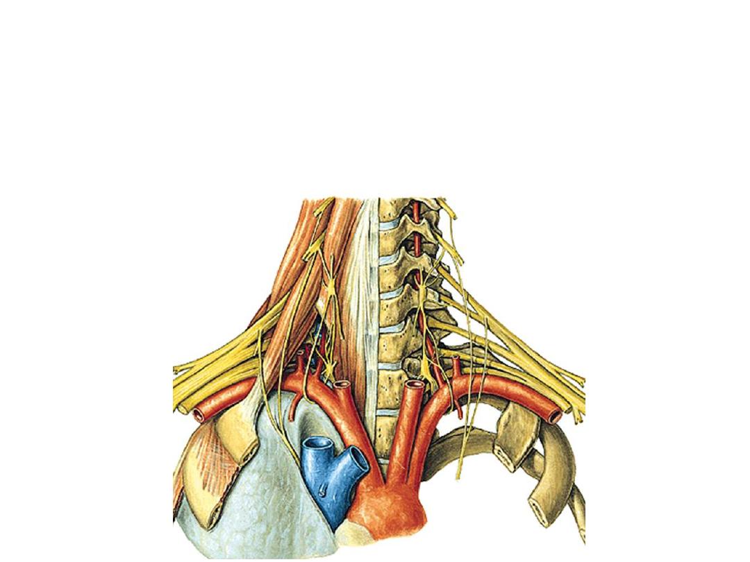
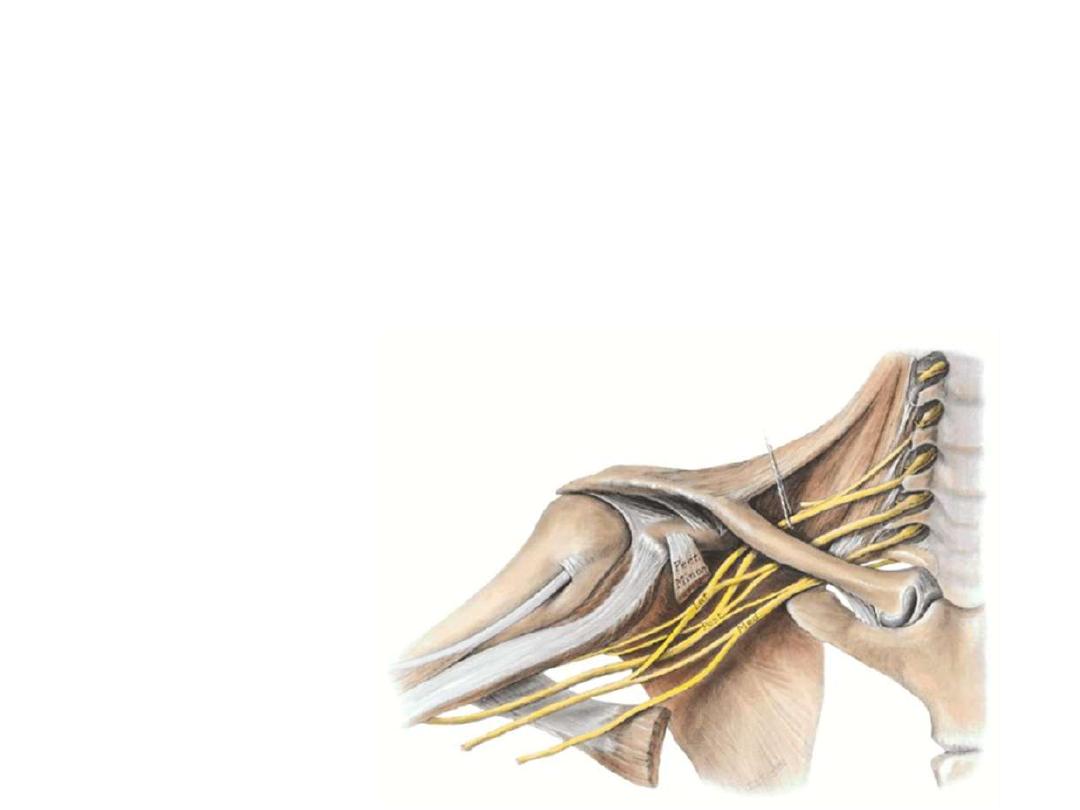
The roots:
-
They are the ventral rami of C5,6,7,8 & T1 nerves
-
They lie on the side of the vertebral column in theneck between
scalenus anterior & medius muscles

The trunks:
As they emerge from between these two muscles, the roots form the trunks
in this way:
•
C5 & C6 unite to form the upper trunk
•
C7 continues as the middle trunk
•
C8 & T1 unite to form the lower trunk

The divisions:
Each trunk divides into:
• Anterior division
• Posterior division
Fibers which formed the anterior divisions will supply flexor muscles
Fibers which formed the posterior divisions will supply extensor muscles
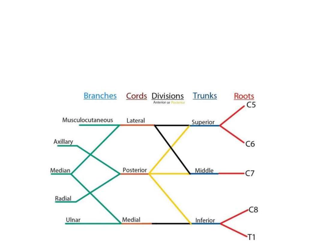
The cords:
The six divisions enter the axillary inlet
They unite to form the cords in this way:
•
Anterior divisions of the upper & middle trunks unite to form the
lateral cord
•
Anterior division of lower trunk continues as the
medial cord
•
The three posterior divisions unite to form the
posterior cord
Cords lie in the axilla
Cords derive their names from their relation to the second part of axillary
artery
Most of UL branches arise from the cords
Root branches:
1- Dorsal scapular n (C5):
Pierces the middle scalene muscle, and
continues deep to levator scapulae and the
rhomboids supplying them (LS, R major &
R minor)
2- Nerve to subclavius (C5&6):
Small nerve supplies the muscle
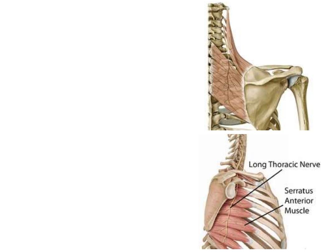
3- Long thoracic n (C5, 6 & 7):
- Leave the lateral surface of scalene
muscles
- Descends behind the axillary artery
- Lies on the side of serratus muscle
supplying its degitations
Trunk branches:
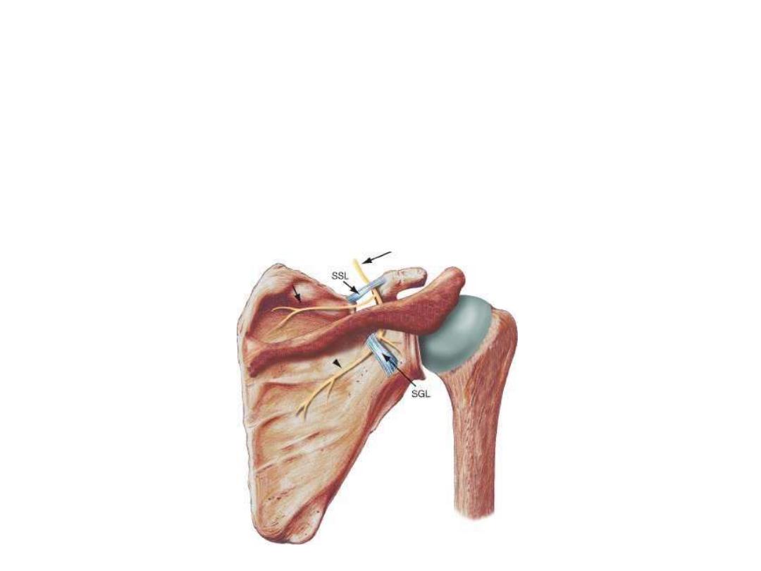
The suprascapular n (C5 & 6):
- Arises from the upper trunk
- Passes in the posterior cervical triangle
- Enters the scapular region below the transverse scapular ligament to supply
supraspinatus & infraspinatus muscles
- Gives sensory branches to the shoulder joint
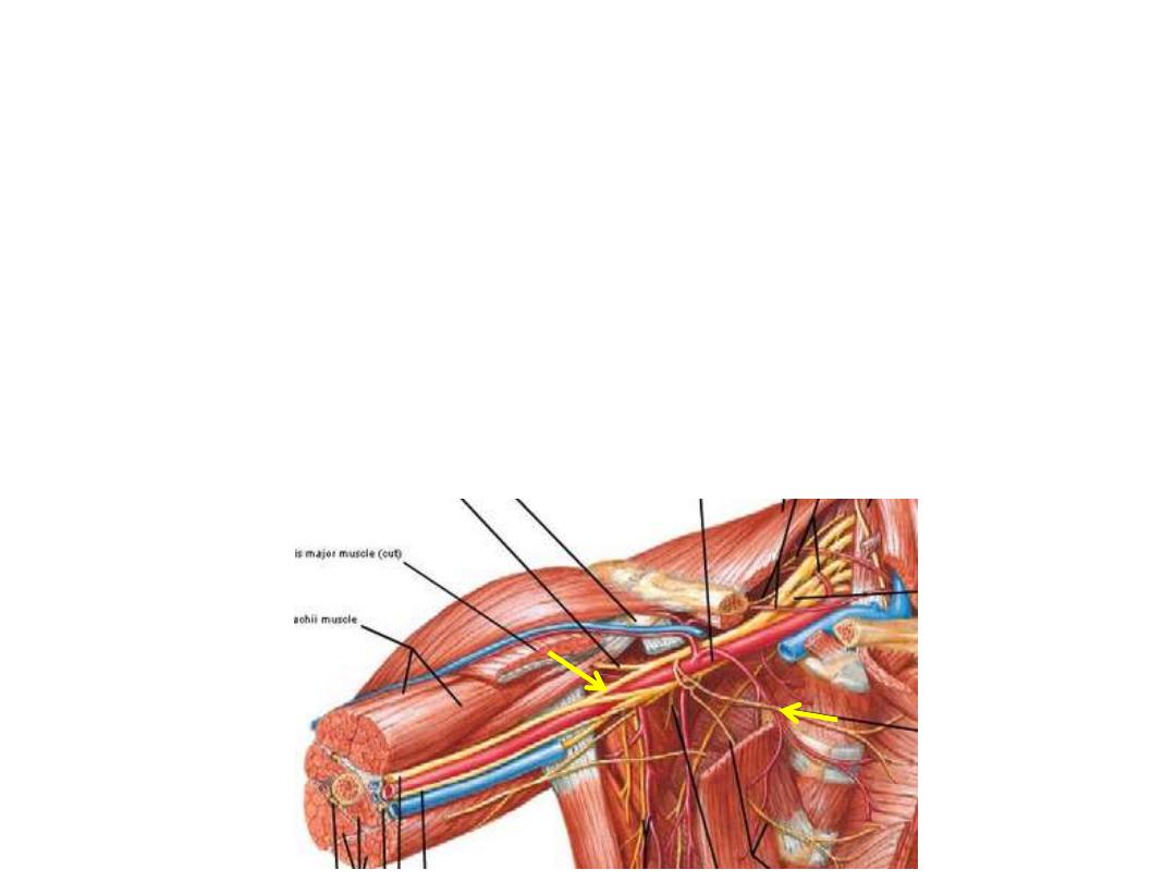
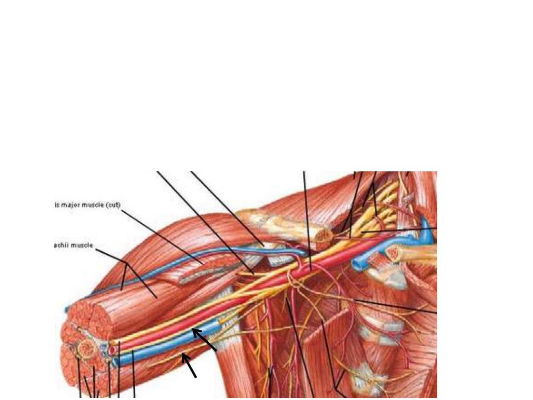
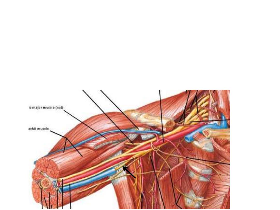
Cords branches:
Lateral cord branches:
1- Lateral pectoral n (C5-7):
- Pierces the clavipectoral fascia
- Enters the deep surface of pectoralis major to supply it
2- Lateral head of median n (C5-7):
Leaves the lateral cord & goes medially to join the medial head in front of the
axillary artery forming the median nerve
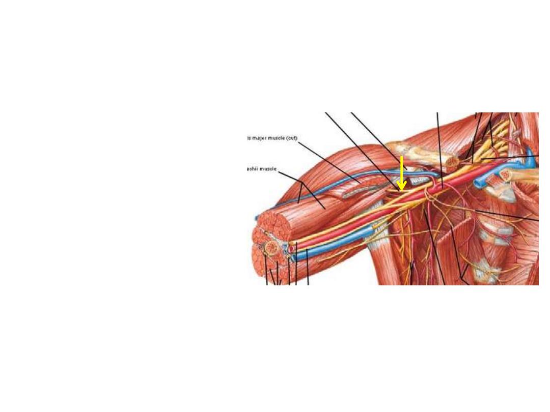
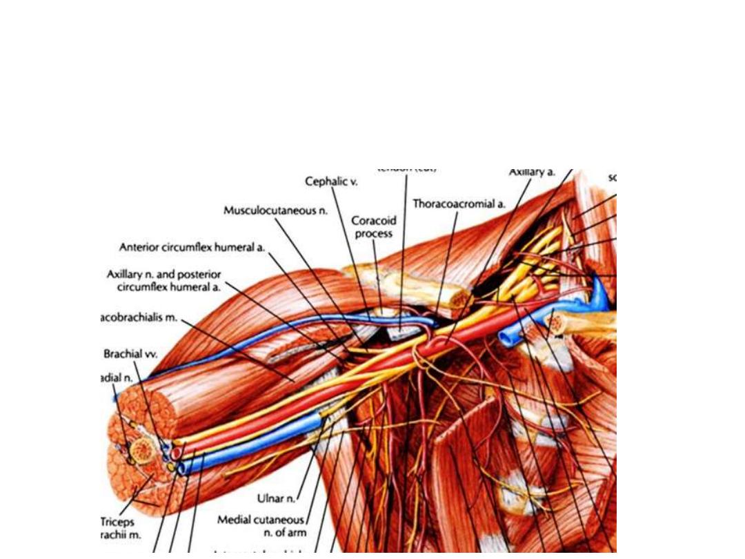
3- Musculocutaneous n (C5-7):
- Pierces coracobrachialis
- Enters the arm to supply
muscles
of
the
anterior
compartment
- Continues
as
the
lateral
cutaneous nerve of the forearm
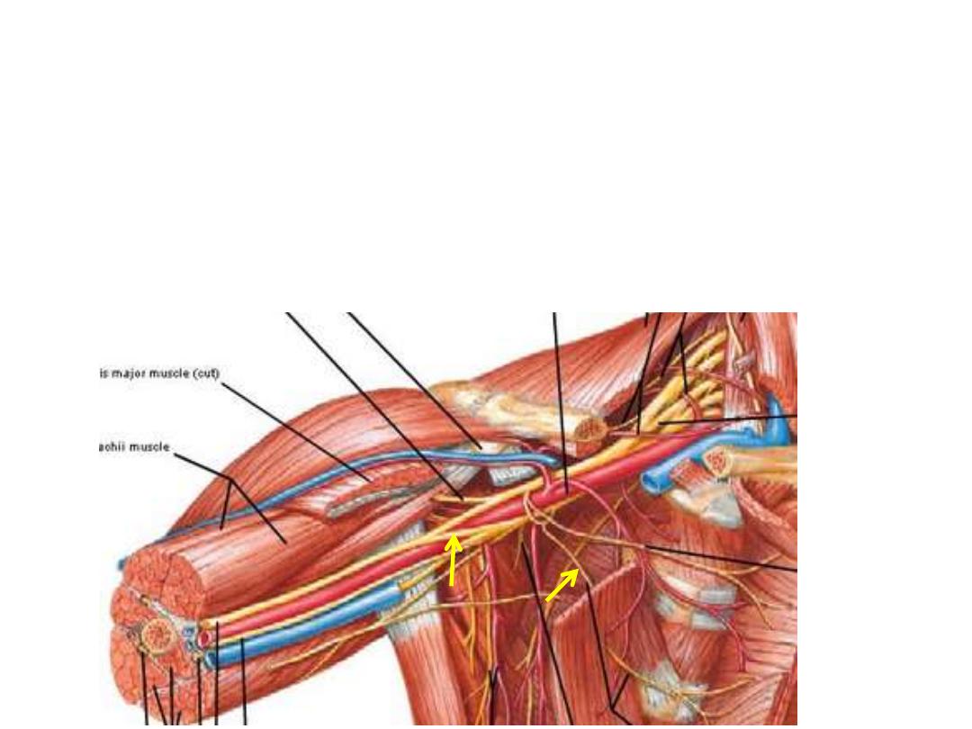
Medial cord branches:
1- Medial pectoral n (C8 &T1):
- Enters the deep surface of pectoralis minor to supply it
- Leaves the minor muscle & enters P major supplying it too
2- Medial head of median n (C8 &T1):
Leaves the medial cord & goes laterally to join the lateral head in front of the
axillary artery forming the median nerve
3- Medial cutaneous n of the arm (C8 &T1):
- Smallest branch of BP, lies medial to the axillary vein
- Descends in the arm & supplies the lower ½ of medial skin of the arm
4- Medial cutaneous n of the forearm (C8 &T1):
- Lies between the axillary vessels
- Accompany brachial artery
- Pierces arm fascia with basilic vein
- Supplies the forearm skin down to the wrist by its anterior & posterior
branches
5- Ulnar n (C8 &T1):
- The continuation of the medial cord
- Receives C7 fibers from the lateral cord
- It is the main nerve of the hand
- Its injury causes claw hand
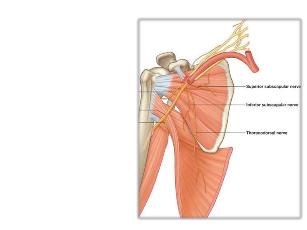
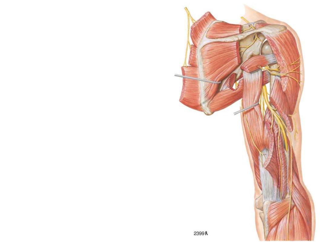
Posterior cord branches:
1 & 2- Subscapular nerves (C5 & 6):
- Upper: pierce the upper part of
subscapularis supplying it
- Lower: supplies the lower part of
subscapularis & teres major
3- Thoracodorsal n (C6-8):
- Descends on the subscapular
muscle
- Enters latissimus dorsi near its
insertion supplying it
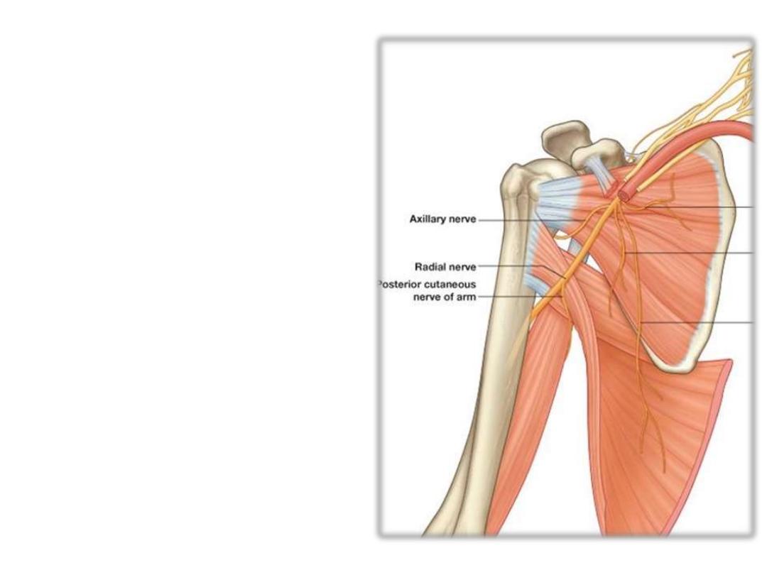
4- Axillary n (C5 & 6):
- Leaves the axilla through the
quadrangular space
- Supplies deltoid & T minor
- Sensory to:
1- Shoulder joint
2- Upper lateral cutaneous n of the arm
5- Radial n (C5
– T1):
- Leaves the axilla through the
triangular interval
- It is the nerve of extensor
compartments of the UL
- Supplies most of the skin of the
back of the UL
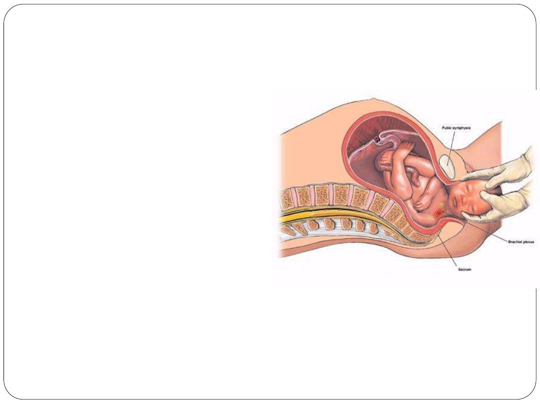
Erb-duchenne palsy:
Are injuries resulting from
excessive displacement of
the head to the opposite
side and depression of the
shoulder on the same
side.
This
causes
excessive
traction or even tearing of
C5 and 6 roots of the
plexus.
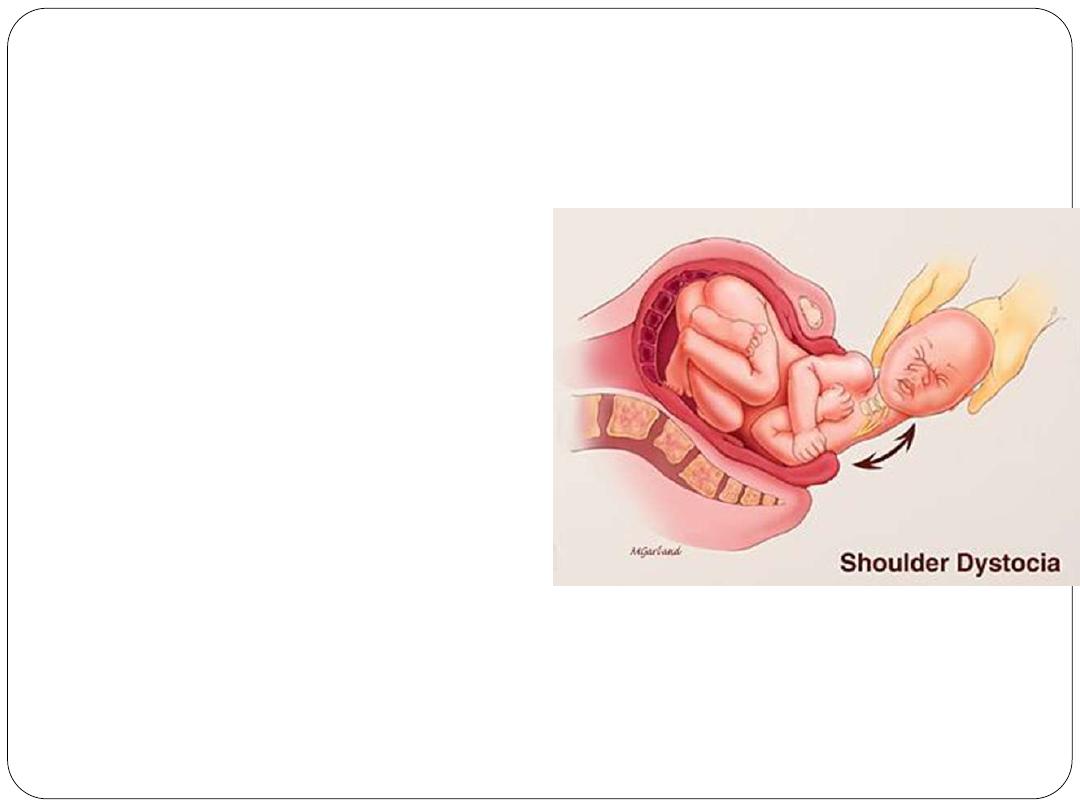
It occurs in infants during a difficult
delivery or in adults after a blow to or
fall on the shoulder.
The suprascapular nerve, the nerve to
the subclavius, and the
musculocutaneous and axillary nerves
all possess nerve fibers derived from
C5 and 6 roots and will therefore be
functionless.
(1) The supraspinatus and infraspinatus, (2)
the subclavius , (3) the biceps brachii and
the greater part of the brachialis and the
coracobrachialis, and (4) the deltoid and the
teres minor.
Thus, the limb will hang limply by the side,
medially
rotated
by
the
unopposed
sternocostal part of the pectoralis major; the
forearm will be pronated because of loss of
the action of the biceps.

Lower root injuries of brachial plexus:
Are usually traction injuries caused by excessive abduction of the arm,
as occurs in the case of a person falling from a height clutching at an
object.
The first thoracic nerve is usually torn. The nerve fibers from this
segment run in the ulnar and median nerves to supply all the small
muscles of the hand.
The hand has a clawed appearance caused by hyperextension of the
metacarpophalangeal joints and flexion of the interphalangeal joints.
loss of sensation will occur along the medial side of the arm.
