بسم الله الرحمن الرحيم
وفوق كل ذي علم عليم
Surgical removal of impacted teeth
Evaluation of the difficulty of the impacted tooth removal
1- the orientation of the tooth2- the depth of the tooth
3-type of impaction like soft tissue impaction , bony impaction , tooth to tooth impaction.
4 –root shape like bulbous root or thin spindly roots.
5 – bone density (texture of the investing bone)
6-relation to the inferior dental canal .
7 – patient age
8 – degree of mouth opening9- general buildup of the patient .
10 – the relation of the impacted tooth to the external oblique ridge and to the internal oblique ridge .
The change in orientation of the occlusal surface from mesial inclination to a vertical inclination occurs primarily during root formation .during this time the tooth rotates from horizontal to mesioangular to vertical . most third molars do not follow this typical eruption sequence and instead become impacted tooth
There may be differential root growth between the mesial and distal root ,which causes the tooth to either remain horizontally inclined or rotates to a vertical position depending on the amount of root formation.
Under development of the mesial root result in mesioangular impaction.
Over development of the mesial root result in over rotation of the third molar into a distoangular impaction
Overdevelopment of the distal root lead to mesioangular or horizontal impaction
Before the surgical procedure the following should be done
1- proper clinical and radiographic evaluation of the condition.2-classification of the impacted tooth to evaluate the difficulty of the surgical
procedure .
3- selection the time for surgical procedure.
4- explain the condition to the patient
5 – select type of anesthesia
Proper radiographic evaluation of the impacted tooth
- periapical radiograph- occlusal radiograph
Panoramic radiograph (OPG)
Lateral oblique view of the mandible
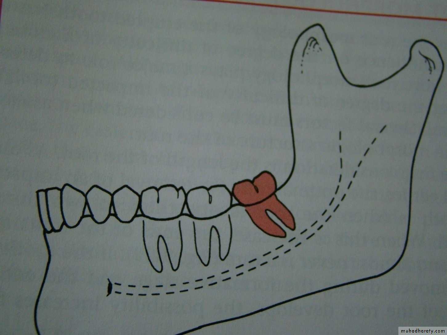
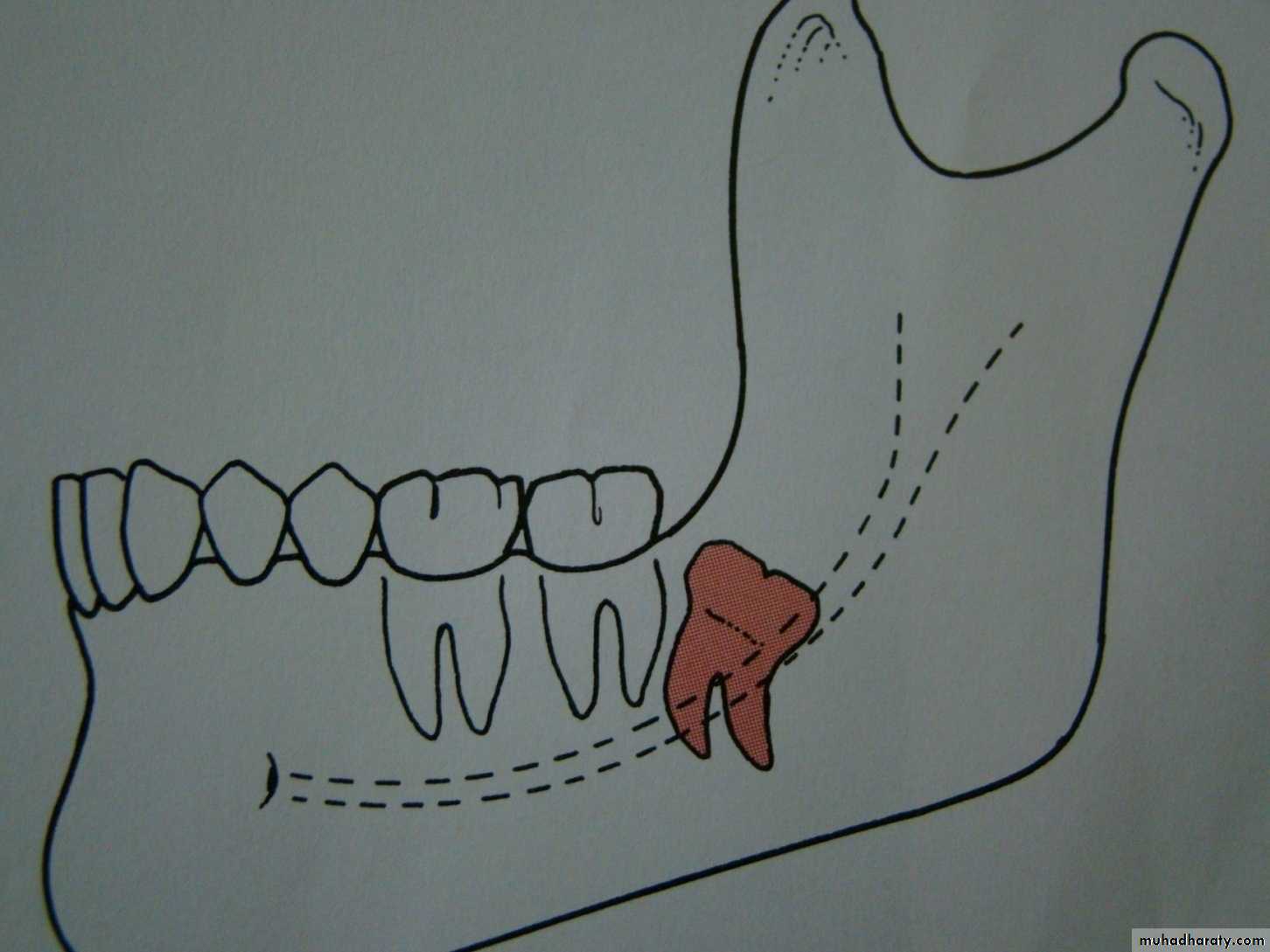
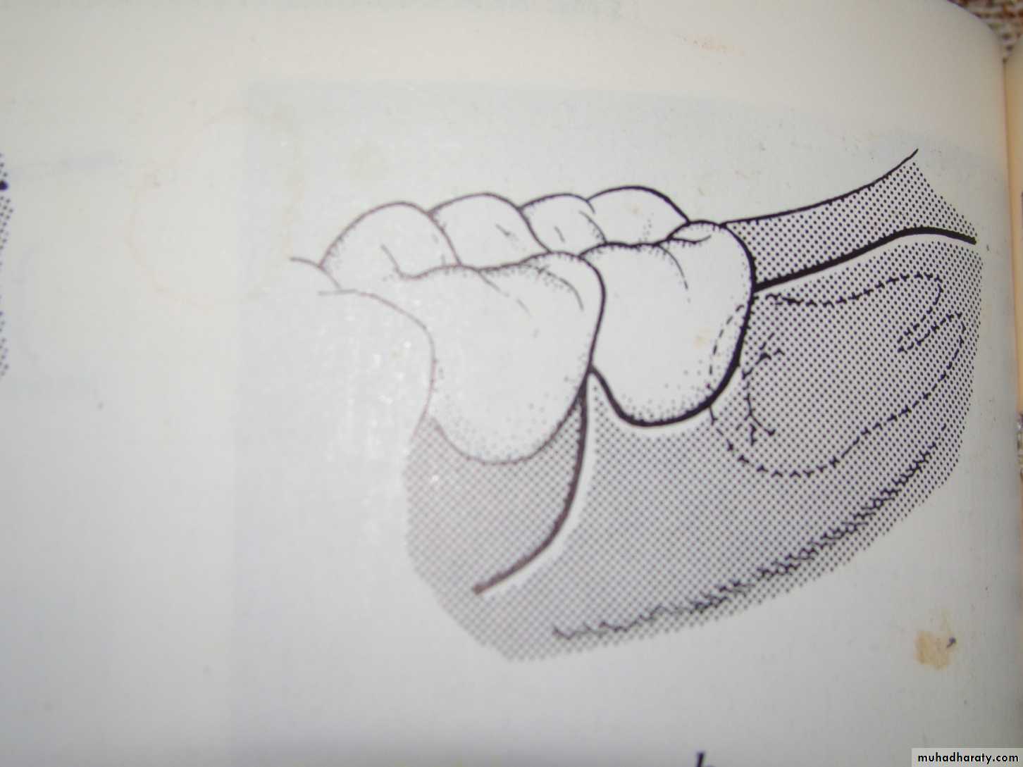
Winter’s lines
1-White line: it is an imaginary line draw along the occlusal surface of erupted first and second molars extend posteriorly over 3rd molar region . Its benefit is to determine the angulations of an impacted tooth and its relationship with occlusal surface of erupted 2nd molar(depth).2- Amber line: It is a line drawn from the surface of bone laying distally to the 8 and to the crest of interdental septum or alveolar septum between 6 and 7. it determine the amount of bone removal
3-Red line : it is draw perpendicular from amber line to an imaginary point of elevator application located mesially to the CEJ except in Disto angular impaction. It is used to measure the depth of the impacted tooth.
Note: Any tooth with red line length more than (5 mm) it is better to remove it under G.A.
White Line
Amber Line
Vertical Impaction with cariesPoint of application
Horizontal Impaction
Red Line
Mesio-angular ImpactionDisto-angular Impaction
Classification of the impactions to determine the difficulty of the procedure
The difficulty index:Angulations Mesioangular 1 , horizontal or transverse 2, Vertical 3, disto angular 4.
Depth : level A value 1 ,level B value 2 ,level C value 3
Ramus relation ship cl 1 value 1, cl 2 value 2 , cl 3 value 3.
summation of the three categories give the difficulty index value
Difficulty index
Very difficult 7-10 , moderately difficult 5-7 , minimally difficult 3-4Example a tooth which is mesioangular B class 2
mesioangular =1
Level B =2
Class 2 = 2
Difficulty score =5
This impaction can be expected to be moderately difficult to remove .

Minimally difficult
Moderately difficult

Very difficult
Selection the time for surgical procedure
Early removal1- the impaction should be documented
2 –ball in a socket syndrome
Late removal increase difficulty due to long root and dense bone.
The best time for removal when only 2/3 of the root had completed .

Development of mandibular third molar
The mandibular third molar tooth germ is usually visible radio graphically by age 9 years and cusp mineralization is completed at age 11 years.At age 11 years the tooth is located within the anterior border of the ramus with it,s occlusal surface facing almost directly anteriorly
.crown formation is usually complete by age 14 years
At age 16 y the roots are approximately 50 % formed
At age 18 years the roots are completely formed with an open apex .
At age 24 years 95% of all third molars that will erupt have completed their eruption.The patient
The condition should be explained to the patient in a simple easy way directing his attention to possible complication that may arise from leaving the tooth in position .Choice the type of anesthesia
Local anesthesia or general anesthesiaThe indications for G.A may include :
1 – procedures lasting more than 45 minutes .
2 – uncooperative patient
3- level C impacted lower 8
4- class 3 impacted lower 8 .
5 – very apprehensive patient
6 – multiple impacted teeth removal
The surgical procedure for extraction of impacted lower third molar
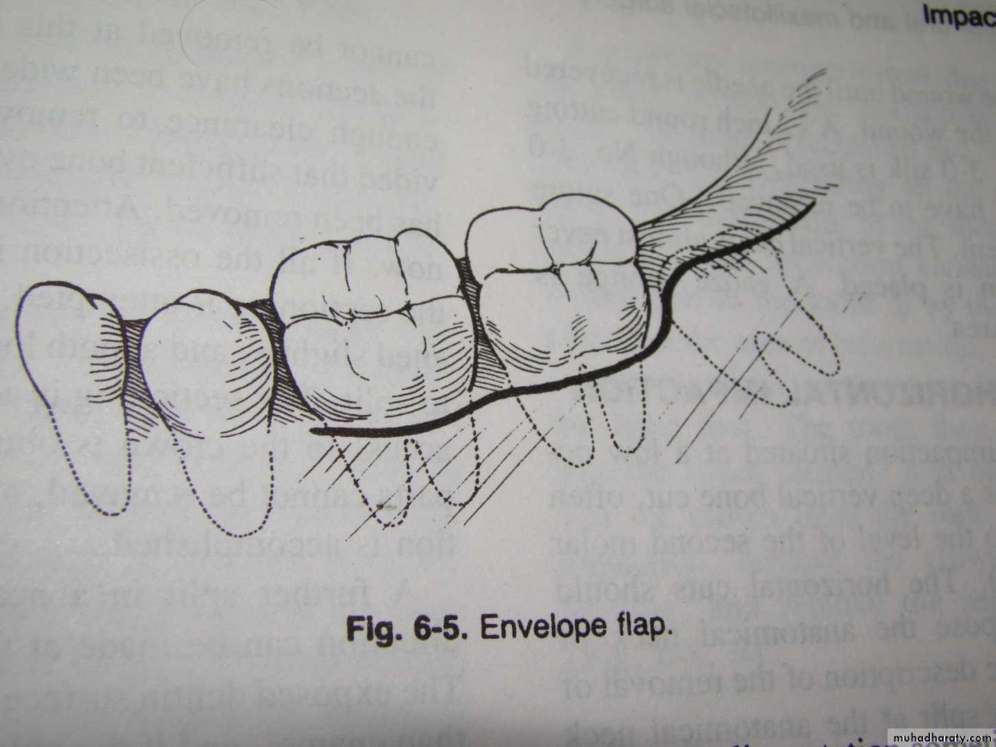
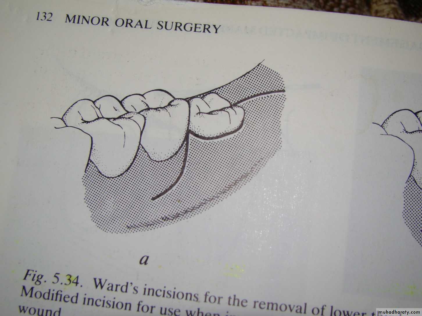
1-elevation of an adequate mucoperiosteal flap
There are 3 types of mucoperiosteal flap design for removal of impacted lower third molar :1- envelop flap : used for soft tissue impaction that need no bone removal. No vertical incision
2- standard flap indicated in all cases of impacted lower third molar . The vertical releasing incision lie distal to the second molar.
3- modified standard flap indicated for impacted tooth that completely covered by bone .the vertical releasing incision lie mesial to the second molar


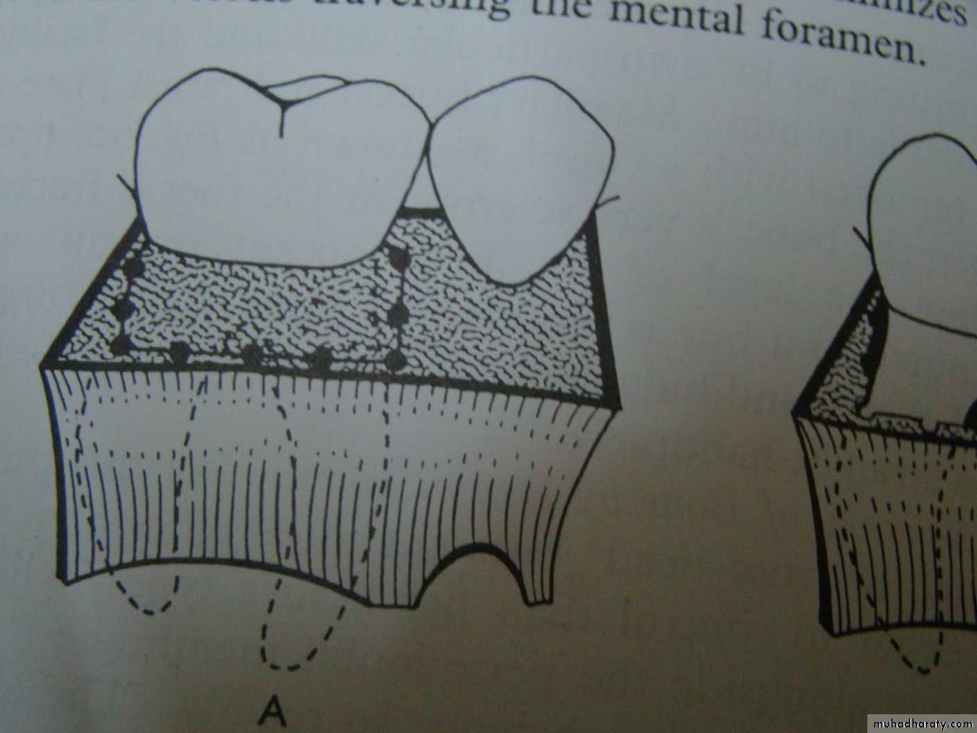
Bone removal
Purpose of bone removal :1- exposure of the impacted tooth
2- reduction of resistance (removal of bone in the path of removal)
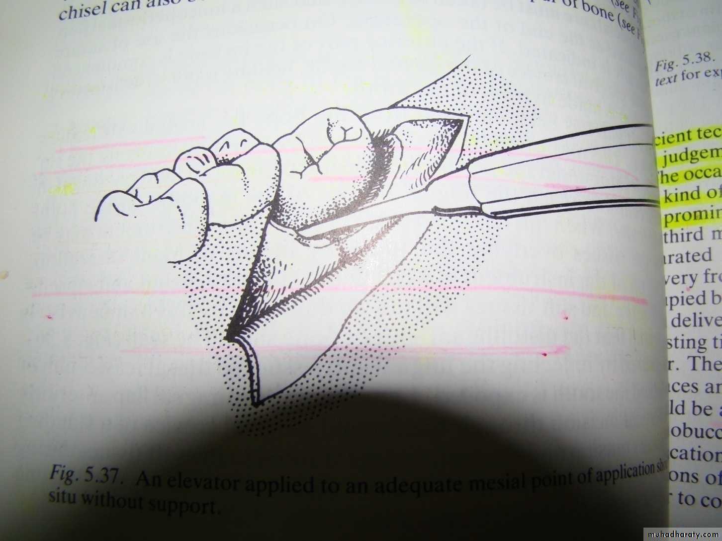
3-exposure of a point for elevator application( at the cemento enamel junction )
Types of bone removal
Guttering with surgical bur
Postage stamp method with surgical bur
Chisel and hammer
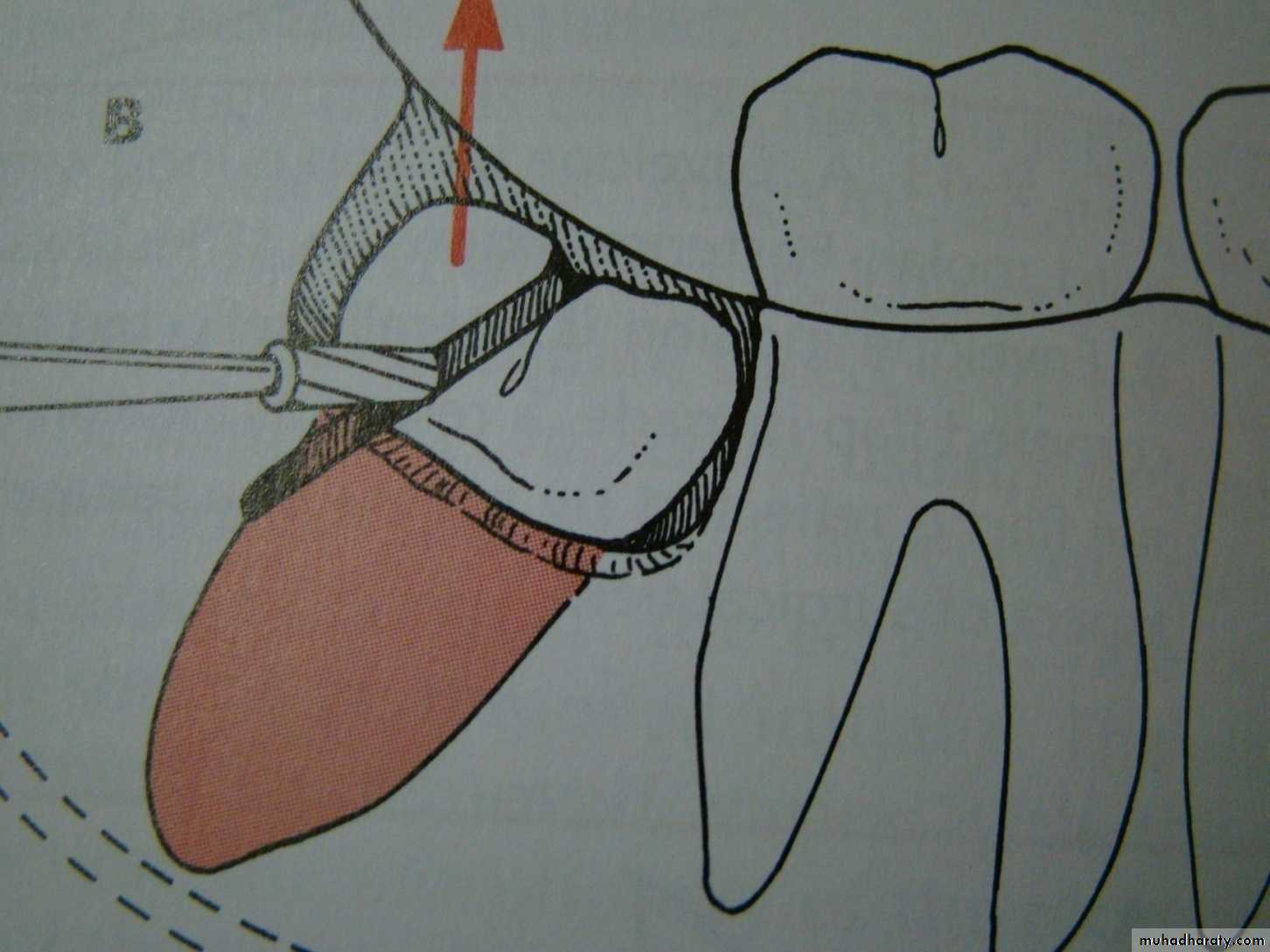
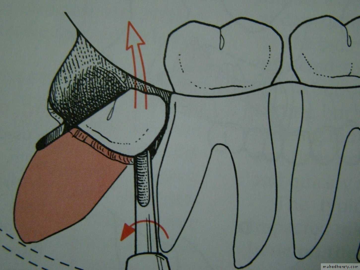
Tooth delivery
1- whole tooth delivery without tooth division . Total delivery by application of force using elevator , mesial or buccal elevator application.2- tooth division : division is indicated to reduce resistance , create space or remove interlocked cusps of the tooth
Tooth division/ 3 types
1- decapitation : division of the crown of the tooth at cervical margin level. It is indicated in horizontal mandibular and maxillary third molar impaction and palataly impacted canine 2 – longitudinal tooth division : indicated when the impacted tooth has a widely divergent straight roots or when one root is straight and the other is curved .


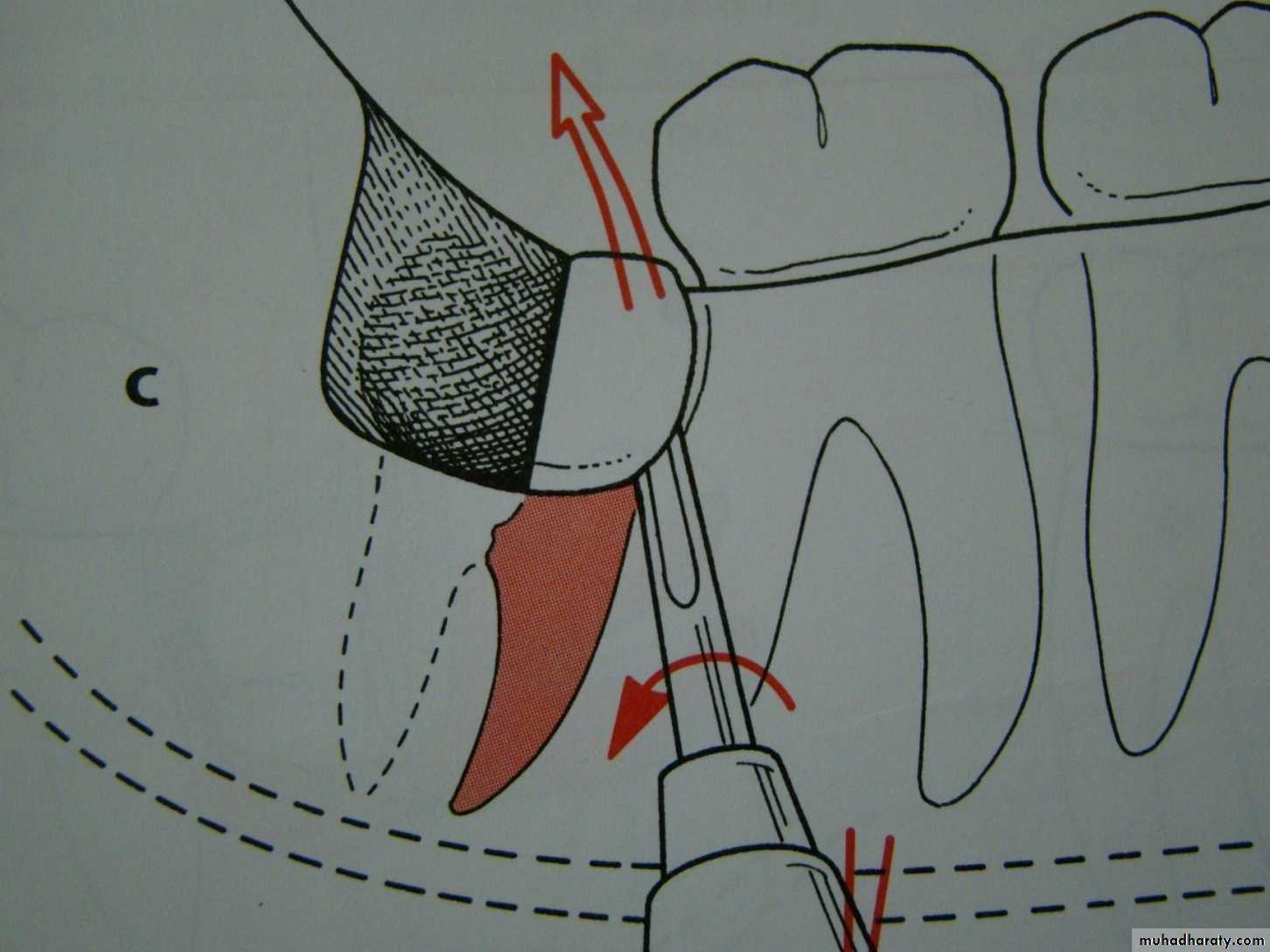
3- division of the interlocking cusp: indicated for mesioangular impaction , removal of the interlocking segment of the tooth ,usually located under the distal surface of the second molar
Toilet of the socket or preparation for wound closure
After removal of the tooth from its socket the wound is gently irrigated with sterile normal saline solution and inspected for :1- Any remnant of the residual tooth sac is removed .
2- Remnants of tooth structure or fragment of bone debris is gently removed.
3- small fragment of detached bone .
4- sharp edges of interseptal or alveolar boneis trimmed and smoothed .then final irrigation and wound now is ready for closure
Closure of the wound
Well designed and properly reflected flap can be easily repositioned into its place ,using 000 black silk suture .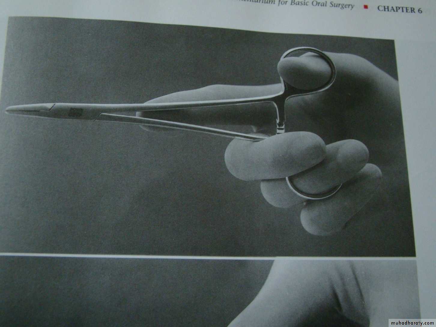
Post operative care
1- a pressure pack is held in place for 1 hour. Usually use wet gauze or wet cotton .2- post operative instruction is given to the patient :
a/ cold packs at the angle of the mandible in case of lower 8 ,or on the infra orbital area in case of upper 3 or on the cheek in case of upper 8 .for 20 minutes 5 times daily.
B / proper antibiotic therapy
c / maintaining good oral hygiene including mouth washes and teeth brushing
D / soft diet
3- appointment for check up after 2 days .
4 – appointment suture removal after 5-7 days
Complications associated with surgical removal of impacted teeth
Complication during the surgery :1 – laceration of the soft tissue flap due to ;
A / improper incision design.
B / improper elevation of the flap and improper retraction , this lead to delayed healing .
2 – fracture of the jaw at the angle of the mandible due to improper use of the elevators with uncontrolled force.
3 – fracture of the tuberosity : this occur with erupted rather than unerupted tooth due to improper use of the force .
4 - Complications related to injury of adjacent structure :
A / injury to inferior alveolar canal occur in deeply seated vertical impaction
B / damage to the nasal floor
C / involvement of the maxillary sinus
D / pushing the maxillary tooth into the maxillary sinus .
E / pushing the maxillary third molar into the pterygopalatine fossaF / pushing of the mandibular third molar into the pterygomandibular space or into the submandibular space.
G / aspiration or swallowing of the impacted tooth .
Post operative complication
1 – post operative pain which is greatly reduced when preemptive analgesia like tramadol injection used .
2- post operative swelling or oedema which is greatly reduced when preoperative decadron (dexamethasone 8 mg ) injection used .
3 - Post operative infection which is greatly reduced when preoperative antibiotic like ampiclox 500 mg injection used .

4 - Post operative bleeding which is greatly reduced if complete homeostasis is secured before suturing .
5- trismus
6- osteomyelitis
7- pain at the TMJ
8 - pain with swallowing (dysphagia) due to edema of the pharynx or hematoma formation
