Nerve structure and function
Lecture 2

1- Define myline sheath and its origin.
2-Recognize the functional and structural
organization of the neuron.
3-Compare between the mylinated and
the unmylinated nerve fibers.
4-Differentiate between the types of
neural communication ?
5-Classify the nerve fibers according to
their basic properties and factors affecting
each type
:
Objectives
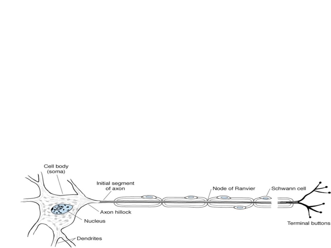
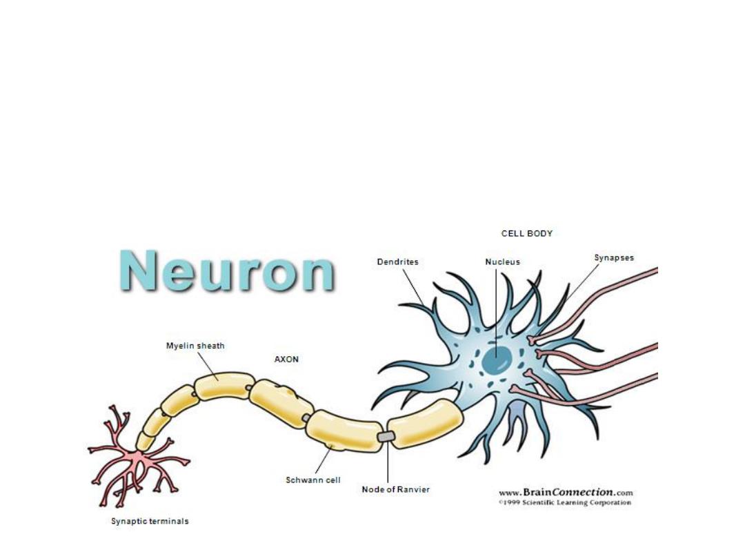
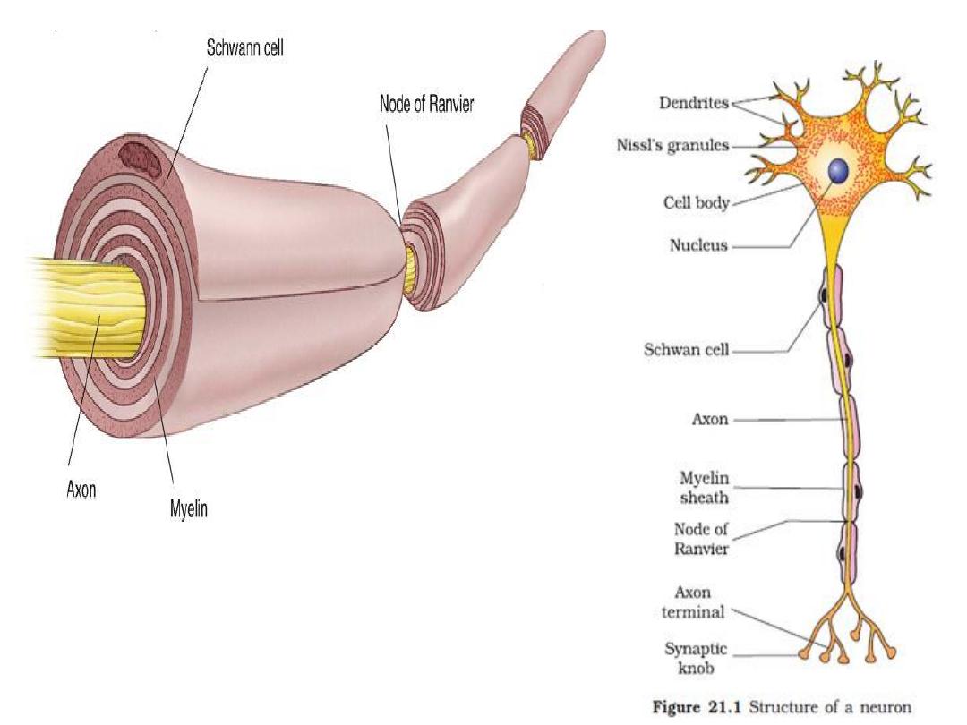
Neurons in general are composed of 3 major parts:
1-The soma.
the main body of the neuron, contains specialized
cytoplasm, single nucleus and other granules
2- Dendrites (2-7). Conduct towards the cell body.
The soma and the dendrites form a large area which is specialized
for reception.
3- Single axon, it conducts away from the cell body. starts from a
thick area called the axon hillock. after that the part of the axon is
called the initial segment (thinner), then the axon terminate in the
(axon knob),where the chemical substance (neurotransmitter ) is
released in response to nerve impulse.

Nerve cells are secretory cells,
but they differ from other
secretory cells in that the
secretory zone is generally at the
end of the axon, far from the cell
body
.
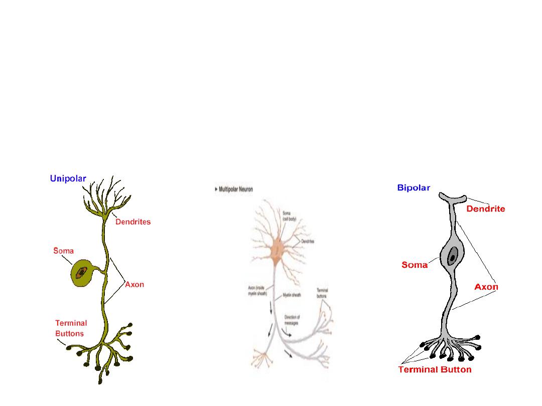
TYPES OF NEURONS:
Structurally divided into;
Unipolar Multipolar Bipolar

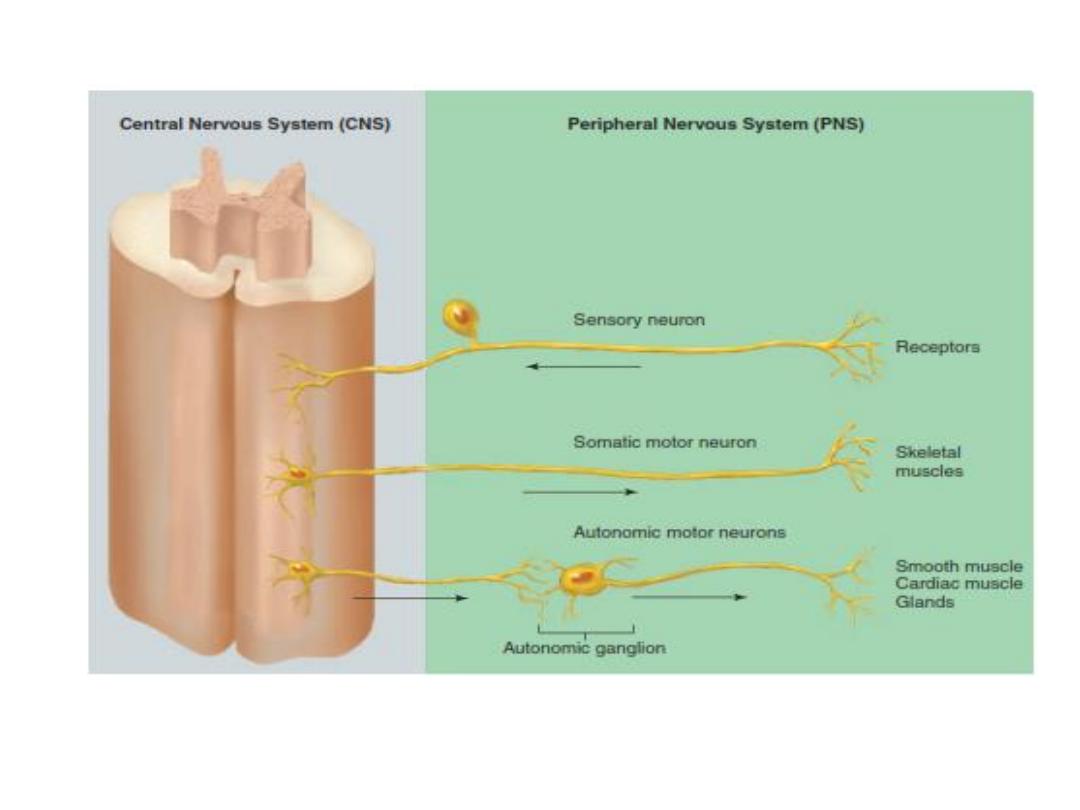
Functionally they are divided in to:
1-Sensory(
conduct impulses from sensory receptors into
the CNS
.
)
2- Motor (
conduct impulses out of the CNS to the organs
(muscles and glands)
)
.
They are 2 types somatic motor (are
responsible for both reflex and voluntary control of skeletal
muscles, and autonomic motor (innervate the involuntary
effectors: smooth muscle, cardiac muscle, and glands)
.
3- Interneuron
(
are located entirely within the CNS and
serve the associative, or integrative, functions of the nervous
system).

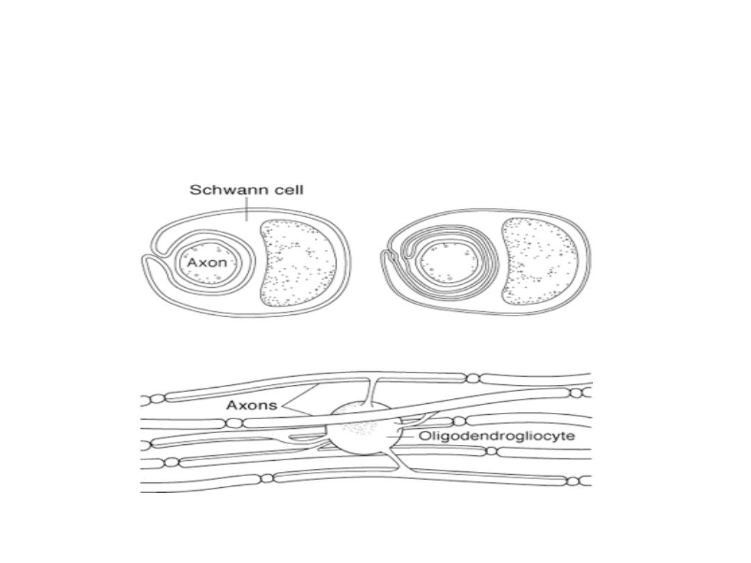
Each axon in the peripheral nervous system after a short
distance from its origin is covered by a series of schwann
cells which are the supporting cells of the peripheral
nervous system; they form the
myline sheath
of the
nerve (
containing the lipid substance sphingomyline which
is an electricalinsulator decreasing the ion flow through the
membrane).
It is not continuous, it is interrupted by a small exposed area
of 1 microne in length which is called "Node of
Ranvier"
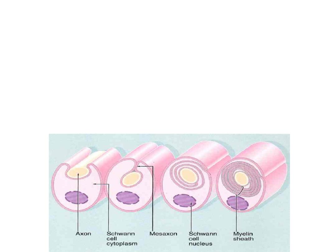
The process of myline sheath forming (mylination)
involves the following:
The Schwann cell membrane first envelops
the axon.
The cell rotates around the axon many
times laying down multiple layers of
Schwann cell membrane.

The function of the myline sheath
The main role in conduction because, it
increases the velocity of conduction 5- 50 times,
so diseases like
multiple sclerosis
causes
demylination and sever nerve defect which
block conduction.
In addition to that myline will conserve
energy for the axon because small area will
have the exchange of ions.

Another demylinating disease in the
nervous
system
is
Guillain–Barré
syndrome
in which the body's immune
system mistakenly attacks the peripheral
nerves
and
damages
their
mylin insulation.
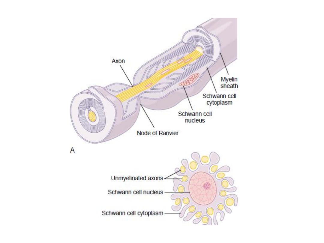
Not all the nerve fibers are mylinated, some are
not mylinated but surrounded by Schwann cells
without the deposition of myline
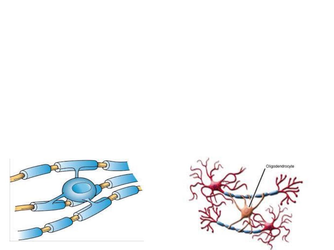
In the CNS the mylination is done by other cells
which are called
the oligodendrocytes
.
.
•
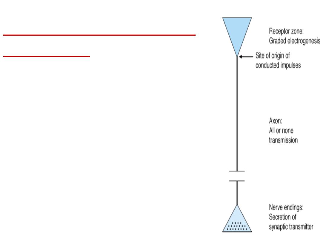
Functional organization of
the neuron:
1- The receptor zone or dendritic zone it
represents the site for the reception of nerve
signals, (local potential are formed in this area).
2- The initial segment zone.
it is the site and origin
of the conducting impulses.
3- The axonal zone.
or called the transmitting zone
where the nerve impulses are propagated and
transmitted
4- The nerve ending zone.
the site where the nerve
impulses causes the release of the neurotransmitter
to affect other neuron or muscle fiber
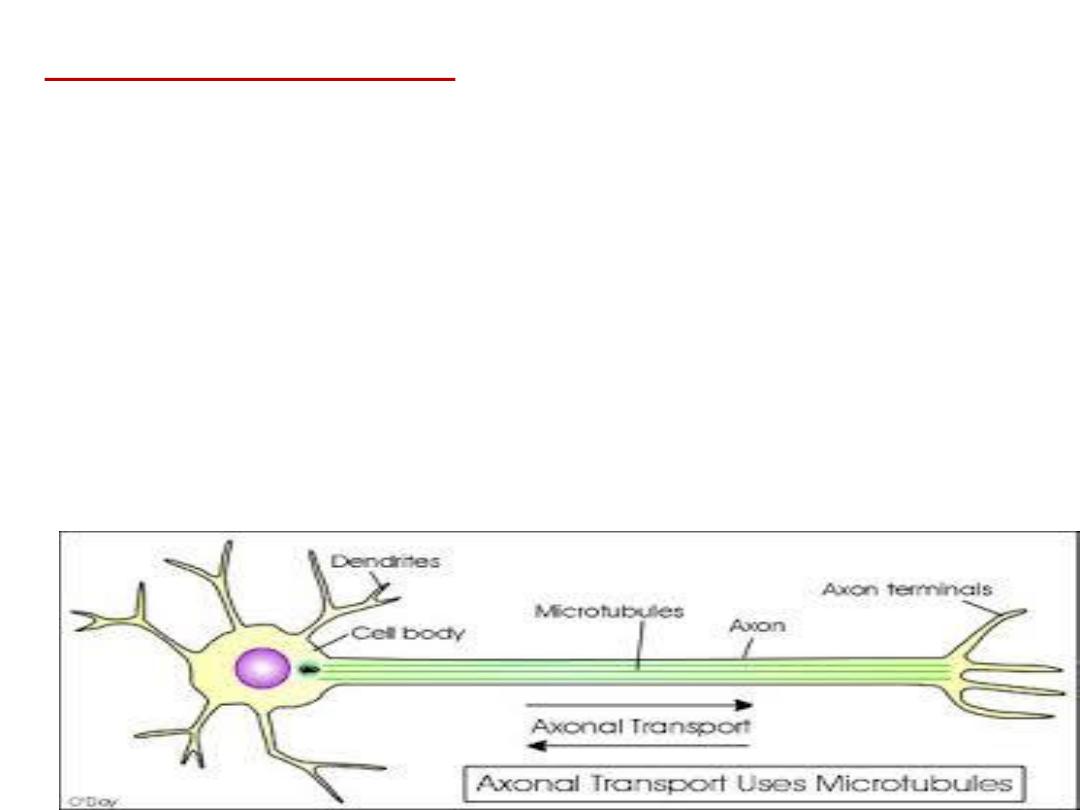
Axoplasmic transport:
-Fast one.
-Slower axoplasmic flow.
- Slowest axoplasmic flow.
-Retrograde transport. this type is for the transport of
substances which are taken by the nerve ending, like
nerve growth factor
and some
viruses
are transported
from the endings to the soma. Indeed, retrograde
transport may be responsible for the movement of
herpes virus, rabies virus, and tetanus toxin from the
nerve terminals into cell bodies

Communication of cells inside the
human body
At cellular level,
communication is based
on
Electrical & Chemical signalling

Neural communication
The neurons communicate with each other by 2
types of communication:
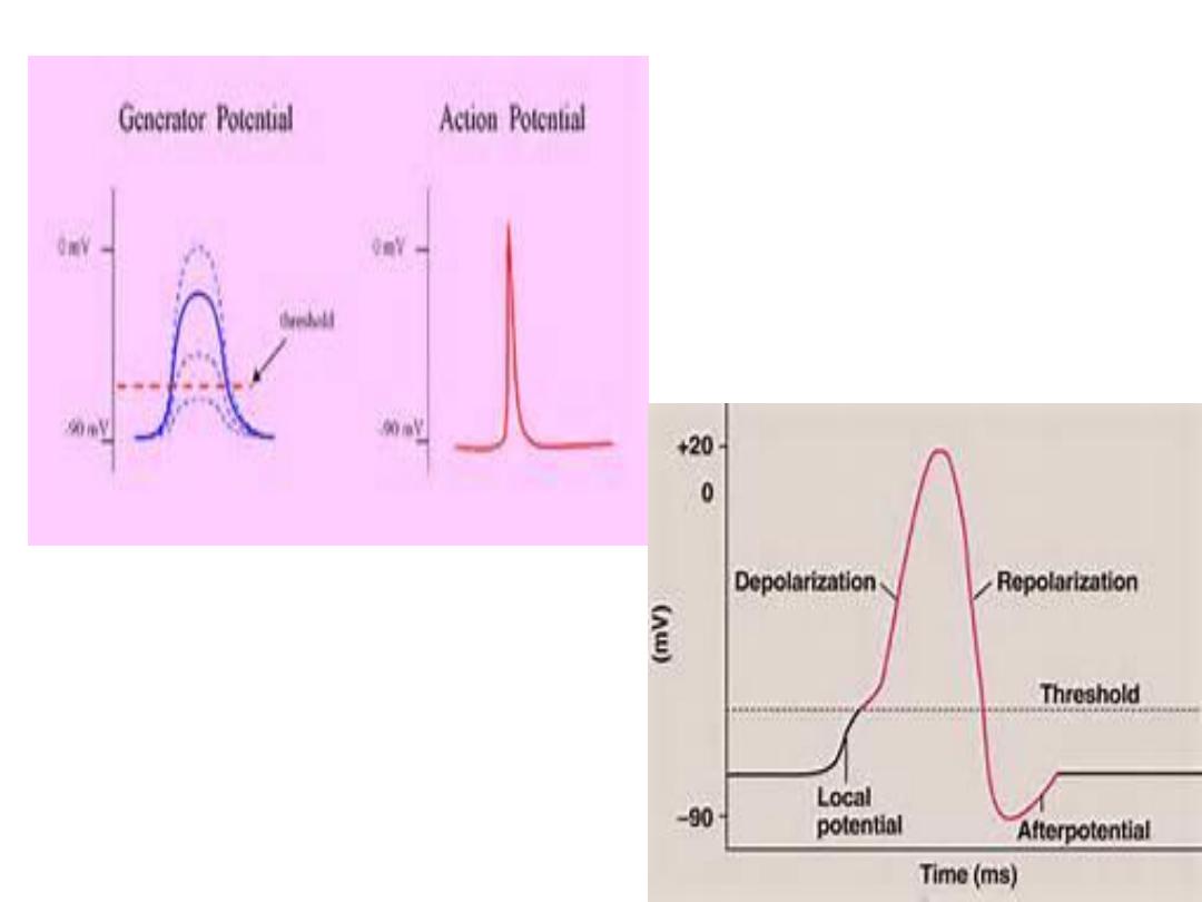
1-The electronic potential (generator potential):
Local,
nonpropagated
potentials
called
synaptic, generator, or electrotonic potentials.
2- The action potential (nerve impulse). is a
propagated
disturbance
used
to
send
information for long distances without any loss
of energy.
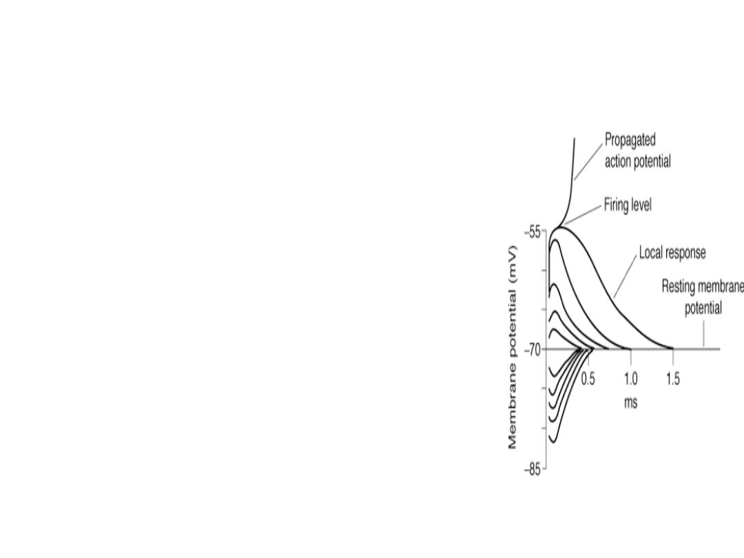
The local potential:
It is a localized depolarizing potential
change that rises sharply and decays
with time. It is proportional to the
magnatitude of the stimulus. So it loses
intensity as it spreads, and
its spread is graded
.
The importance of this potential is that
in the CNS, the information is
exchanged between adjacent cells by
this type.
(
used for communication between
neurons which are very close to each
other e.g. the brain and the eye
).

The action potential
Keeps its size and shape all along its
way.
Transmitted for long distances.
Not graded .
Used to send information for long
distances without any loss of energy
.
Can not be produced subthreshold
.

NERVE FIBER TYPES & FUNCTION
:
Conduction differs due to:
1 -The difference in diameter: (the greater the diameter the
faster the speed of conduction).
2- Presence of myline sheath
So the nerve fibers are classified into different types by 2
systems of classifications:
A –General system: 3 types according to the peaks produced
during compound action potential:
A, B, and C groups, further subdividing the A group into α(the
fastest), β, γ, and δ(the slowest) fibers.
B- Numerical system (Ia, Ib, II, III, IV).

Fiber Type Function Fiber Diameter(μm) Conduction Velocity(m/s) Spike Duration Absolute refractory period
A
Proprioception;
12-2 70-120
somatic motor
α
B Touch, pressure 5-12 30-70 0.4-0.5 0.4-1
γ Motor to muscle spindles 3-6 15-30
Δ Pain, cold, touch 2-5 12-30
B
Preganglionic autonomic
<3 3-15 1.2 1.2
C
Dorsal root
Pain, temperature
,
some mechano-reception,
reflex responses 0.4-1.2 0.5-2 2 2
Sympathetic
Postganglionic sympathetics 0.3-1.3 0.7-2.3 2 2
A and B fibers are myelinated; C fibers are unmyelinated
.

when we give anesthesia, there will be loss of sensation first. During
hypoxia there will be loss of autonomic function first then motor
actions, then sensation
Susceptibility to: Most Susceptible Intermediate Least
Susceptible
Hypoxia B A C
Pressure A B C
Local anesthetics C B A

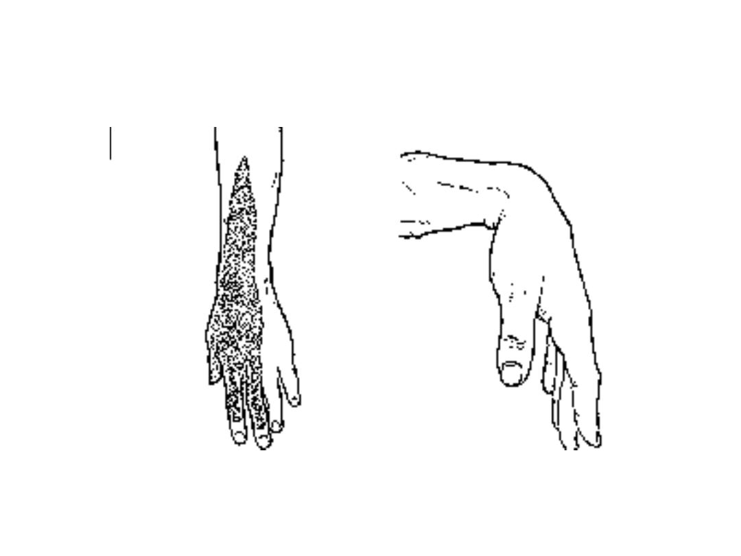
When sleeping with the arms under the head
for long period there will be pressure on the nerve
in the arm causing loss of motor activity while the
sensation is preserved this is called Sunday
morning or Saturday night syndrome, and when
we give anesthesia, there will be loss of sensation
first. During hypoxia there will be loss of
autonomic function first then motor actions, then
sensation.

Summary :
-
There are 2 types of axons mylinated which
are faster than the other type which is the
unmylinated.
-
-There are 2 types of neural communication:
local and action potential
- Different types of nerve fibers are affected
more or less by different types of stimuli
.