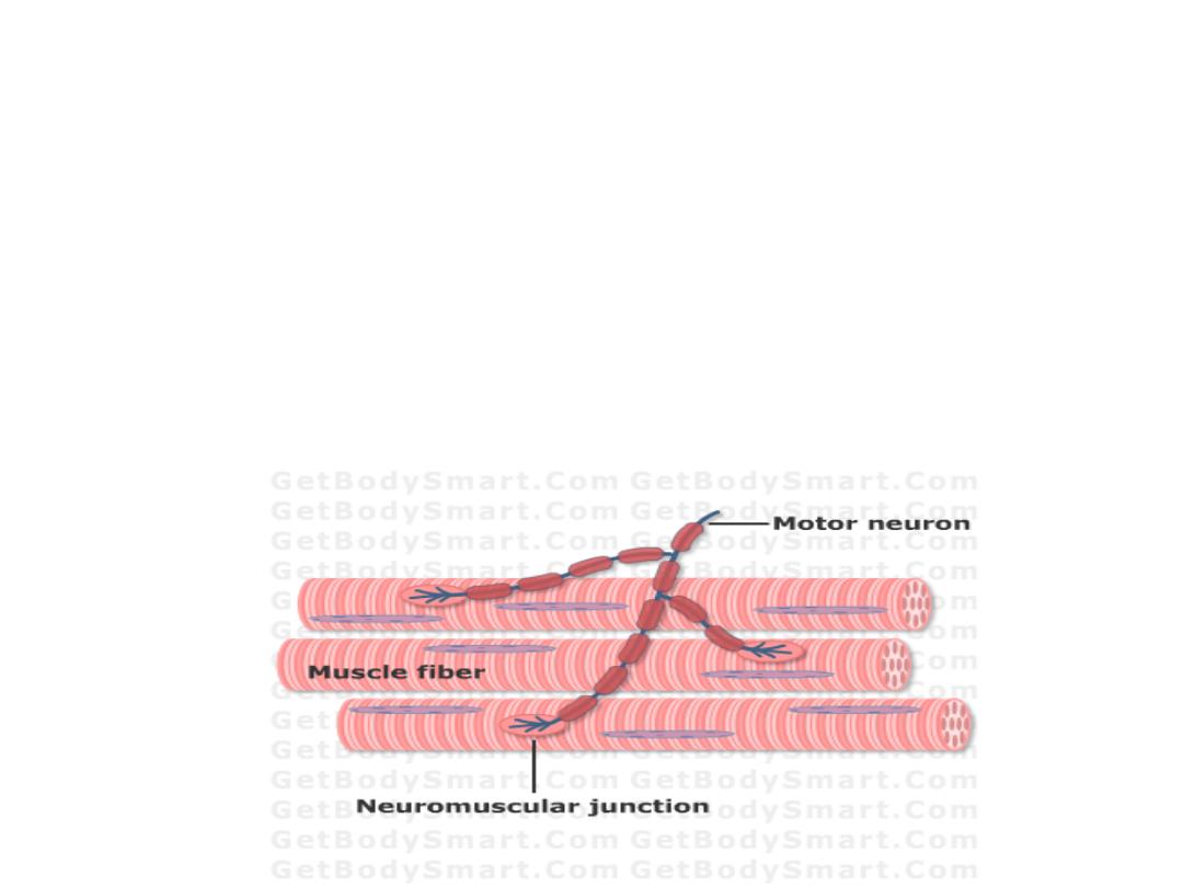
The neuromuscular
junction
The muscle

Objectives:
1- Describe the neuromuscular junction and the
sequence of events during neuromuscular transmission
2-Recognize the basic structure and morphology of
skeletal muscle.
3-Define sarcotubular system and determine
its function.
4- State the electrical characteristics of
skeletal muscles.
5- State the principles of walk along theory of Skeletal
muscle contraction
6- List the sources of energy in skeletal muscle.
.
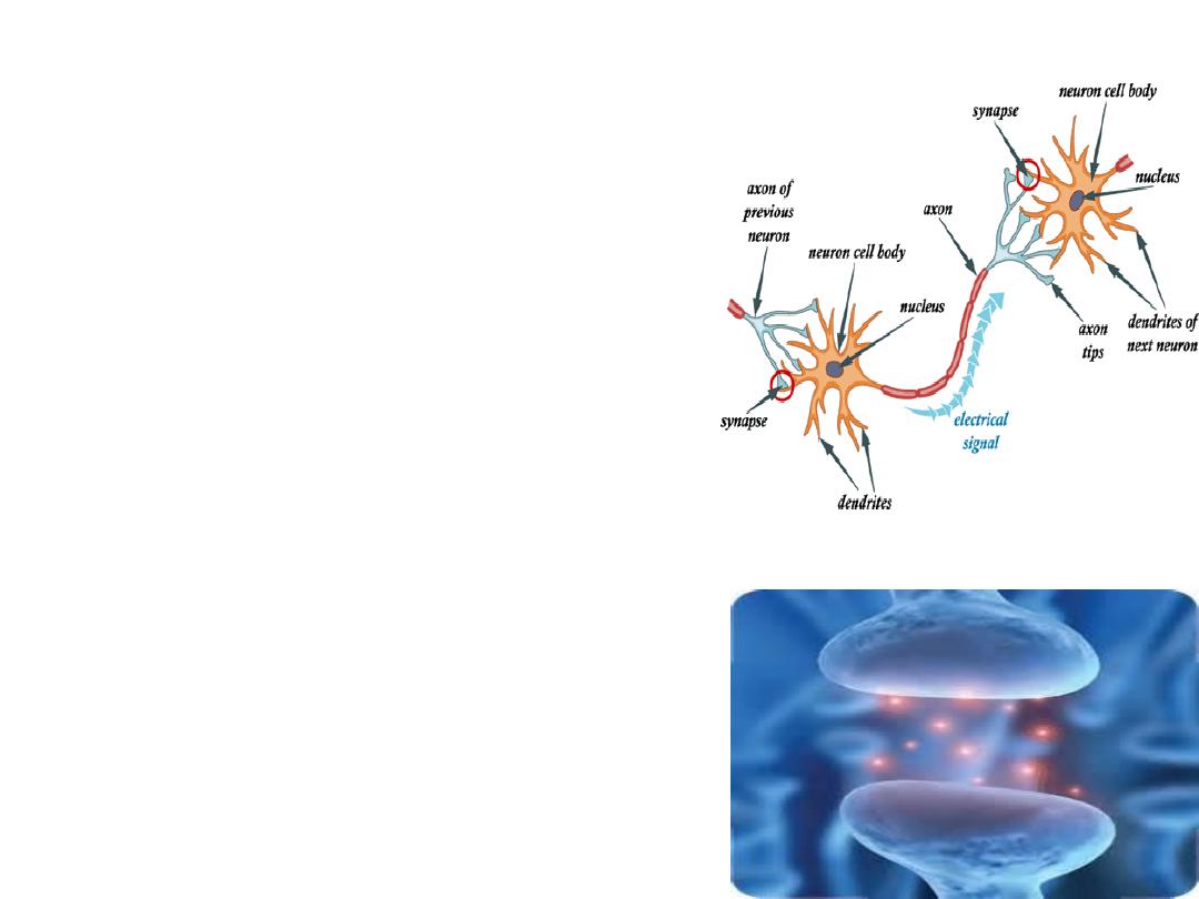
Junction is the connection
between a nerve cell and
another (muscle fiber or gland).
Synapse is the connection
between 2 nerve cells.
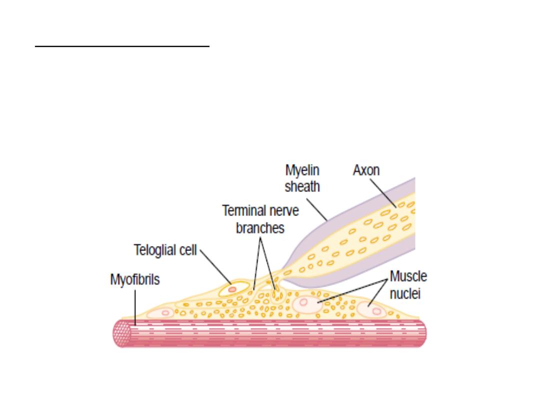
Each nerve ending makes a
junction with the muscle fiber
near its midpoint, called the
neuromuscular junction.
The nerve fiber with its
branching plus the thickened
muscle surface is called the
motor end plate.

The invaginated membrane is called the
synaptic gutter, and the space between the
nerve terminal and the fiber membrane is
called the synaptic space or synaptic cleft
(which
contains
the
enzyme
acetylcholinesterase,
which
destroys
acetylcholine .
At the bottom of the gutter are numerous
smaller folds of the muscle membrane called
subneural clefts, which greatly increase the
surface area at which the synaptic transmitter
can act, and there are also large numbers of
Acetylecholine (Ach) receptors.
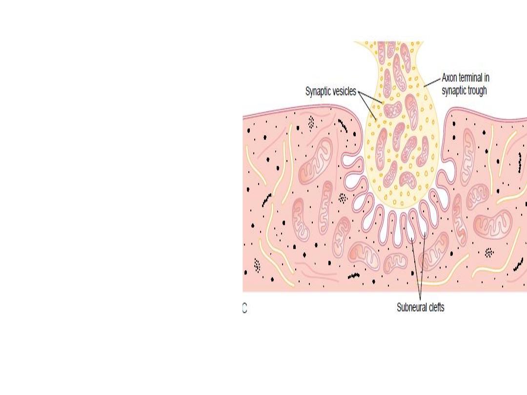
In the axon terminal there
are many mitochondria that
supply
adenosine
triphosphate (ATP).
The acetylcholine that excites
the muscle fiber membrane
is
synthesized
in
the
cytoplasm of the terminal,
and it is absorbed rapidly into
many small synaptic vesicles.
There
are
also
protein
particles that penetrate the
neural membrane; these are
voltage
gated
calcium
channels.
.
The NMJ consists of:
- Axon terminal.
-Synaptic gutter or synaptic trough.
-The synaptic space or synaptic cleft.
-Subneural clefts, Acetylecholine (Ach) receptors

Neuromuscular transmission
1-
Action potential spreads over the terminal; →
channels open and allow calcium ions to diffuse
to the interior of the nerve terminal.
2-
The calcium ions ,exert an attractive influence
on the acetylcholine vesicles, drawing them to
the neural membrane adjacent to the dense bars.

3-
The vesicles then fuse with the neural
membrane and empty their acetylcholine
into the synaptic space by the process of
exocytosis (Botulinum and Tetanus toxins
block
the
transmitter
release)
4-
Ach will diffuse to the synaptic cleft and
binds with the receptors(
Nicotinic
receptors
)
, causing activation of the Na and
K ionic channels resulting in local
depolarization and firing level is reached
,and action potential is initiated.

Curary
is an arrow poisoning used by the
American Indians to paralyze their victims .The
poison
binds to the Ach receptors
reducing the
number of receptors for the binding with Ach and
so they reduce the reaction of the released Ach .
Acetylcholine Receptors
The varying responses of postsynaptic cells to the
same chemical can be explained, in part, by the fact that
different postsynaptic cells have different subtypes of
ACh receptors.
Historically, acetylcholine receptors have been divided
into two main types on the basis of their pharmacologic
properties.1- Muscarinic, found in the plasma
membrane of smooth muscle cells, cardiac muscle cells,
and the cells of particular glands and 2- Nicotinic
receptors, found in specific regions of the brain, in
autonomic ganglia and in skeletal muscle fibers.
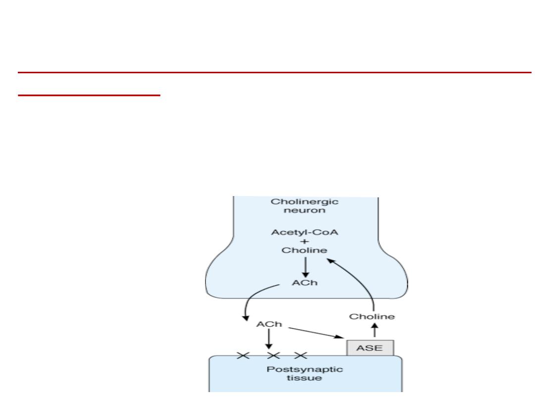
Destruction of the Released Acetylcholine by Acetyl
:
cholinesterase
1-Most of the acetylcholine is destroyed by the
enzyme acetylcholinesterase (which is present in
the synaptic space), into choline and acetate.
2- A small amount of acetylcholine diffuses out of
the synaptic space.
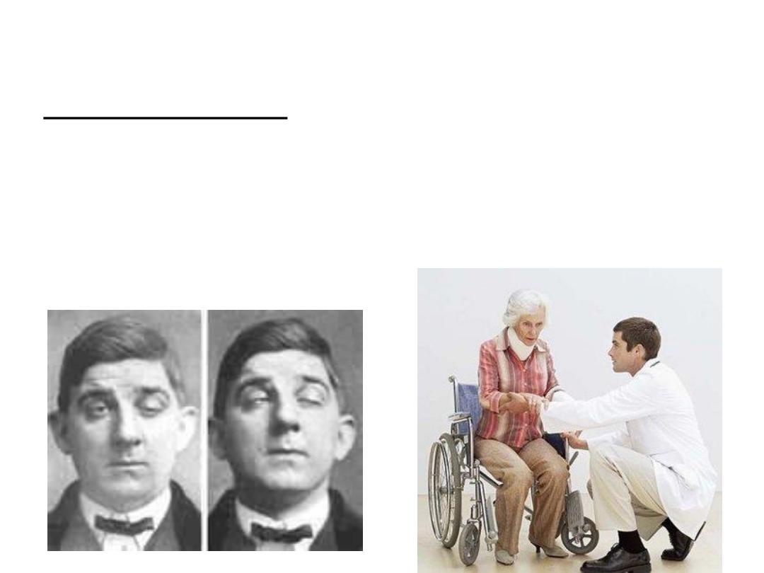
Mysthenia Gravis :an autoimmune (i.e
antibodies against your own tissues) disease
in which there is sever voluntary muscle
weakness. Antibodies that destroy the Ach
receptors

Summary :
-Nuromuscular junction consists of presynaptic part
(axon), synaptic space ,postsynsptic part (muscle).-
-
-Nuromuscular transmission starts from arriving of
action potential to the presynaptic axon ,releasing of
the neurotransmitter ,acting on the postsynaptic
muscle and generation of local potential by opening of
ion channels.
-
-Nuromuscular transmission is affected by many
factors
.
The muscle

The muscles are excitable cells; they are
machines to convert the chemical energy
to mechanical energy
.
It differs from the nervous system by the fact
that
it has a contractile
mechanism which is
activated by A.p.
The muscle can be excited electrically,
mechanically, chemically → action potential
(A.p.).
Types of muscle:
Skeletal muscles: These are voluntary muscles attach to bone.
Smooth muscles: Involuntary muscle. It is Muscle of the viscera
(e.g., in walls of blood vessels, intestine, & other 'hollow'
structures and organs in the body).
Cardiac muscles: Muscle of the heart. Involuntary.
40% of the body is skeletal muscles and 10% are smooth and
cardiac muscles.
Characteristics of muscle:
1-excitability - responds to stimuli (e.g., nervous
impulses)
2-contractility - able to shorten in length
3-extensibility - stretches when pulled
4-elasticity - tends to return to original shape & length
after contraction or extension
Functions of muscle:
1-motion
2-maintenance of posture
3-heat production

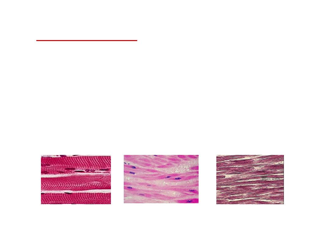
The skeletal muscle
:
It is that type of the muscles that is attached to
bones & moves skeleton, also called striated
muscle.
It lacks anatomical and functional connection
between individual muscle fibers.
It is voluntary
.

Morphology
:
It is composed of numerous fibers.
Each muscle fiber extends along the length of
.
the muscle.
The muscle fibers are arranged in parallel between
the two tendon ends, so that the force of
contraction is additive, also this allows each fiber
to be controlled individually so we can contract
fewer or more fibers and the strength of
contraction will be graded. .

The muscle fiber is a cylindrical single cell
containing:
-multiple nuclei.
-Cell membrane (sarcolemma).
-Sarcoplasm (intracellular fluid fills the spaces
between the myofibrils).
-Other organ cells
-Small muscle fibrils, which consist of filaments
that are made up of contractile proteins (actin
and myosin).
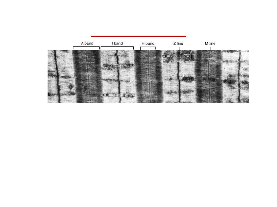
The striations
The myosin and actin filaments interdigitate and cause the
myofibrils to have alternate light and dark bands.
The light bands are only Actin filaments called I band
.
The dark bands contain Myosin (overlapping with actin
filaments) called A bands.
So the striations are due to difference in the refractive index
of the parts of the muscle fibers.
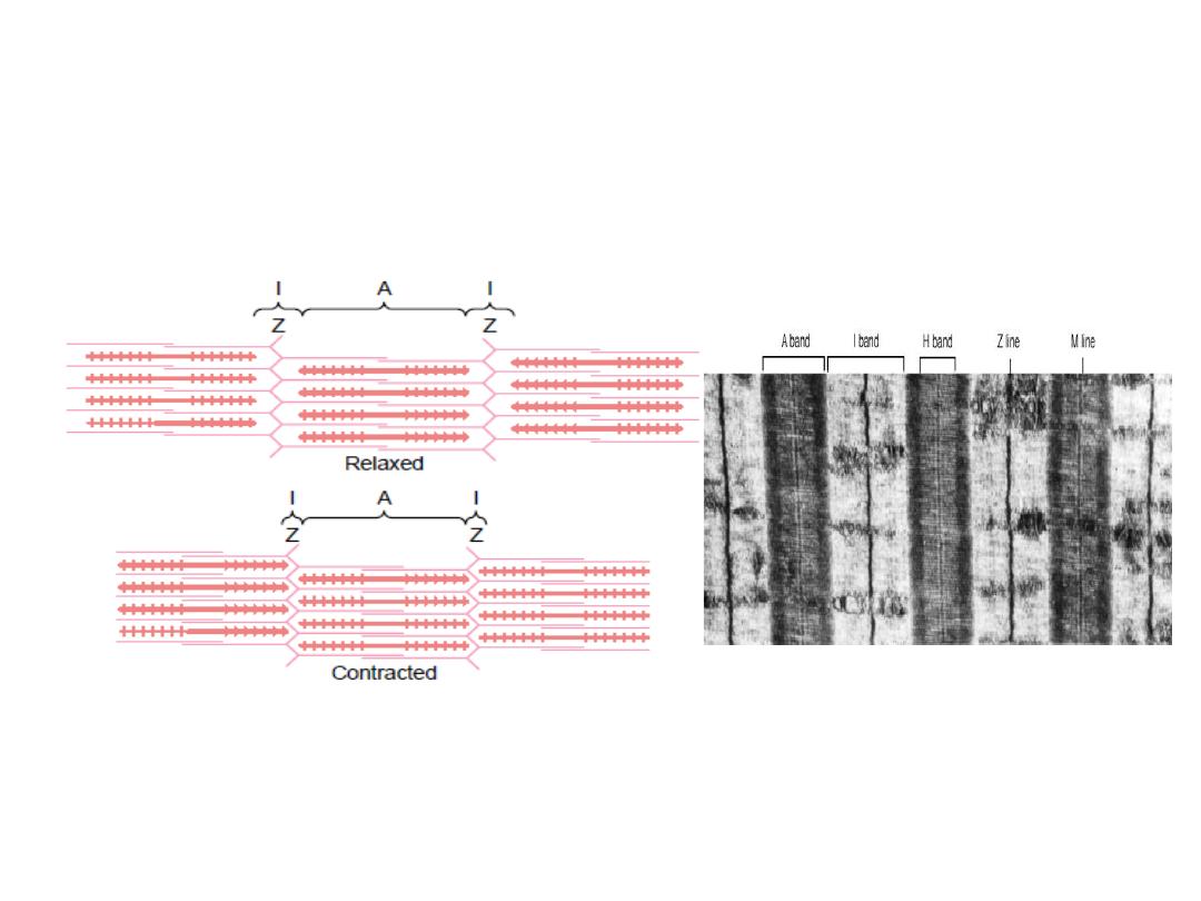
-
In the middle of The I band there is darker
Z
line. The portion of
the myofibrils that is between 2 successive Z discs is called
Sarcomere
which is
the smallest functional unit of the muscle
.
-The A band is divided by the lighter
H
band, in the middle of it a
there is a line called
M
line.
On the ends of the myosin filaments are small projections called
the cross bridges, which interact with the actin filaments to cause
contraction.
Molecular charectristics of the contractile
filaments
Myosin filaments:
It is composed of 6 polypeptide chains,
a/ 2 heavy (wrap spirally around each other to form
double helix
called tail, one end of these chains is folded bilaterally into a
structure called head (2 heads), they contain actin binding sites and
a catalytic site that hydrolyse ATP and,
b/ 4 light chains (are parts of the head (help control the function of
the
head during contraction).
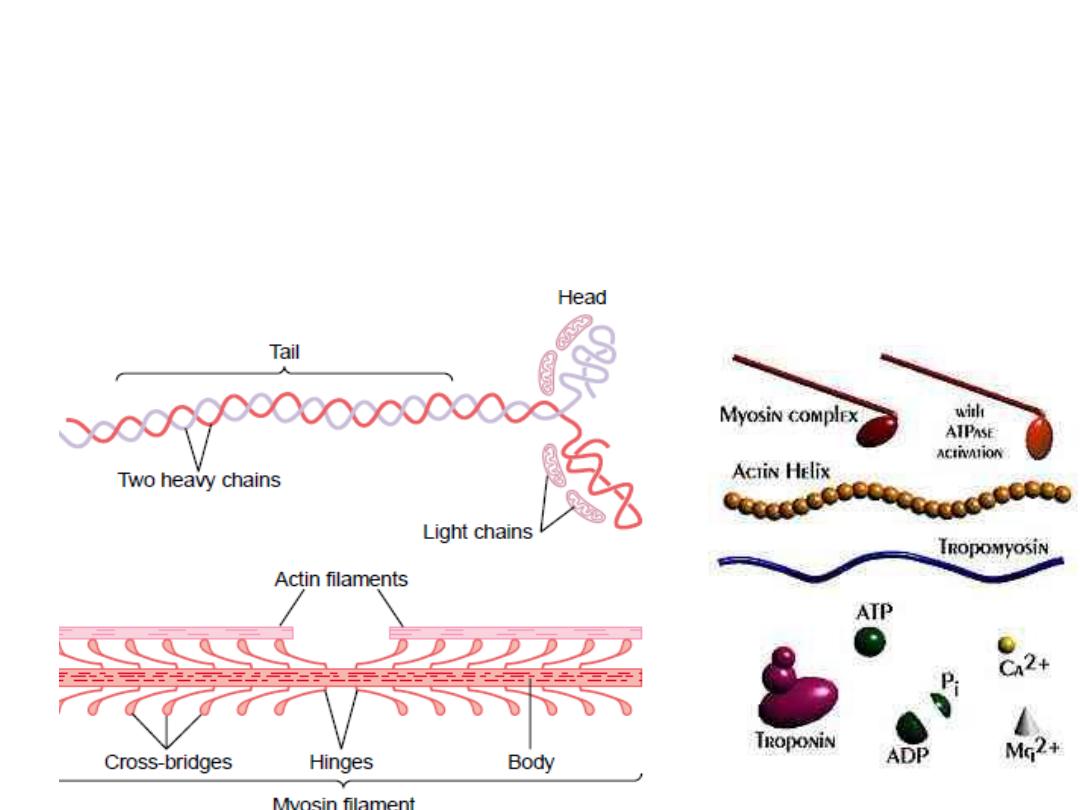
Part of the body + the head extends to form arms (called the
cross bridges
).
The cross bridges are flexible at 2 points one where the arm leaves the body,
the other where the head attaches to the arm, these called hinges.
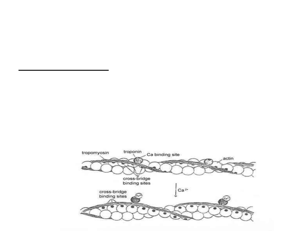
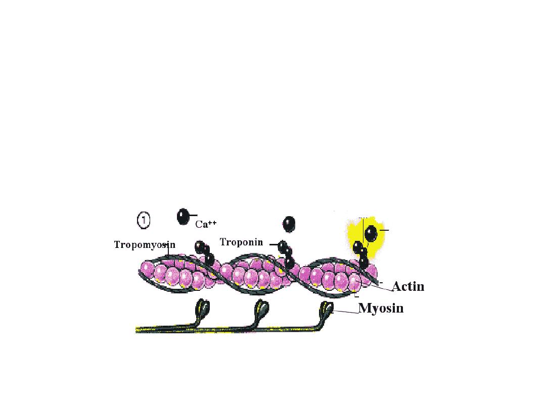
The thin filament is made of
actin
,torponin and tropomyosin
.
Actin molecules:
Actin filament is made up of 2 chains of globular unit that form
a long double helix and contain binding sites for myosin.
Each strand is composed of polymerized G actin molecule
,attached to each one molecule of ADP , these are the active
sites with which the cross bridges of myosin interact.
:
Tropmyosin molecule
They are located in the groove between the two chains forming
long filaments overlying the binding sits of myosin. So in the
resting state they lie on the top of the active sites of the actin
strands so no attraction between actin and myosin.

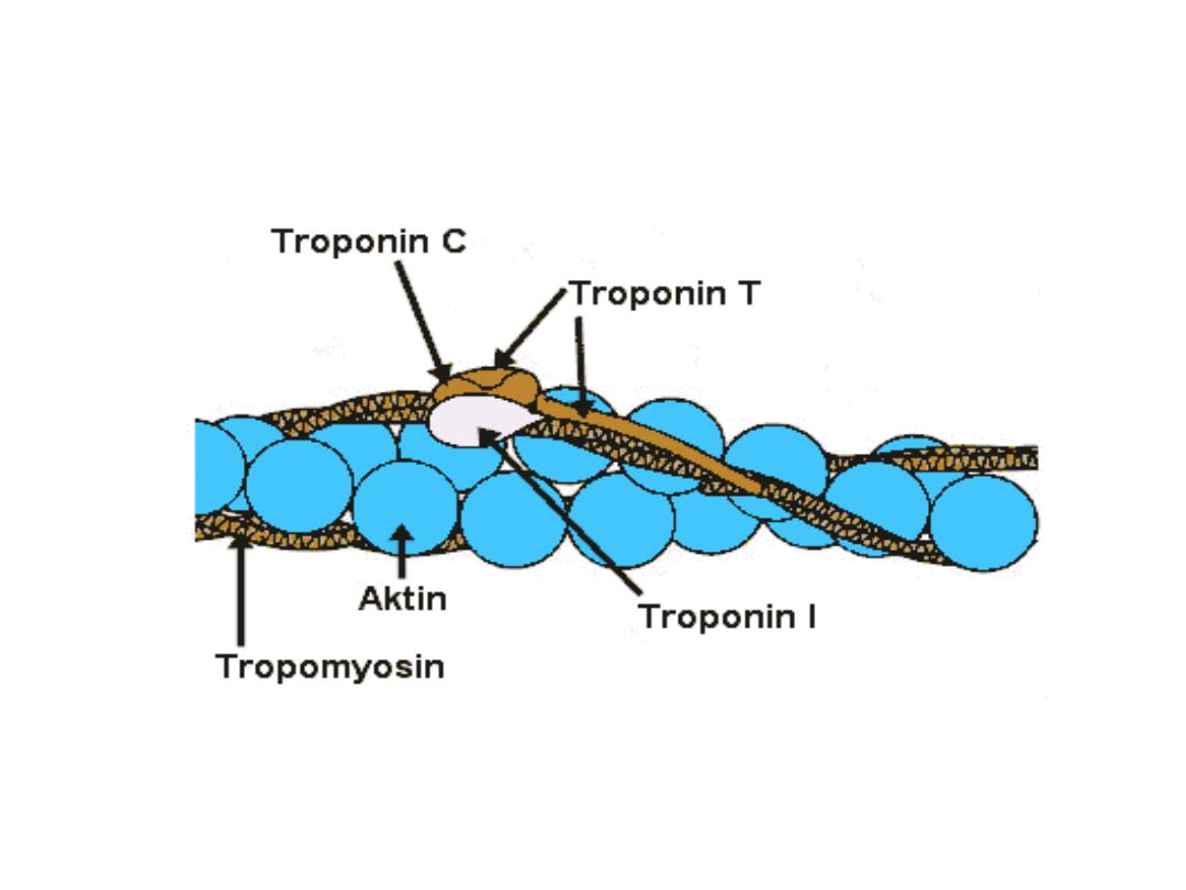
Troponin
It is a protein attached intermittently at regular intervals
along the sides of tropomyosin molecules. It is a
complex of 3 loosely bound protein subunits:
Troponin I
has a strong affinity for actin inhibits the
interaction between myosin to actin
Troponin
T
binds troponin to tropomyosin.
Troponin C
contains binding sites for calcium ions that
initiate
contraction.
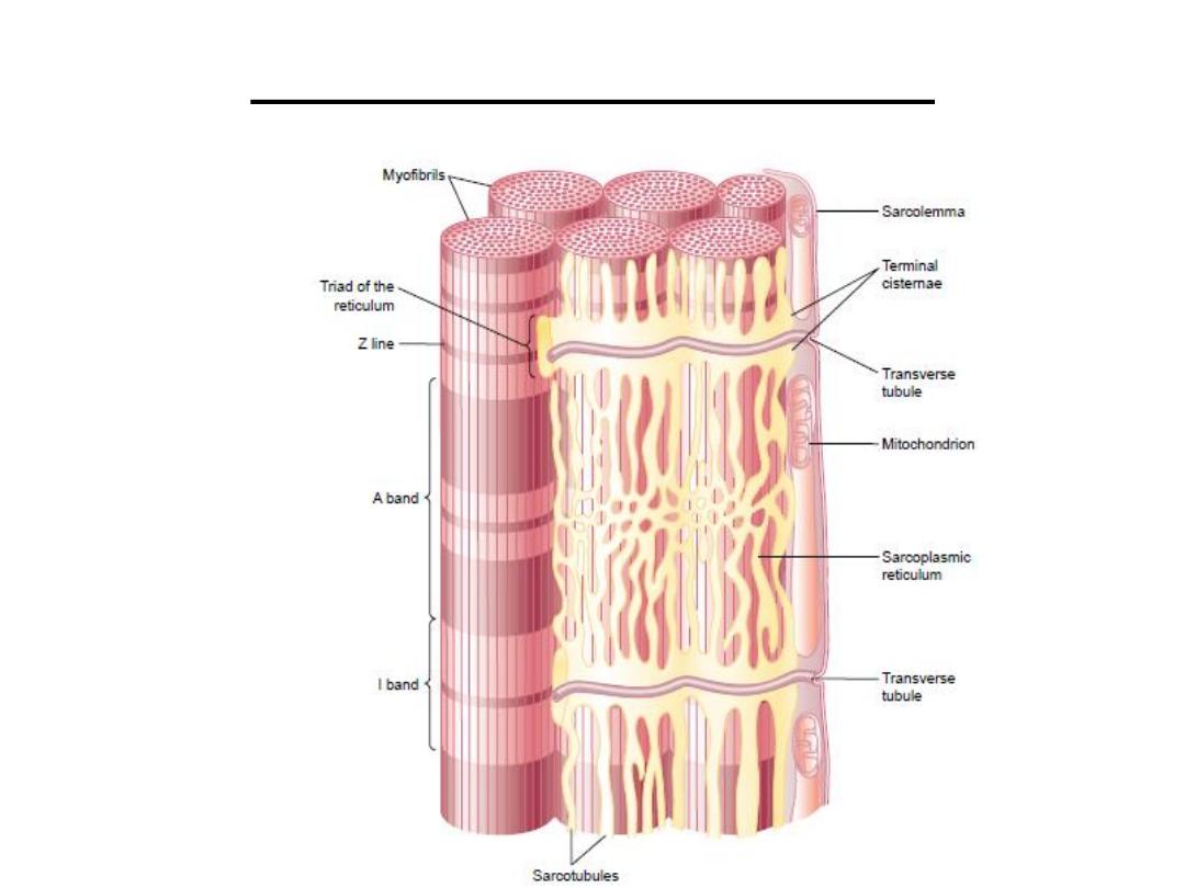
system:
sarcotubular
The

The T system (transverse tubules):
it is a system of
transverse tubules in the form of letter T which is
continuous with the membrane of the muscle fiber.
It starts from one side of the cell membrane to the
opposite side, so it is
continuous with the
extracellular
space, and they contain extracellular fluid
inside ; they are present along the whole length of the
muscle fiber and is responsible for spreading of action
potential from the cell membrane to the interior of
the muscle fiber (because the muscle is large and the
action potential can not flow deep), the electrical
currents around them create the muscle contraction.
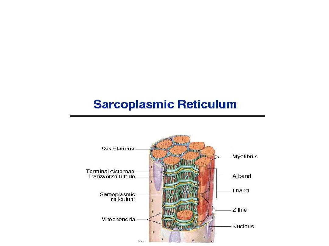
The sarcoplasmic reticulum : it forms an irregular system of
tubules surrounding the myofibrils it has an enlarging ends or
chambers called terminal cisterns .
The arrangement of the T system with the ciatern of the
endoplasmic reticulum at either side called Traid

The sarcoplasmic reticulum contains excess
amounts of calcium ions (in the cistern) in high
concentration which are released when the
action potential occurs in the adjacent tubules.
After the contraction has been occurred ,
active
calcium pump located in the walls of
sarcoplasmic reticulum
pumps calcium back
to
the sarcoplasmic tubules
Electrical characteristics of skeletal
muscles:
1- The resting membrane potential is – 80 to – 90
mill volt in skeletal muscle fiber (same as in
large mylinated nerve fiber).
2- The electrical changes of the ion fluxes are
similar to those of the nerve fiber during action
potential.
3- Duration of the action potential is longer than
that in mylinated nerve fiber.
•

4- The conduction velocity is less than that in
large mylinated nerve fiber.
5-Do not obey all or none law for the whole
muscle but not for a single muscle fiber which
obey this law.
6- Each single contraction is followed by a
single relaxation in response to a single action
potential (simple muscle twitch).
Sources of energy for the rephosphorlation
(Skletal muscle energy source)
:
1- Substance called phosphocreatine (high energy
phosphate bond). This compound synthesized
during resting conditions .
During exercise
this
compound hydrolyse releasing energy.
2-
Glucose
: it is supplied by the blood and
undergoes series of reactions forming finally Co2,
H2o and Energy.
3-
Glycolysis of glycogen
stored in the muscle cells:
4-
The free fatty acids(FFA
) (gives double the energy
that glucose gives) the use of FFA mainly
at rest
and during recovery after contraction