Types of Connective Tissues
Prof. Dr. Malak A. Al-yawer
Department of Anatomy/ Histology Section

Objective
At the end of this lecture, the 1
st
medical student
will be able to
Define the different types of connective tissue
State the general organization and functions of
each type of connective tissue
Distinguish between the different types of
connective tissues
Describe the components of adipose tissue
and compare between its two types
State the histogenesis of adipose tissue
Draw a diagram illustrating the types of
connective tissue
State some related disorders
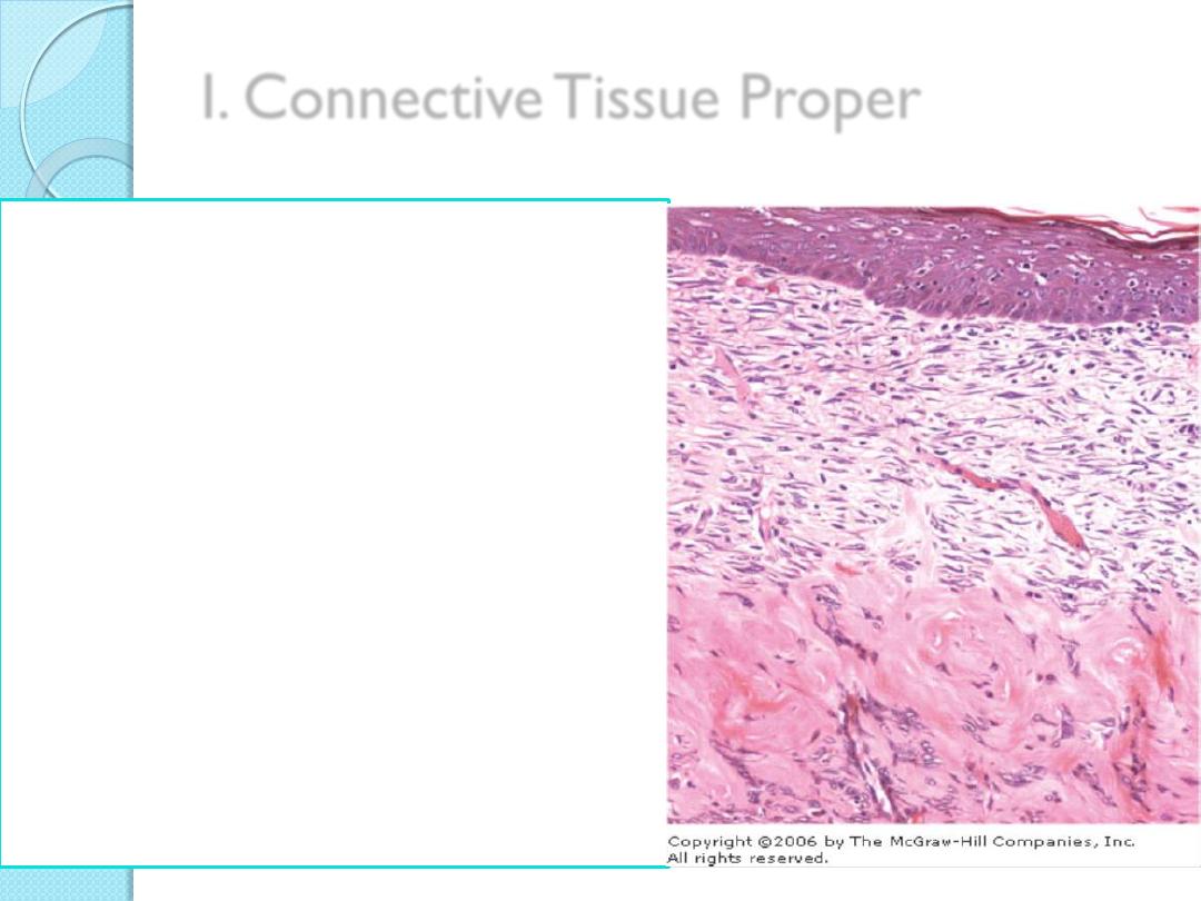
I. Connective Tissue Proper
Two classes
Loose connective tissue:
has a delicate consistency;
it is flexible
not very resistant to stress
Dense connective tissue:
is less flexible
more resistant to stress than
loose connective tissue
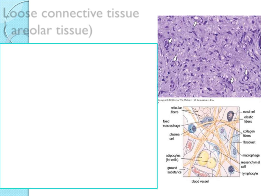
Loose connective tissue
( areolar tissue)
General Organization
Much ground substance
Many cells and little collagen
Randomly distributed
Usually well vascularized
Major Functions
Supports microvasculature,
nerves, and immune defense cells
Examples
Lamina propria beneath epithelial
lining of digestive tract
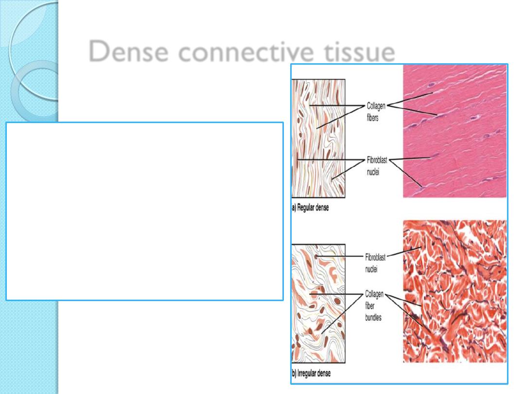
Dense connective tissue
1.
Dense regular connective
tissue
2.
Dense irregular connective
tissue
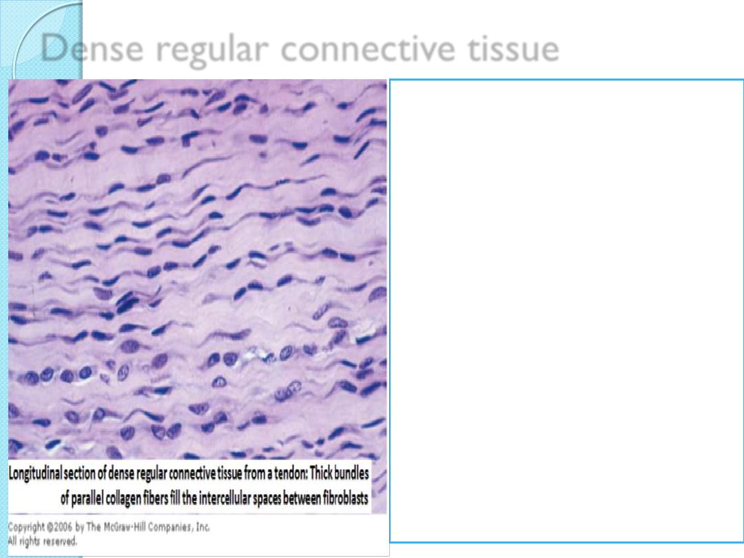
Dense regular connective tissue
General organization
o
Almost completely filled with
parallel bundles of collagen;
o
Few fibroblasts, aligned with
collagen
Major functions
Provide
o
strong connections within
musculoskeletal system;
o
strong resistance to force
Examples
o
Ligaments
o
Tendons
o
Aponeuroses
o
Corneal stroma
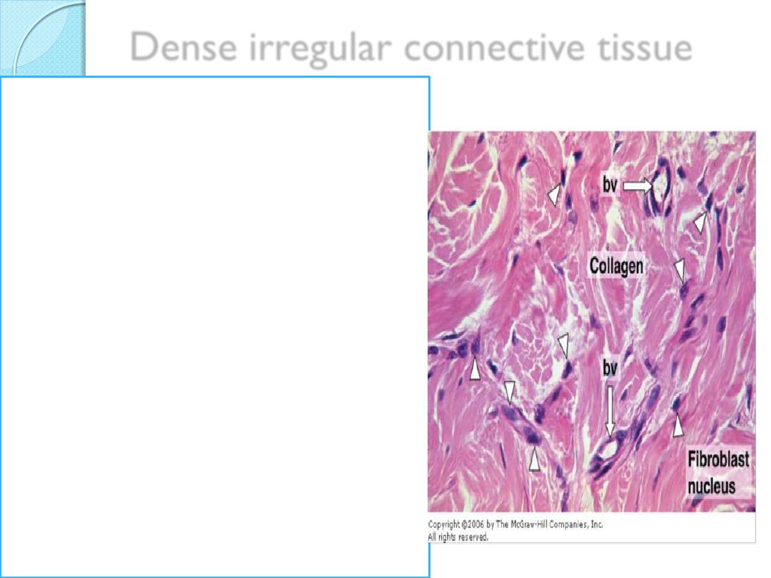
Dense irregular connective tissue
General Organization
Little ground substance;
few cells (mostly fibroblasts);
much collagen in randomly
arranged fibers
Major functions
Protects and supports organs;
resists tearing
Examples
Dermis of skin,
organ capsules,
Submucosa layer of digestive
tract
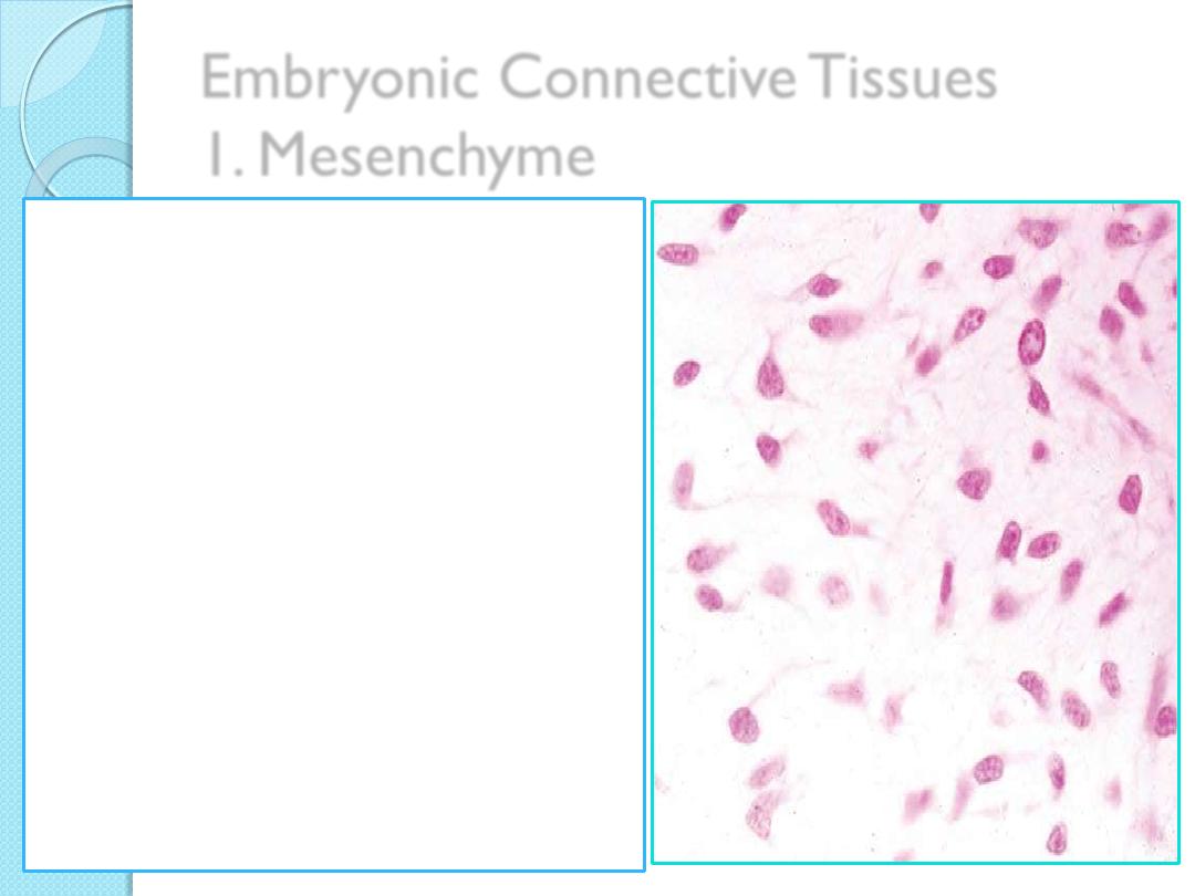
Embryonic Connective Tissues
1. Mesenchyme
General organization:
Sparse, undifferentiated cells,
uniformly distributed in matrix
sparse collagen fibers
Functions:
•
Contains stem/progenitor cells
for all adult connective tissue
cells
Examples:
•
Mesodermal layer of early
embryo
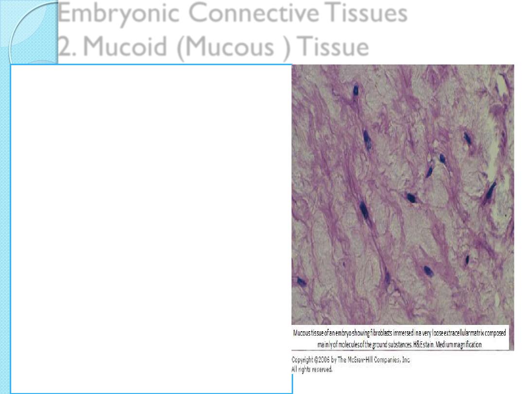
Embryonic Connective Tissues
2. Mucoid (Mucous ) Tissue
General organization:
sparse collagen fibers
scattered fibroblasts
abundance of ground
substance
Functions:
Supports and cushions large
blood vessels
Examples:
•
umbilical cord,
•
the pulp cavities of young
teeth

Specialized Connective Tissues
1.
Reticular connective tissue
2.
Adipose Tissue
3.
Cartilage
4.
Bone
5.
Blood
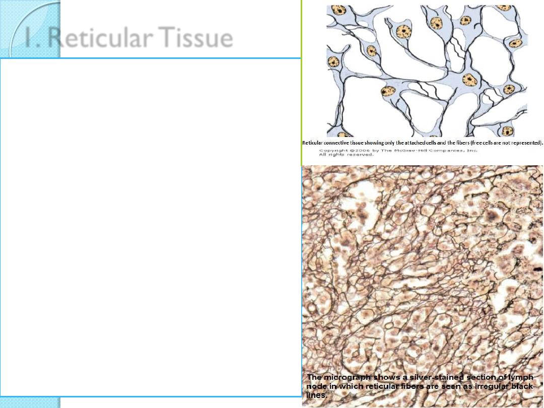
1. Reticular Tissue
General organization:
o
Delicate network of reticulin
/collagen III with
o
fibroblasts (reticular cells)
Functions:
o
Supports blood-forming cells, many
secretory cells, and lymphocytes in
most lymphoid Organs
Examples:
o
Bone marrow,
o
liver,
o
pancreas,
o
adrenal glands,
o
all lymphoid organs except the
thymus
2. Adipose Tissue
Adipose cells can be found
isolated or in small groups
in large aggregates
adipose tissues
.
Adipose tissue represents
15-20% of the body weight in men of normal
weight
20-25% of body weight in women of normal
weight
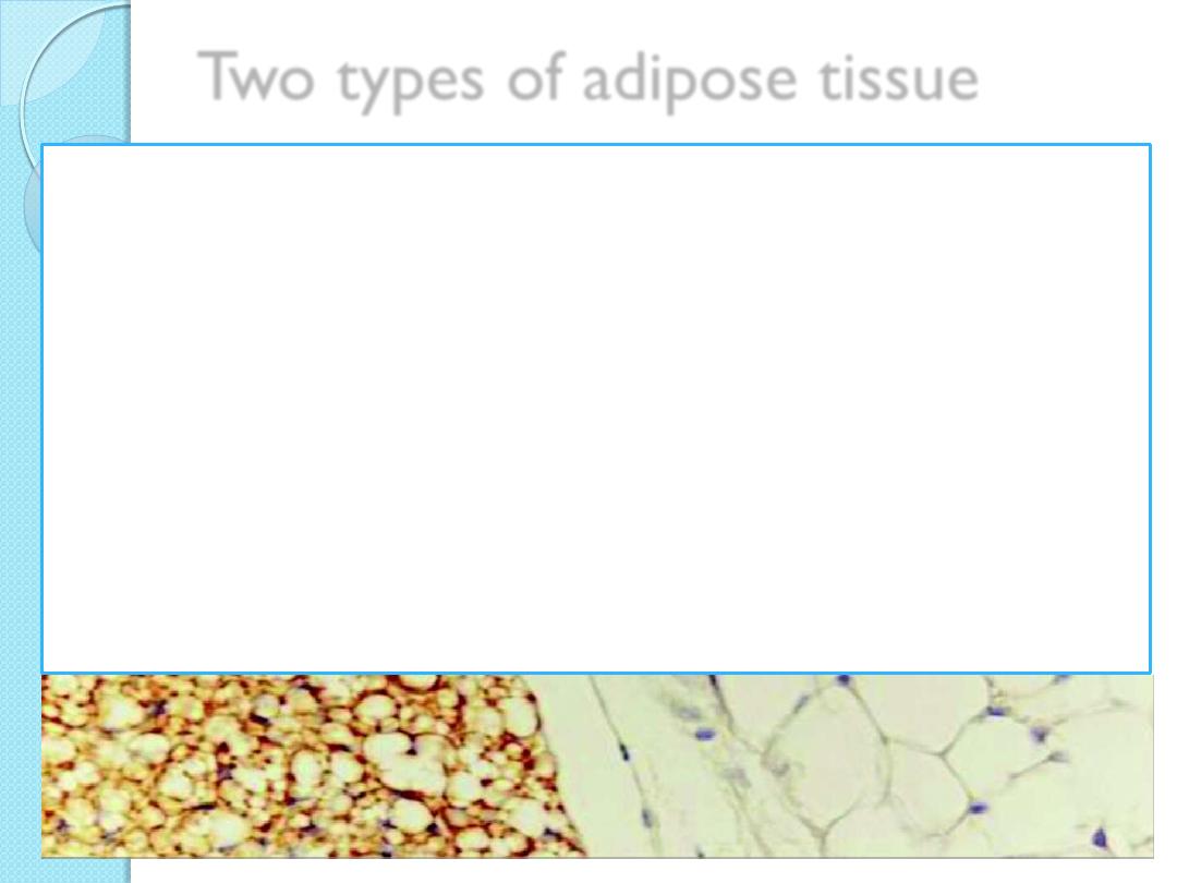
Two types of adipose tissue
1.
White adipose tissue:
the more common type,
is composed of cells that contain one very large
droplet of whitish-yellow fat in their cytoplasm.
2.
Brown adipose tissue
contains cells with multiple lipid droplets interspersed
among abundant mitochondria, which give these cells
a darker appearance.
Both types of adipose tissue have a rich blood supply.
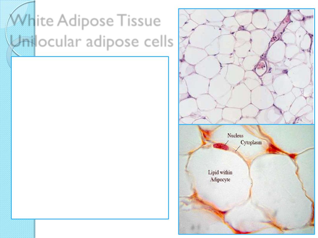
White Adipose Tissue
Unilocular adipose cells
spherical (isolated),
polyhedral (closely packed)
The cells are sometimes
said to have a signet-ring
appearance, with the lipid
droplet displacing and
flattening the nucleus
against the cell membrane

Obesity
Adult-onset obesity
mainly involves increased size of existing
adipocytes (hypertrophic obesity).
Childhood obesity
can involve increases in both adipocyte size and
numbers due to differentiation of more pre-
adipocytes from mesenchymal stem cells
(hyperplastic obesity)
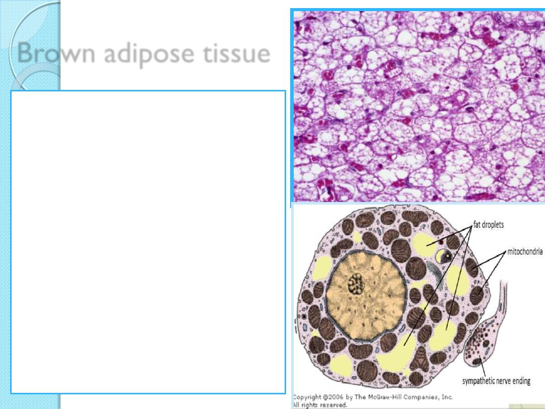
Brown adipose tissue
Cells of Brown adipose
tissue are
polygonal and generally
smaller than cells of white
adipose tissue;
their cytoplasm contains a
great number of lipid
droplets of various sizes
nuclei are often centrally
located
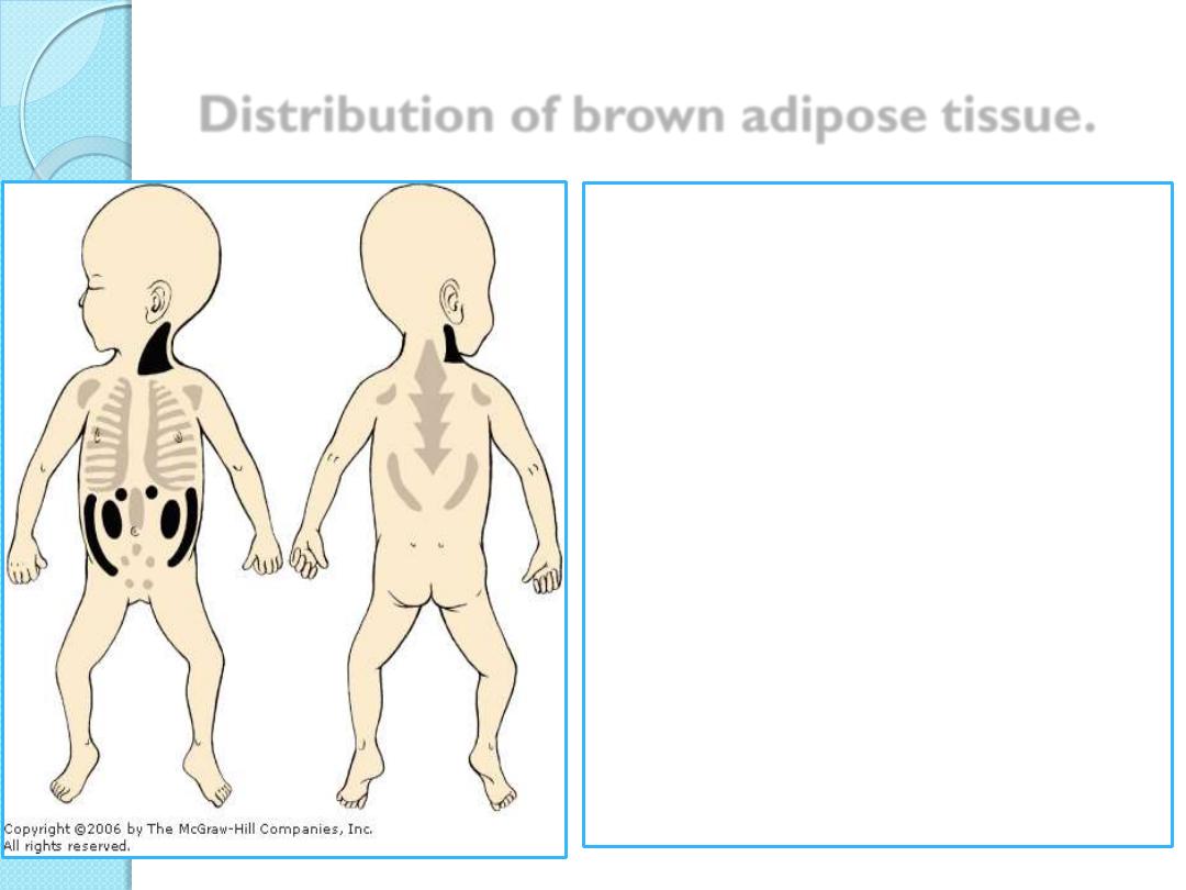
Distribution of brown adipose tissue.
Brown adipose tissue
constitutes 2% to 5% of the
newborn body weight,
located mainly in the back,
neck, and shoulders,
but it is greatly reduced
during childhood and
adolescence.
In adults it is found only in
scattered areas, especially
around the kidneys, adrenal
glands, aorta, and
mediastinum.
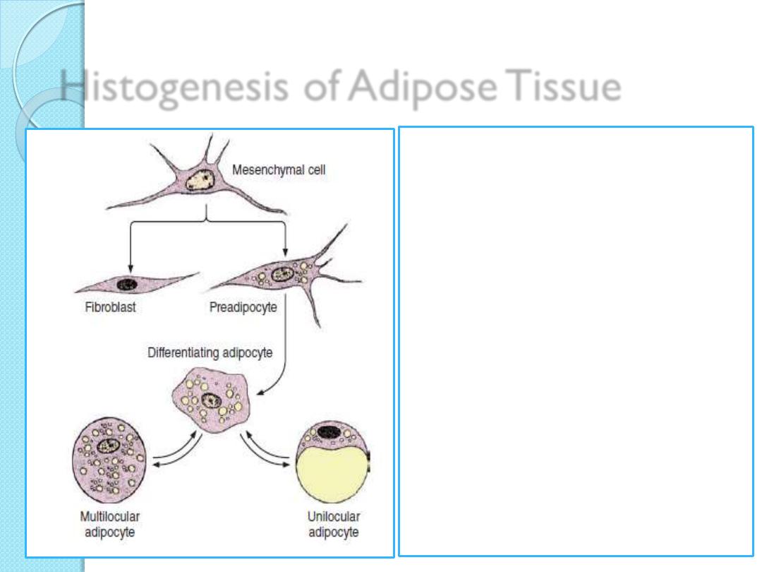
Histogenesis of Adipose Tissue
Mesenchymal cells
differentiate initially as pre-
adipocytes and then
develop further as
adipocytes as they
accumulate fat and thus
give rise to mature
unilocular or multilocular
fat cells.
When large amounts of
lipid are mobilized,
mature fat cells may
return to the lipoblast
stage.
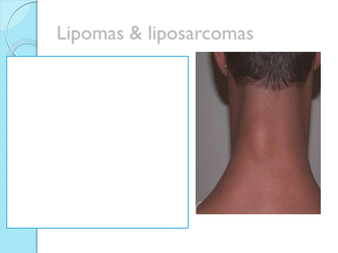
Lipomas & liposarcomas
Unilocular adipocytes can
generate very common
benign tumors called
lipomas.
Malignant adipocyte-derived
tumors (liposarcomas) are
not frequent in humans.

QUIZ

Summary
Connective tissue consists of CT proper, embryonic CT and
specialized CT
Two types of CT proper can be recognized Loose & dense
Two types of embryonic CT can be recognized Mesenchyme and
mucoid tissue
Reticular tissue, adipose tissue, cartilage, bone and blood are
specialized CT
Two kinds of adipose tissue can be recognized: white & brown
adipose tissues