Dr.Eman Al.Hadethy
Al-Faluoja.College of
medicine/CVS Physiology

Cardiovascular System
Heart
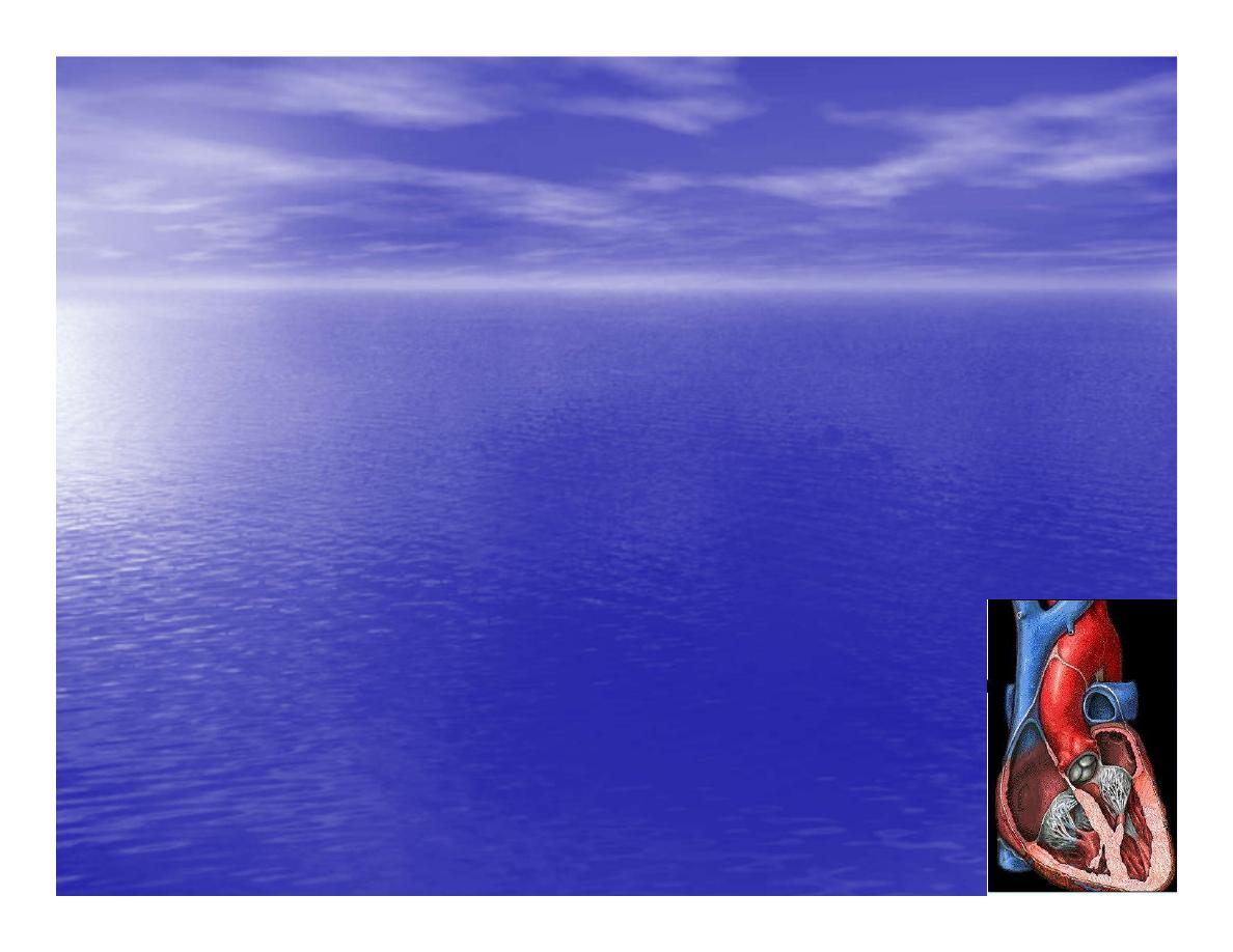
The heart
•
Is the transport system pumps: the
delivery routs are the blood vessels.
Using blood as the transport
medium, the heart propels
oxygen,nutrients,wastes and other
substances to and past the body
cells.
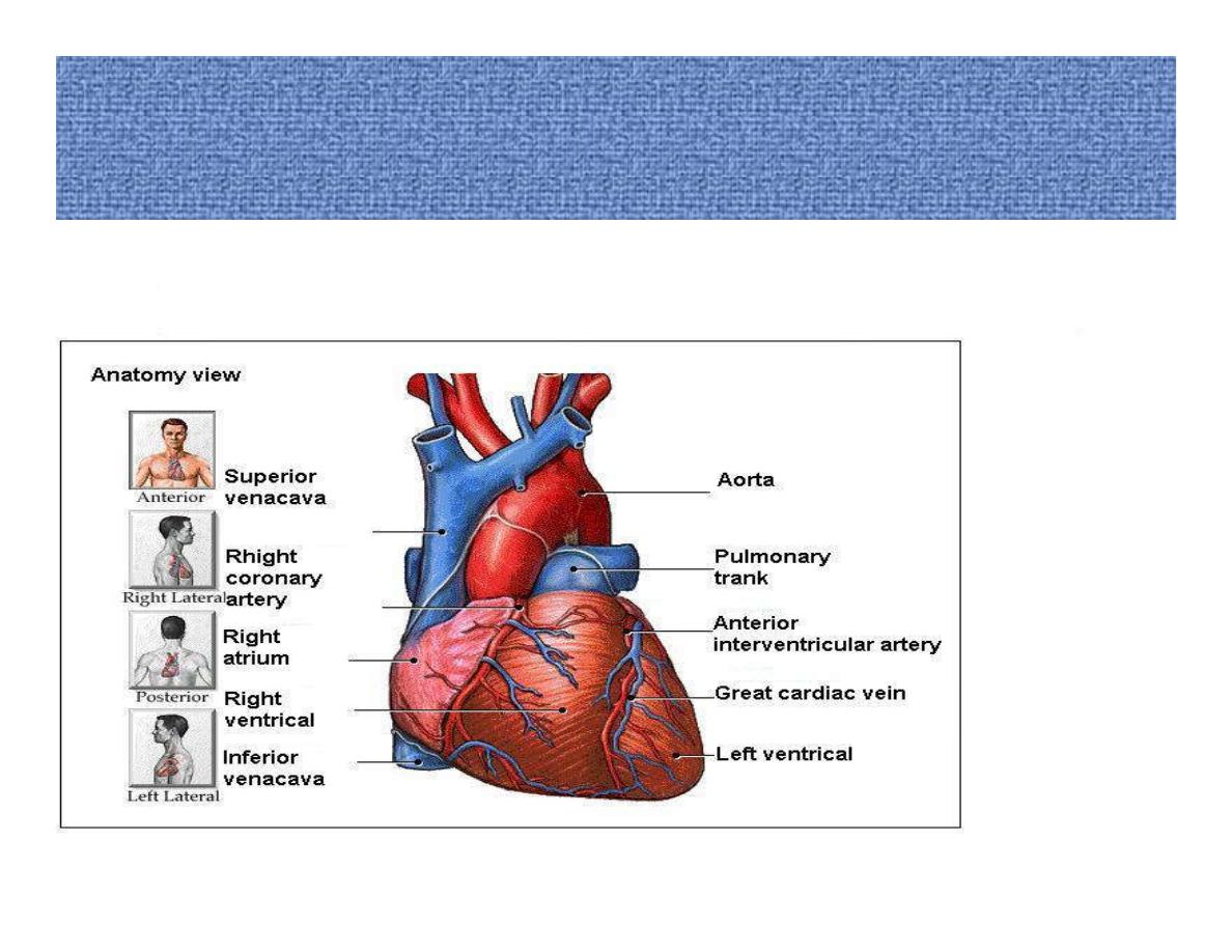
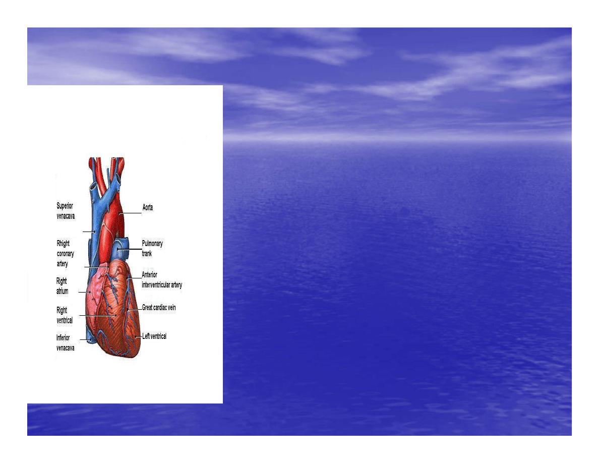
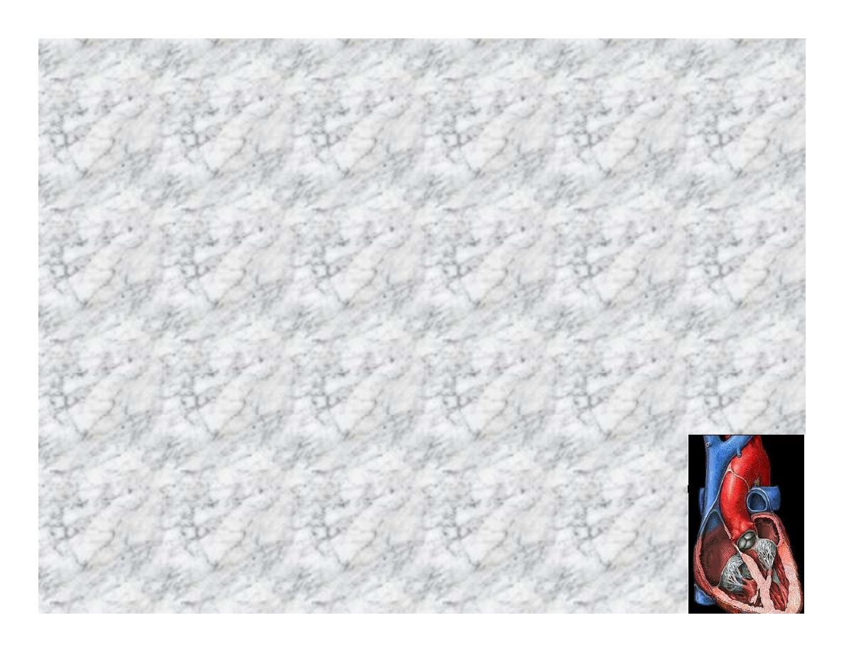
Anatomy View
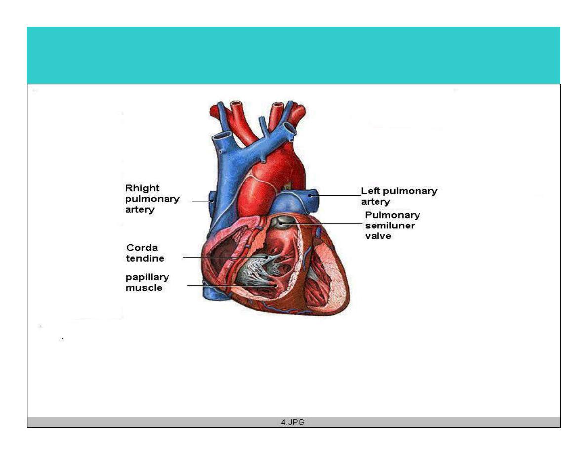
Deep cut away-view
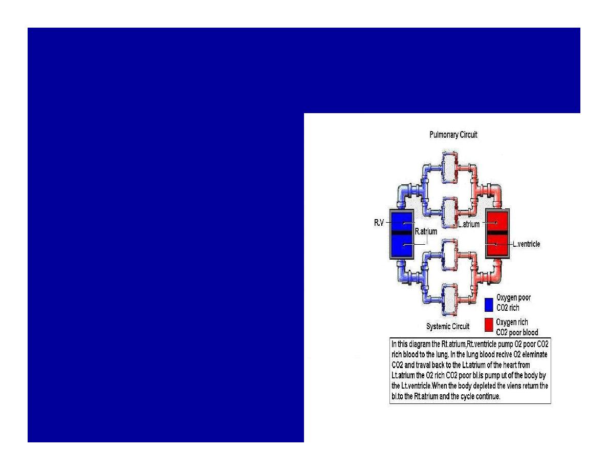
Pulmonary and systemic circulation
•
The heart consist of two
side-by- side pumps.
•
The bl.v are the pipes
carry blood through
the body.
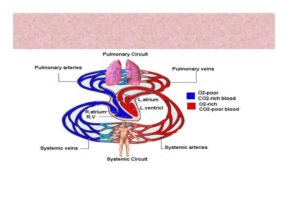
Pulmonary and Systemic Circuit
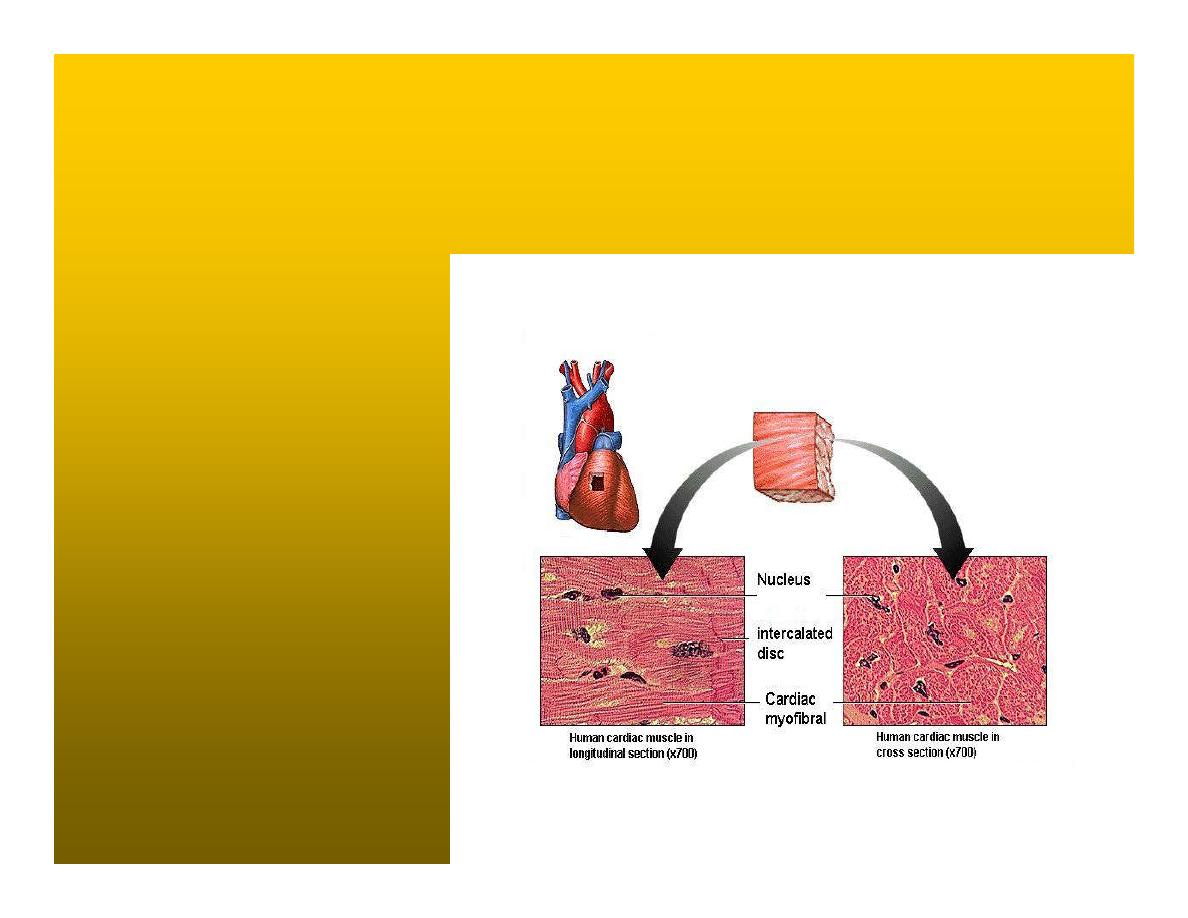
Magnified view of the heart cell
•
Nucleus
•
Intercalated disc
•
Cardiac myofibril
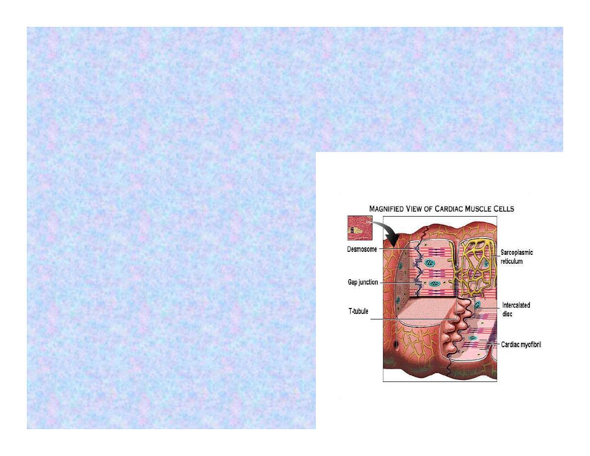
Cell junction
•
Desmosome adjacent
cell together when
m.cell contract they
pull each other.
•
Gap junction imp. for
different reason allow
stimulating impulse to
move across the heart
from cell to cell so the
heart beat as entire
unit if each cardiac m.
cell allow to do
something the heart
useless as a pump.
Intrinsic conduction system
•
Intrinsic conduction system sets the basic
rhythm of the beating heart. It consists of
autorhythmic cardiac cells that initiate and
distribute impulses (action potentials) through
out the heart.
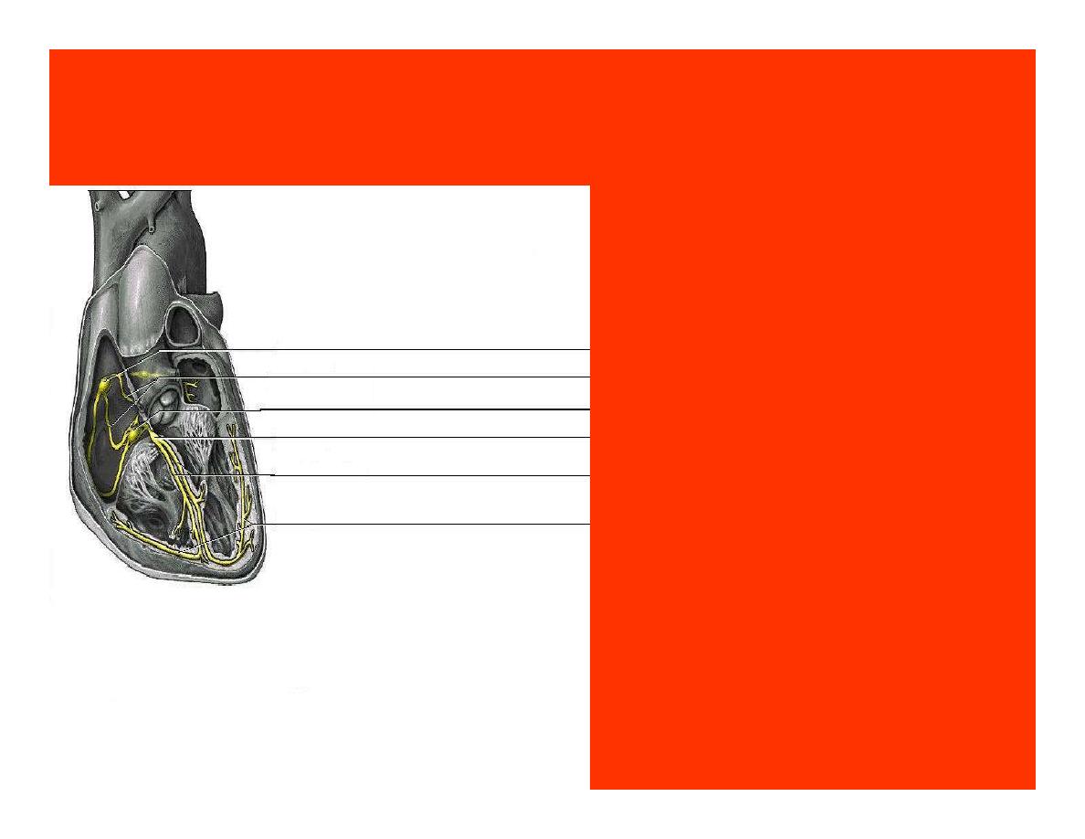
Location of autorythmic cell
*SA node
*internodal pathway
*AV node
*AV bundle
*Bundle branches
*Purkinje fibers
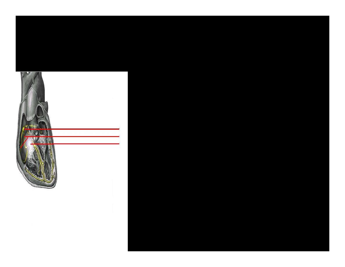
PATH WAY OF DEPOLARIZATION
SA Node
Internodal pathway
AV node
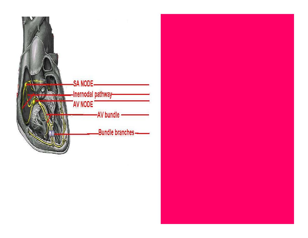
The SA node initiate the depolarization impulse
which in turn generate an action potential that
speared through out the atrium to the AV
node here the impulse delay breafilly before
continue on to the ventricle to
•
To the AV node
•
AV bundle
•
Bundle branches and
•
Purkinje fibers.
Action potential which
spread from the
autorhythmic cells of
intrinsic conduction
system to the contractile
cells, are electrical
events. Subsequent
contraction of the
contractile cells is a
mechanical event that
causes heart beat
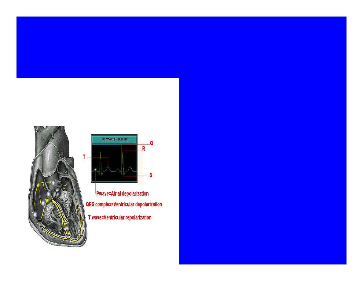
Correlation between heart electrical
activity and ECG wave tracing
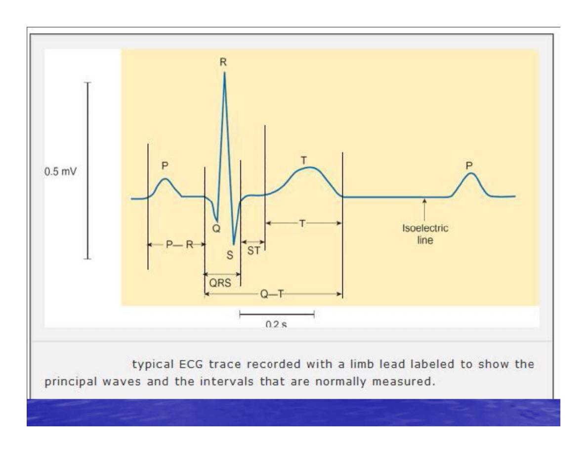
*P wave=Atrial depolarization
followed by atrial contraction.
*QRS wave=V.depolarization
followed by V.contraction.
*T wave=V.Repolarization
followed by V.relaxation.
* Atrial repolarization is hidden
by QRS complex.
SUMMARY
•
The intrinsic conduction system
of the heart initiates
depolarization impulses.
•
Action potentials spread
throughout the heart, causing a
coordinated heart contraction
•
An ECG wave tracing records the
electrical activity of the heart.
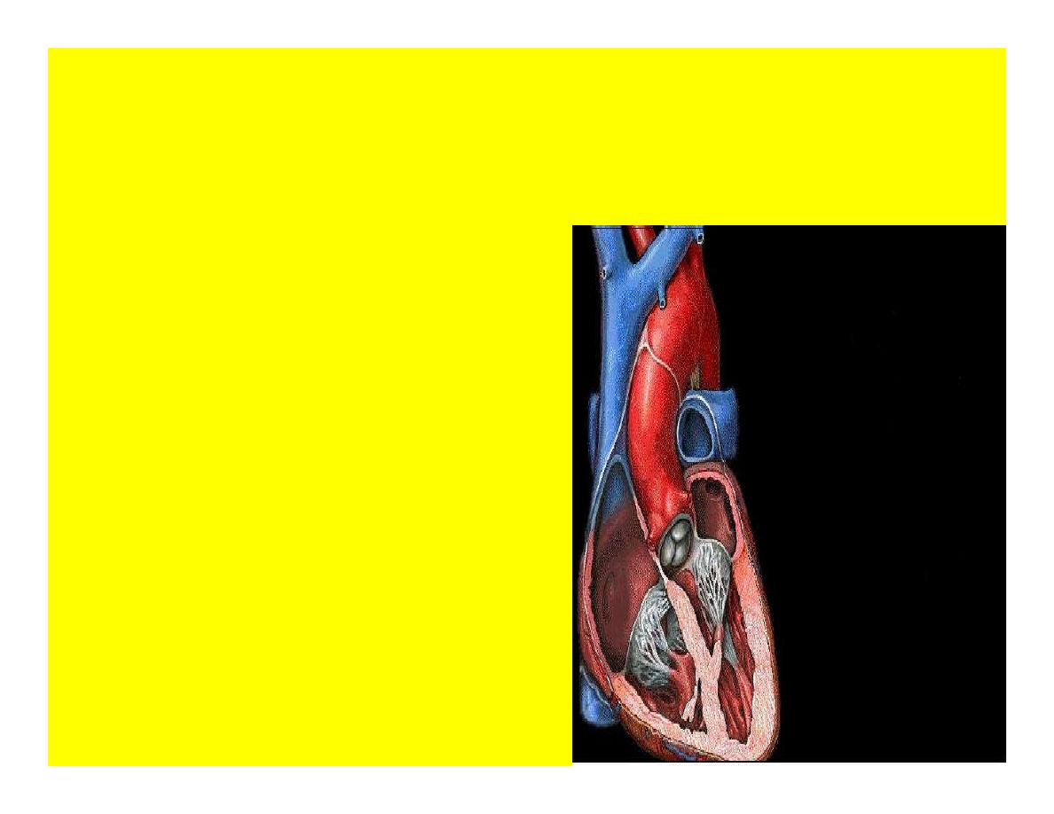
CARDIAC ACTION POTENTIAL
The coordinated contractions
of the heart result from
electrical changes that take
place in cardiac cells

GOALS
*
To understand the ionic basis of the
pacemaker potential and action potential
in a cardiac autorhythmic cell
* To understand the ionic basis of an action
potential in a cardiac contractile
(ventricular) cell.
* To understand that autorhythmic and
contractile cells are electrically coupled by
current that flows through gap junctions.
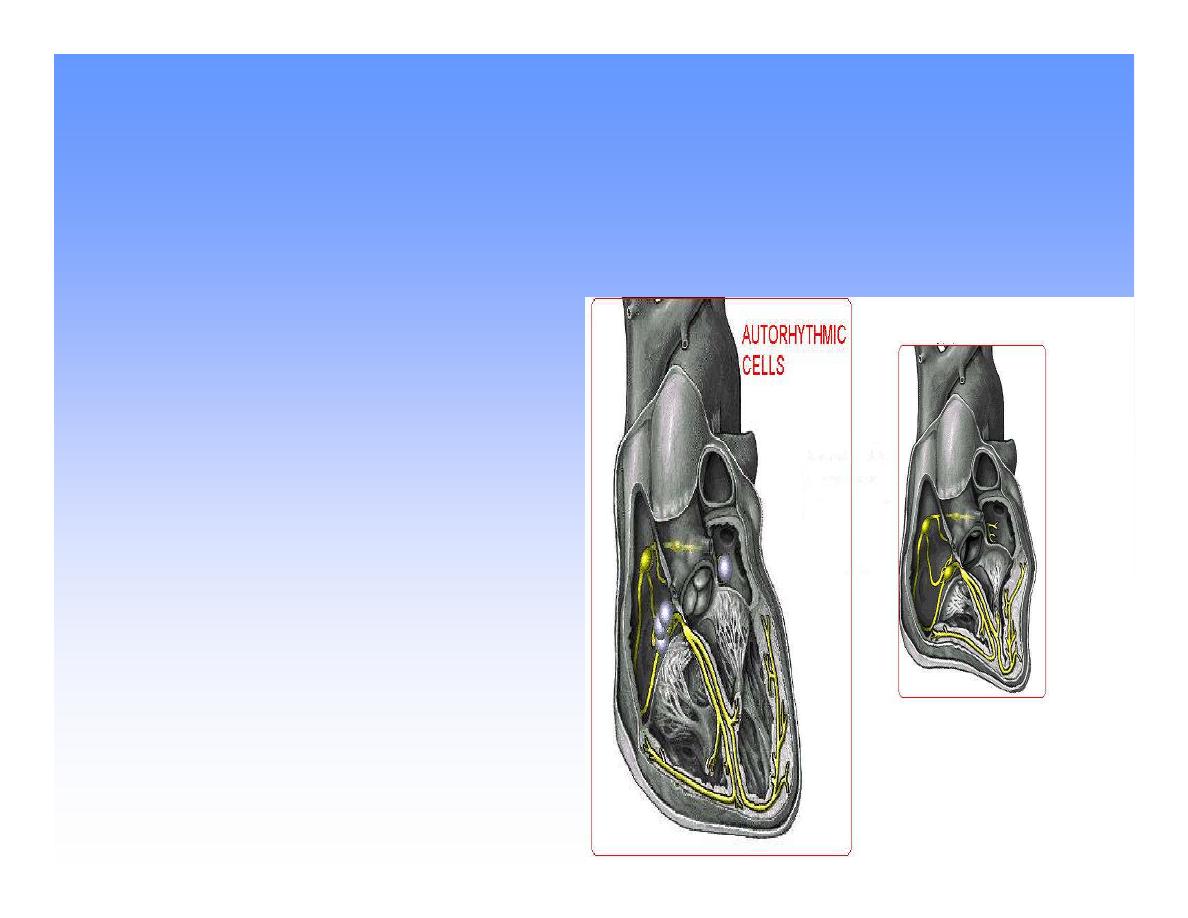
AUTORHYTHMIC CELLS
In the intrinsic conduction
system generate action
potentials that spread
throughout the heart,
triggering contractions in
the contractile cells.
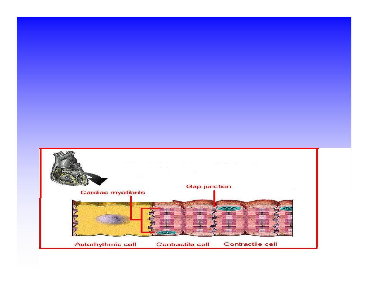
Action Potential
•
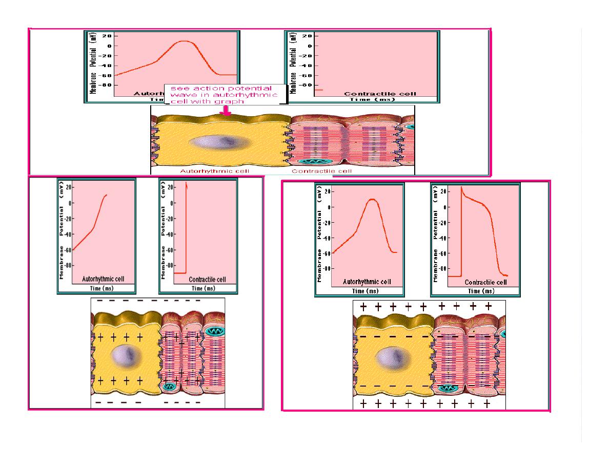
Action potentials generated by autorhythmic cells
create waves of depolarization that spread to
contractile cells via gap junctions
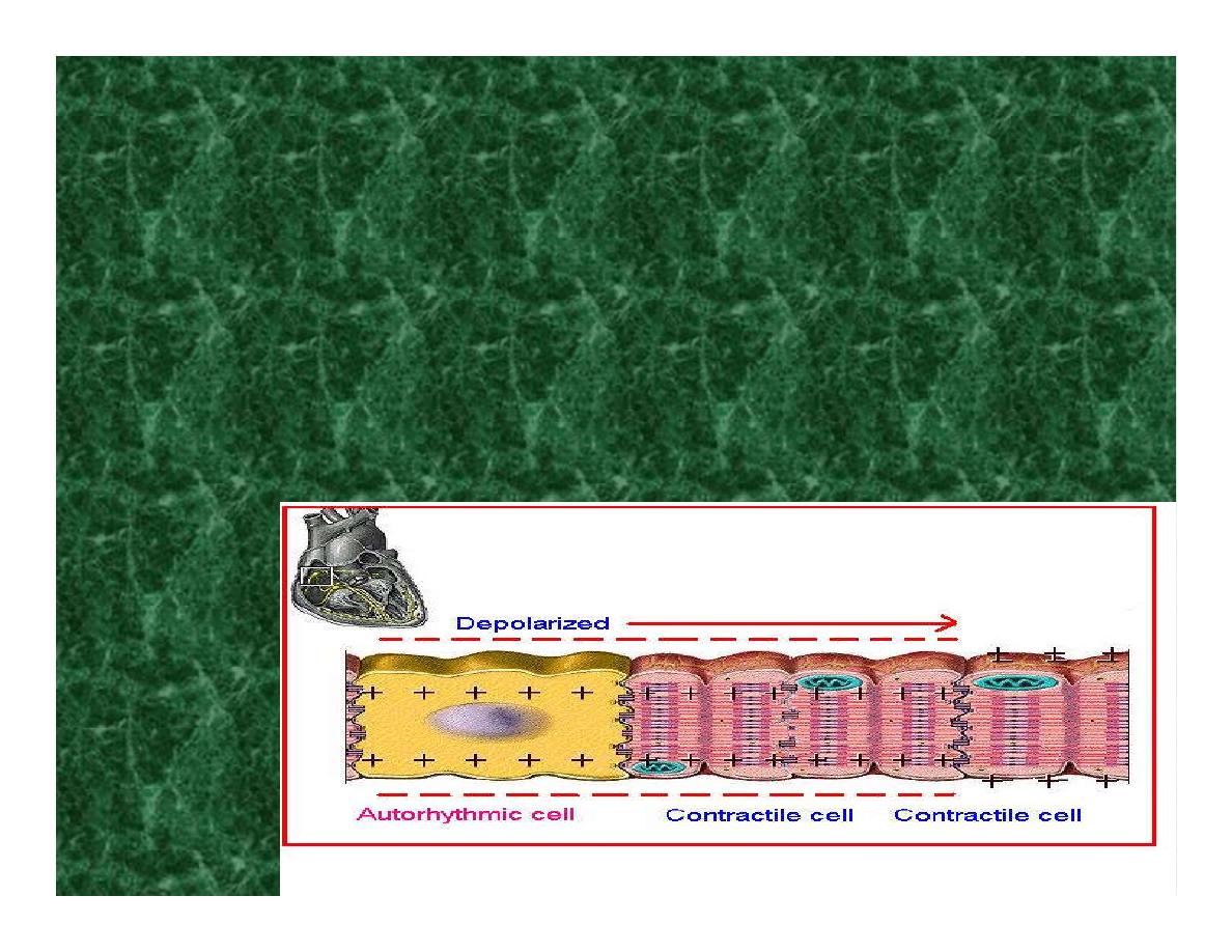
*If depolarization reaches
threshold the contractile cells in
turn generate action potentials
first
depolarizing
then
repolarizing.
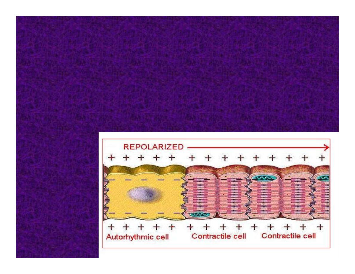
*Contractile cells contract after
depolarization and relax after
repolarization
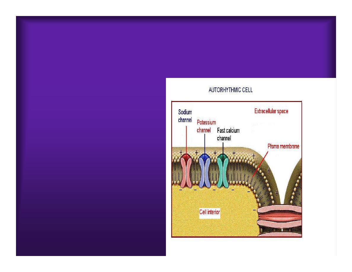
AUTORHYTHMIC CELL
Plasma channel
•
Sodium channel
•
Fast calcium
•
Potassium channel
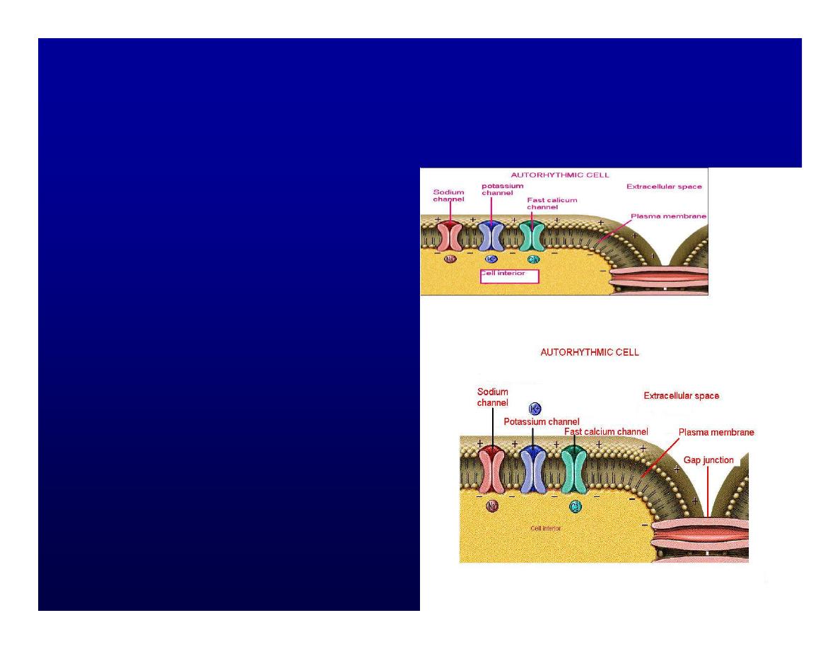
Protein Channel
*Sodium and fast
Ca
++
channels allow
Na
+
and Ca to enter
the cell
* where as potassium
channel allow K
+
to
leave the cell.
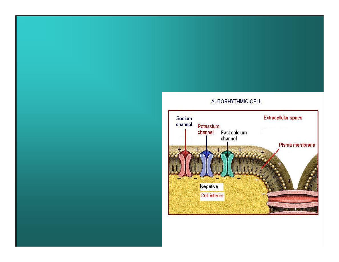
Membrane potential
*The movement of ions
effect the membrane
potential the voltage
Crosse the membrane
potential is result of the
relative the
concentration of the
ions inside or outside of
the plasma membrane
* if there are more +
ions outside the cell
then inside the cell is
relatively _ as shown
here.
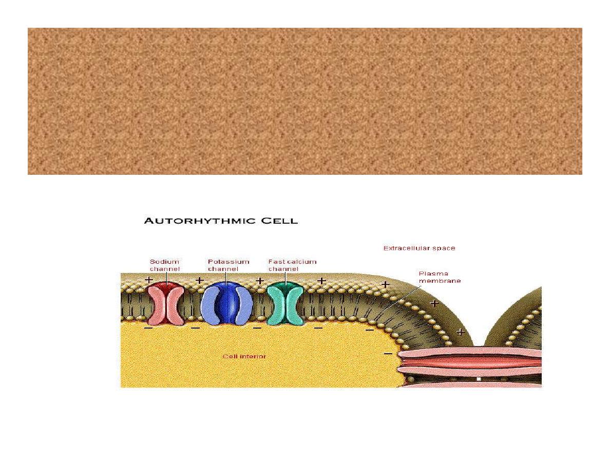
Many transport channel are voltage
regulated that is open and close in
response to specific voltage level across
the membrane.
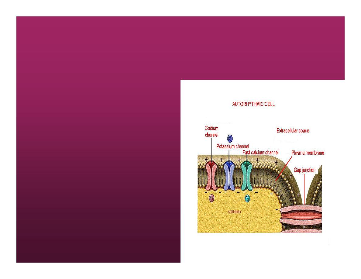
GAP JUNCTION
*
The gap junction
adjacent the cell this
allow the ions to pass
between the cell
allowing of rapid effect
initiate depolarization in
one cell and another and
so on.
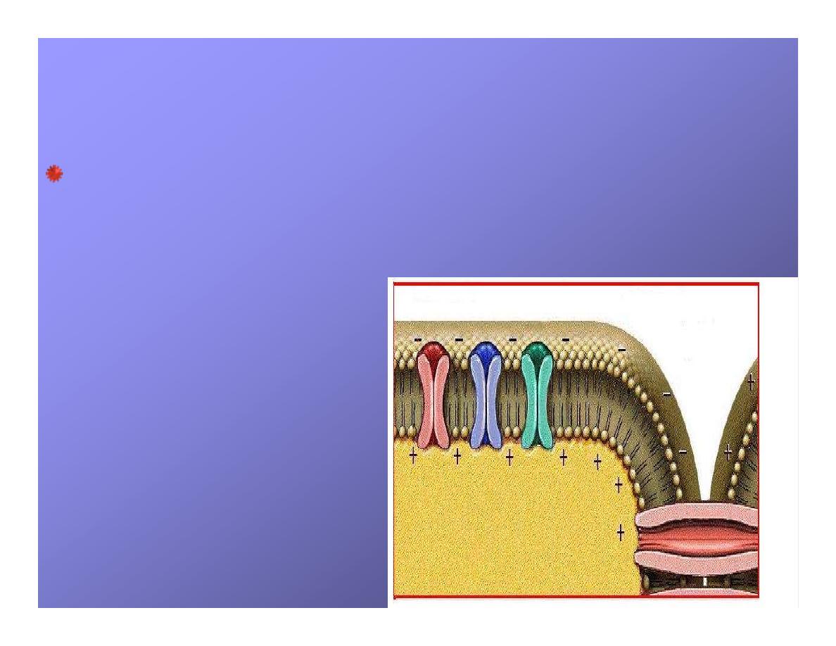
OVERVIW:INTIATION OF ACTION POTENTIALS
IN AUTORHYTHMIC CELLS
Autorhythmic cells has unique
ability to depolarized
spontaneously resulting
1.
Pacemaker potential
once
threshold is reach an action
potential initiated which become
with further
2.Depolarization and
reversal of membrane
potential
3.Repolrization
return the cell
to resting membrane potential.
the cell spontaneous begin to
slow depolarize once again and
sequence is repeated.
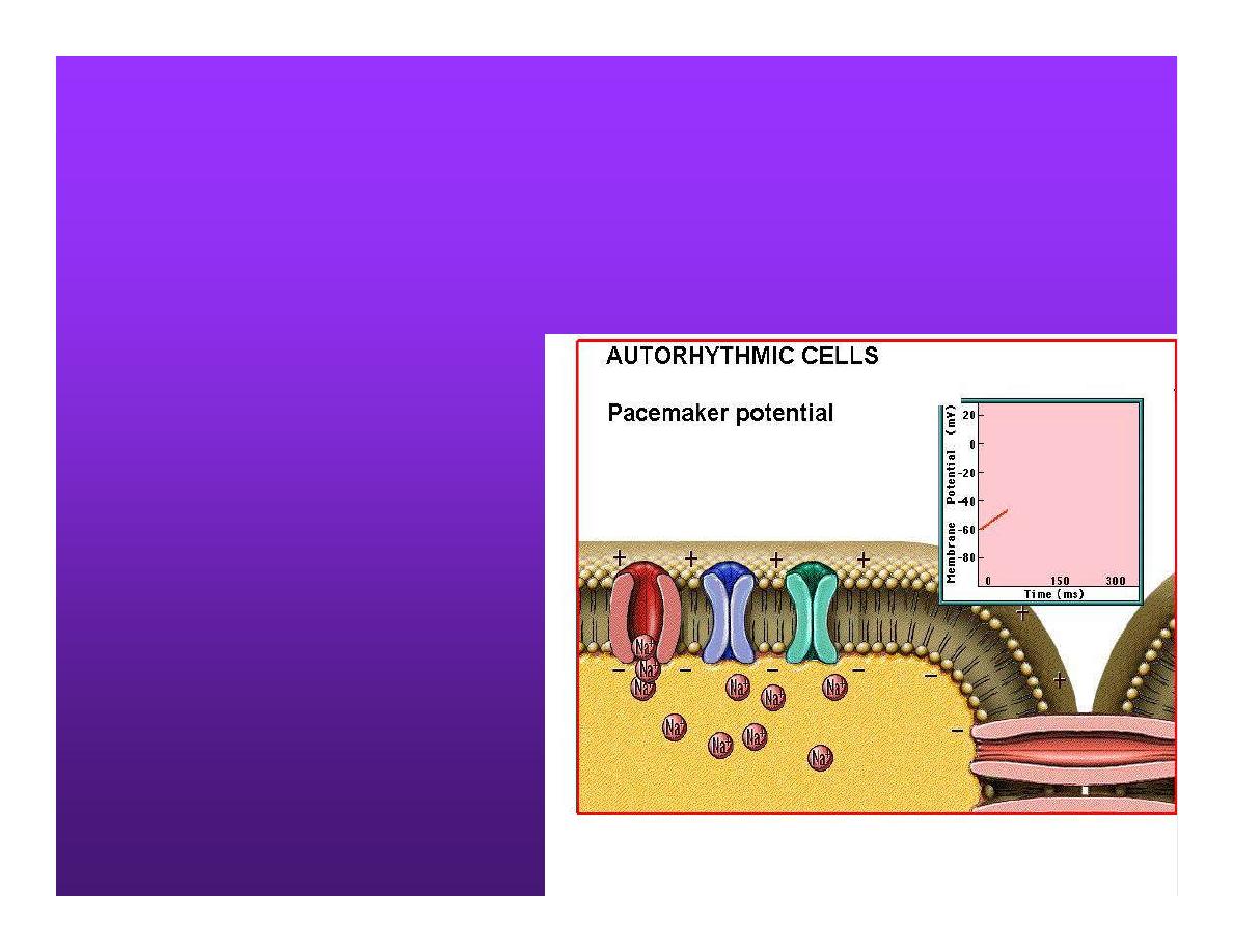
AUTORHYTHMIC CELLS
1.Pacemaker potential
Autorhythmic cells
begin depolarizing
due to slow
continuous influx of
Na+ & reduced
efflux of K
+
.
ACTION POTENTIAL
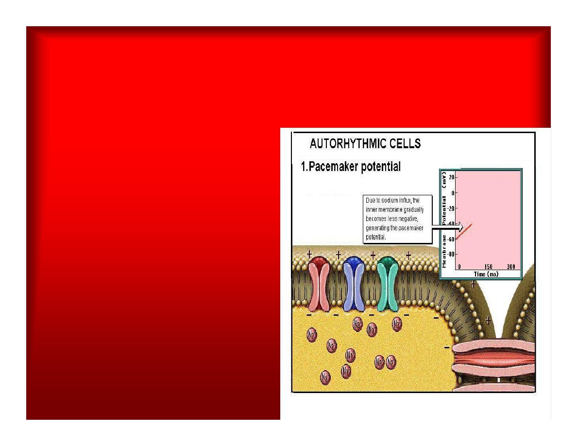
AUTORHYTHMIC CELLS
*
Due to Na+ influx,the
inner membrane
gradually become
less negative,
generating the
pacemaker potential
.
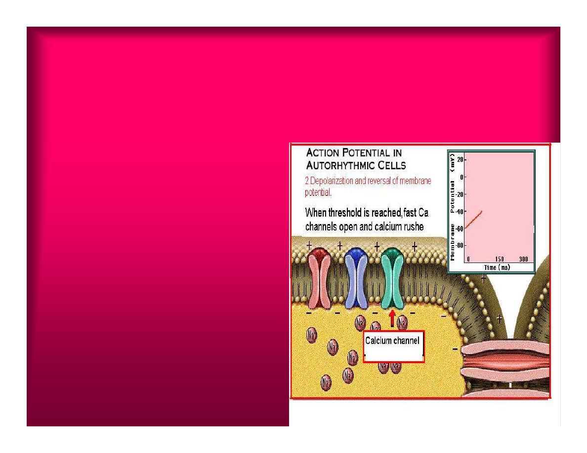
ACTION POTENTIAL IN
AUTORHYTHMIC CELLS
2.Depolarization and
reversal of membrane
potential
When membrane
potential gets to(-40)
mv.It is reached
threshold for initiated
an action potential,
fast calcium channels
open and calcium
rushes in.
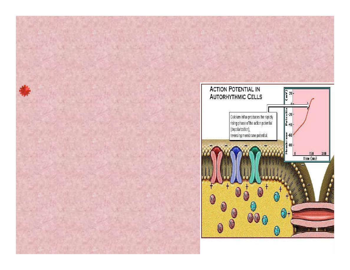
ACTION POTENTIAL IN
AUTORHYTHMIC CELLS
Calcium influx produces
the rapidly rising phase of
action potential
(depolarization), reversing
membrane potential from
negative to positive
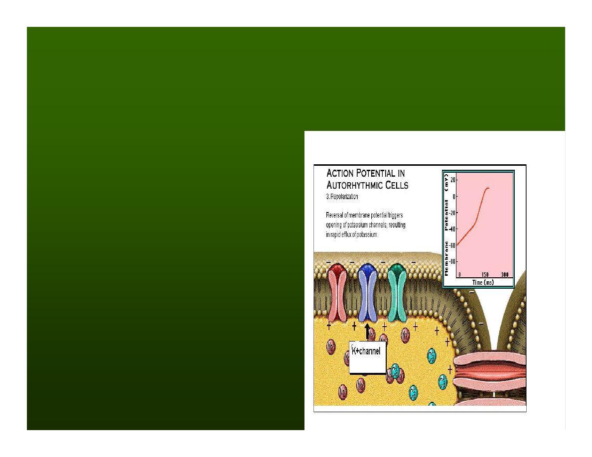
ACTION POTENTIAL IN
AUTORHYTHMIC CELLS
3.Repolarization
Reversal of membrane
potential triggers
opening of K+
channels, resulting in
rapid efflux of
K+.(rapidly K+ leave
the cell).
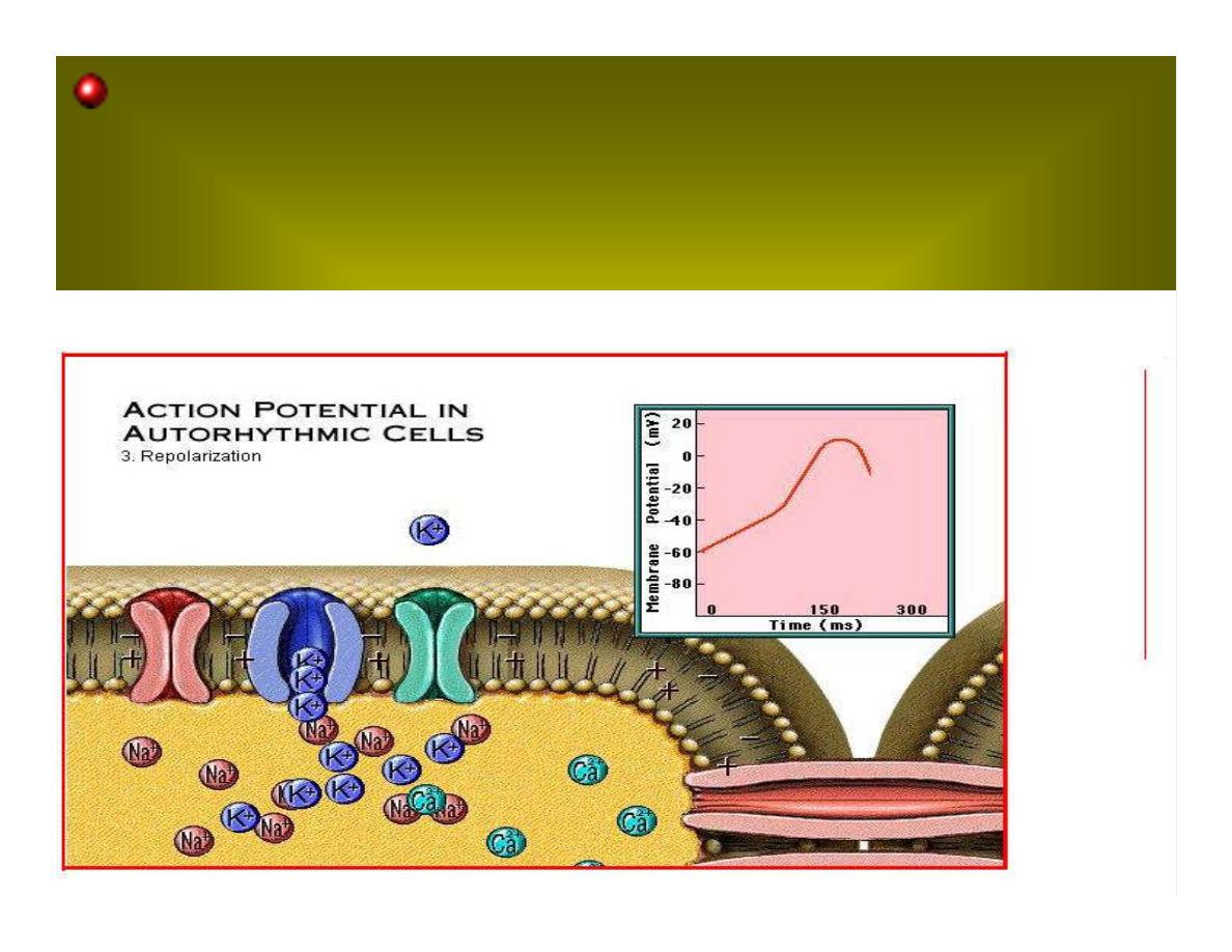
Potassium efflux produces repolarization bring
the membrane potential back down to resting
potential
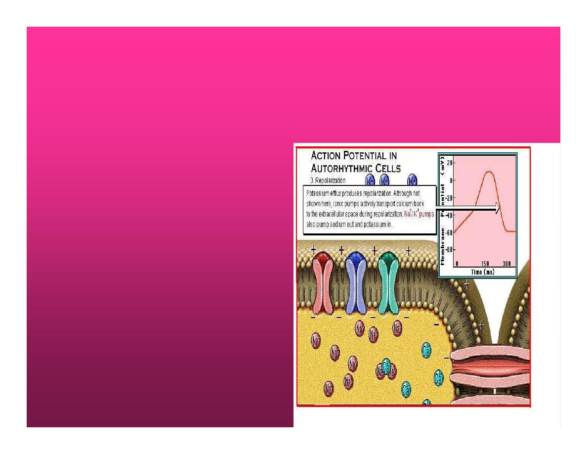
Action potential in
Autorhythmic cells
Although not shown
her, ionic pumps
actively transport
calcium back to the
extra- cellular space
during repolarization
Na+/K+ pumps also
pump sodium out
and K+ in.
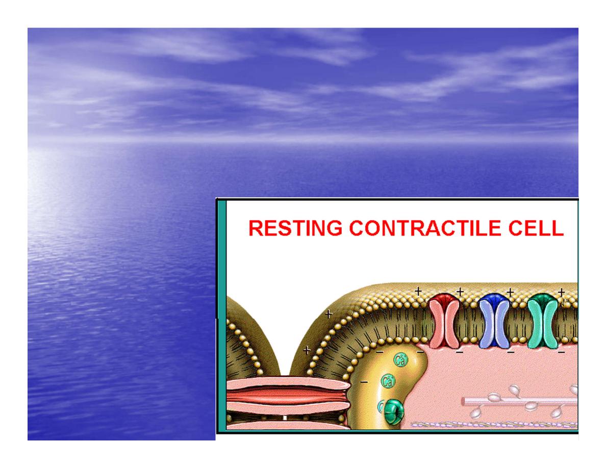
Cardiac contractile cell
•
This cell like
autorythmic cell to
generate action
potential
•
&pass the impulses
down align before
the cell contract
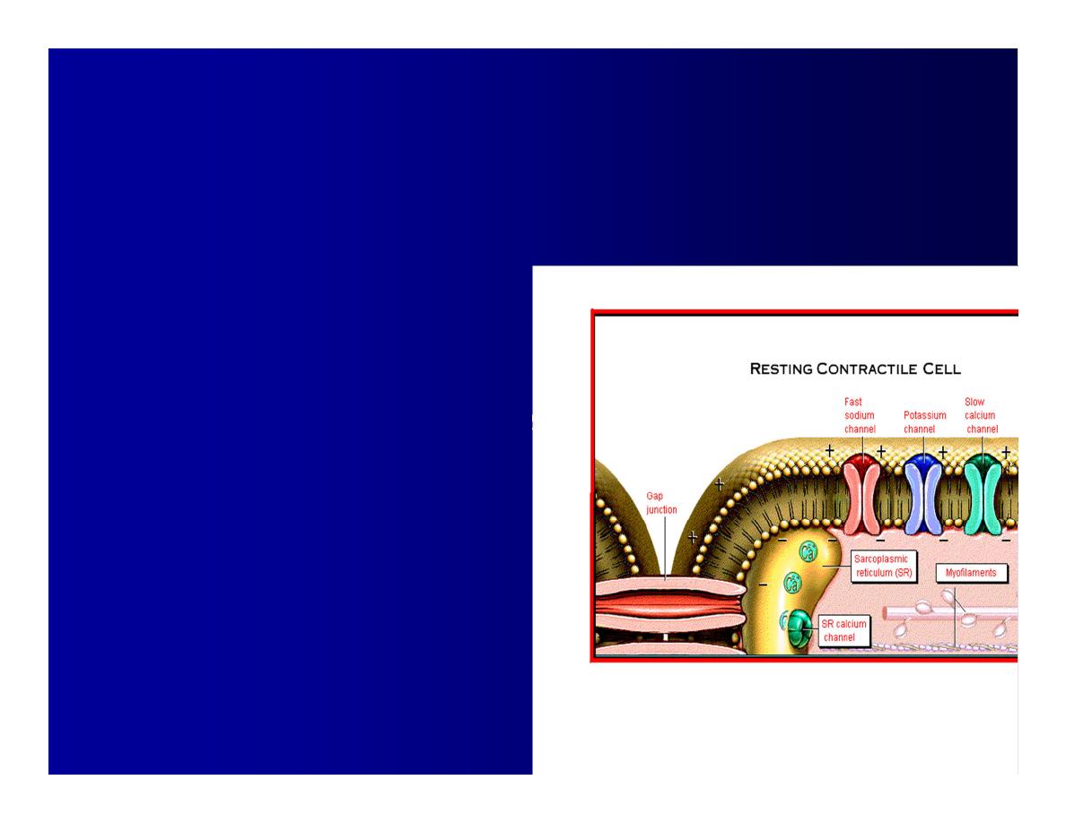
Cardiac Contractile Cell
•
Like the autorhythmic cell it
has protein transport
channels but slightly
different.
•
Gap junction:-link
•
autorhythmic & contractile
cell & contractile with each
other
•
SR which is storage site of
Ca ion &Channel with SR
allow the Ca to the cell.
•
Myofilaments are the
contractile of the C.m.cell.
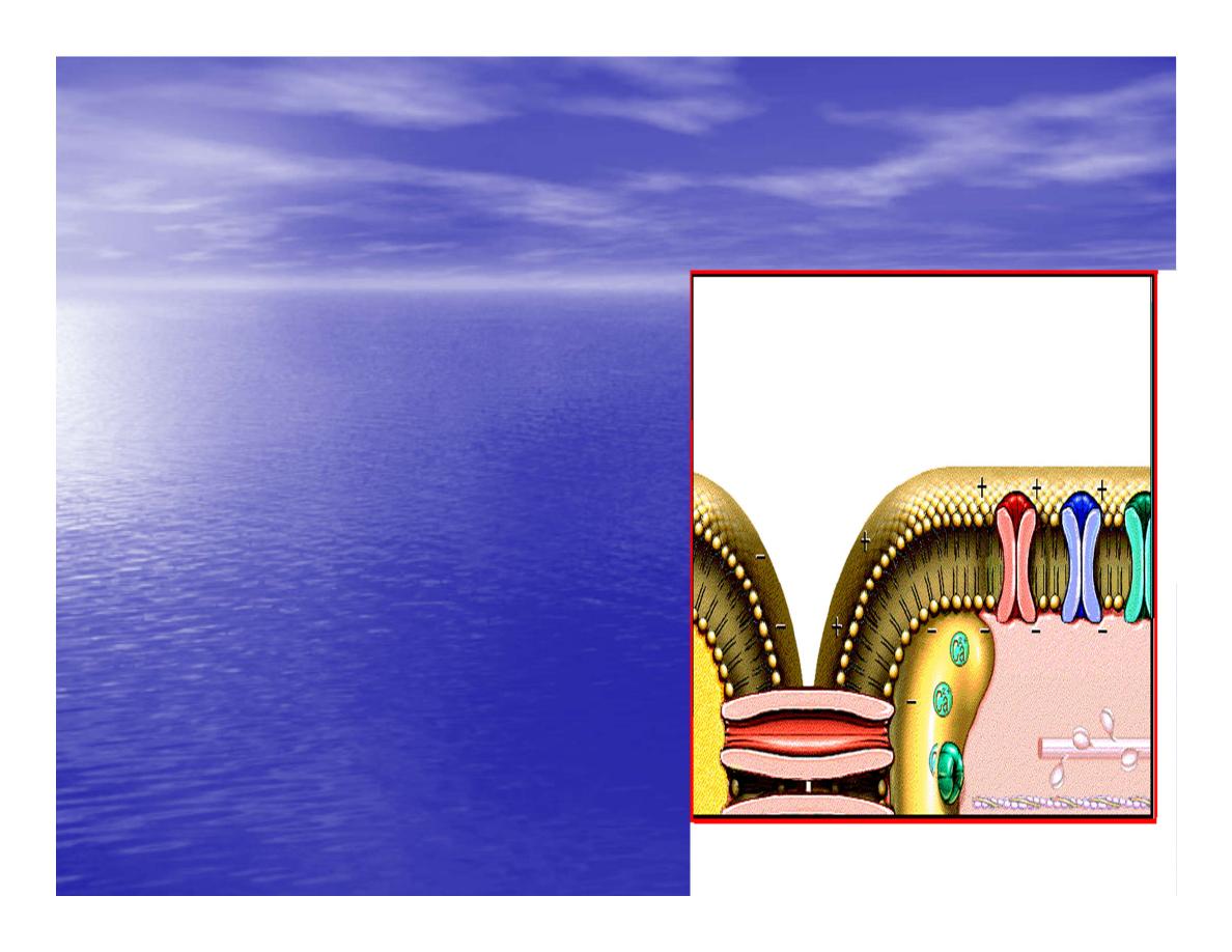
Action Potential in Contractile
cells
•
1- Depolarization
•
2- Plateau
•
3- Repolarization
•
Once threshold is reached the
action potential start with
depolarization
•
during plateau period ions
movement balances out &
memb.potential return to resting
state
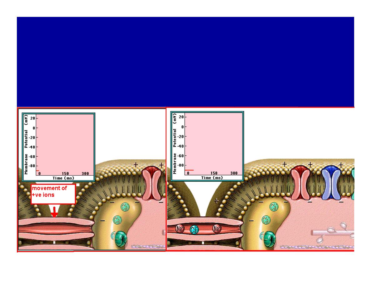
1-Depolarization:
during depolarization.in autorhythmic
cells,+ve ions move through gap junctions
to adjacent contractile cells
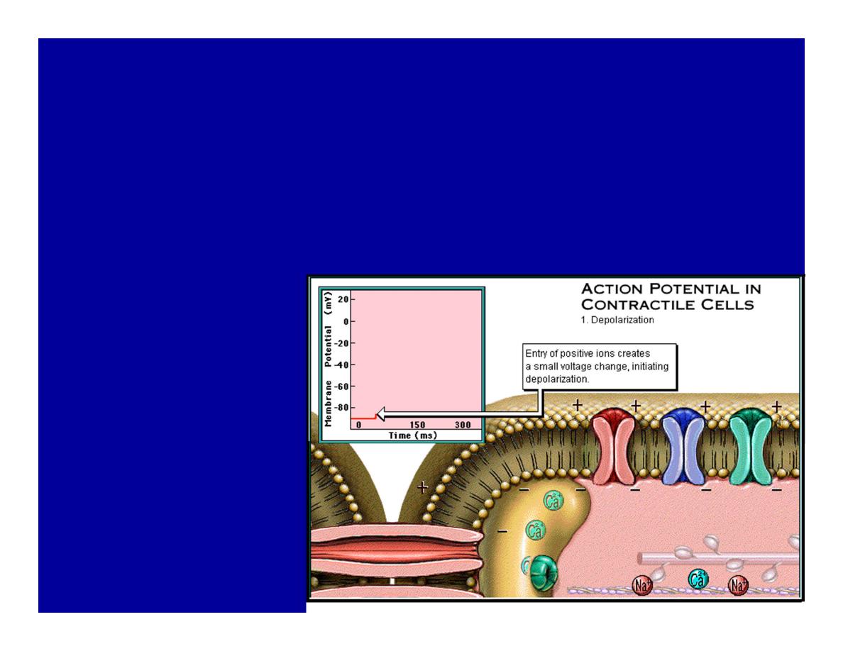
Depolarization
•
The entry of +ve ions (Na+,Ca+)
creates a small voltage change initiating
depolarization.
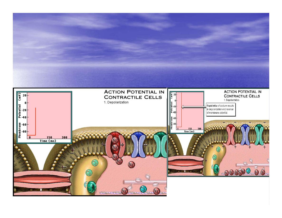
Depolarization
•
Voltage change stimulates opening of voltage-
regulated fast sodium channels.
•
Rapid influx of Na results in dep.of memb.potential.
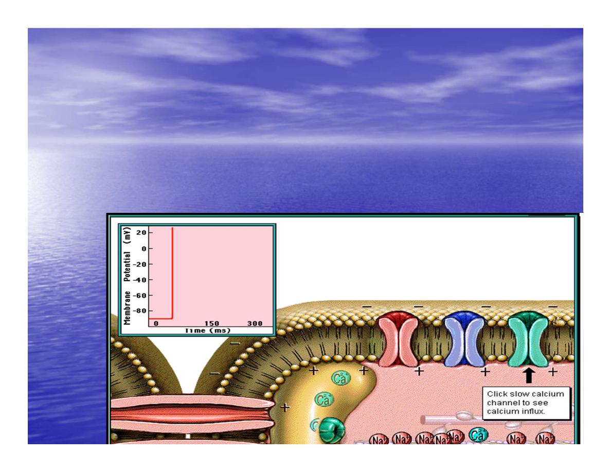
2-Plateau:
Depolarization also triggers opening
of slow Ca channels,allowing Ca
entry from EC space & SR.
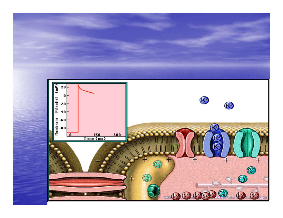
efflux begins
+
At the same time K
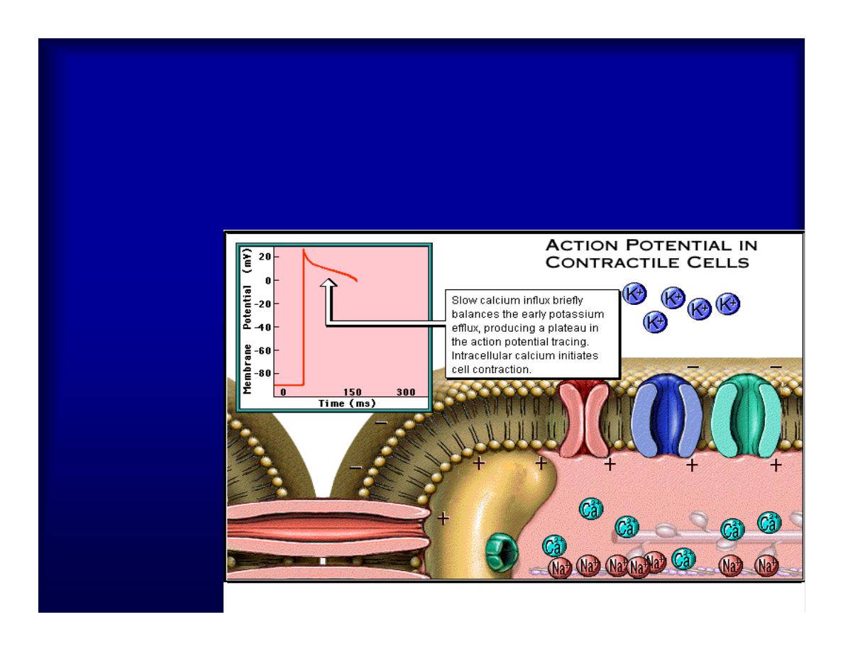
Slow ca
+
influx briefly balances the early
K
+
efflux,producing a plateau in the
action potential tracing intracellular Ca
+
initiates cell contraction.
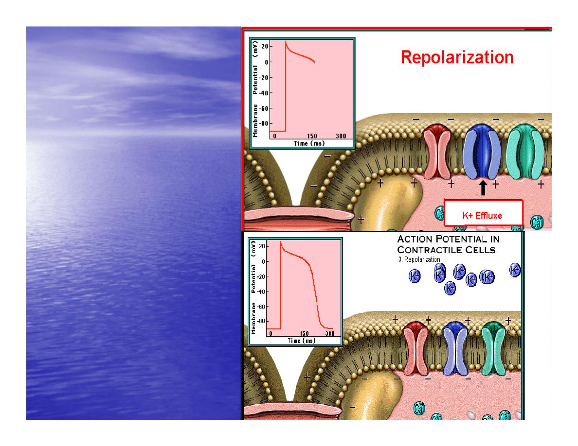
REPOLARIZATI
ON
•
Ca+ channels
close while more
K+ channels
open,
•
thus rapidly
repolarizing the
membrane
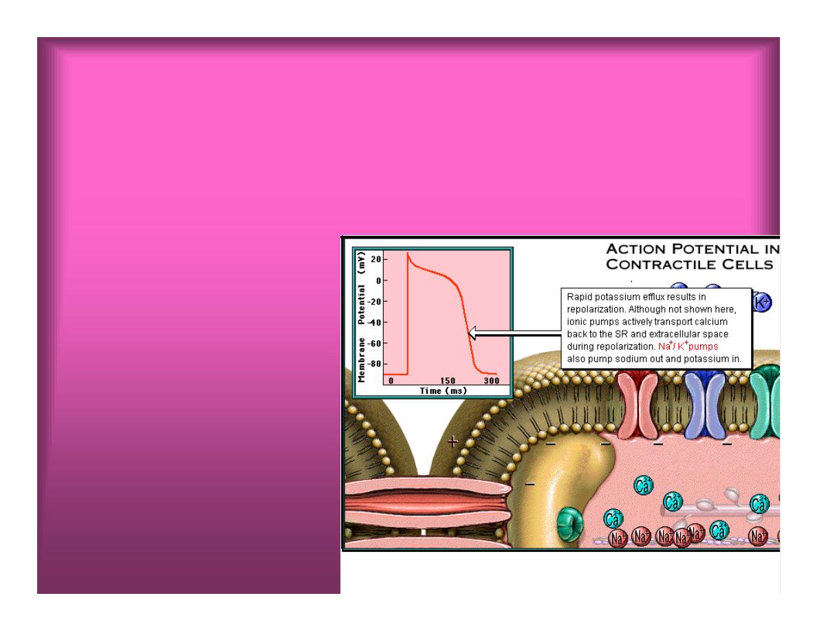
•
Rapid K
+
efflux results in repolarization.
Although not shown here,ionic pumps actively
transport Ca+ back to the SR & extracellular
space during repolarization.
•
Na+/K+ pumps
•
also pump Na+out
•
&K+ in.
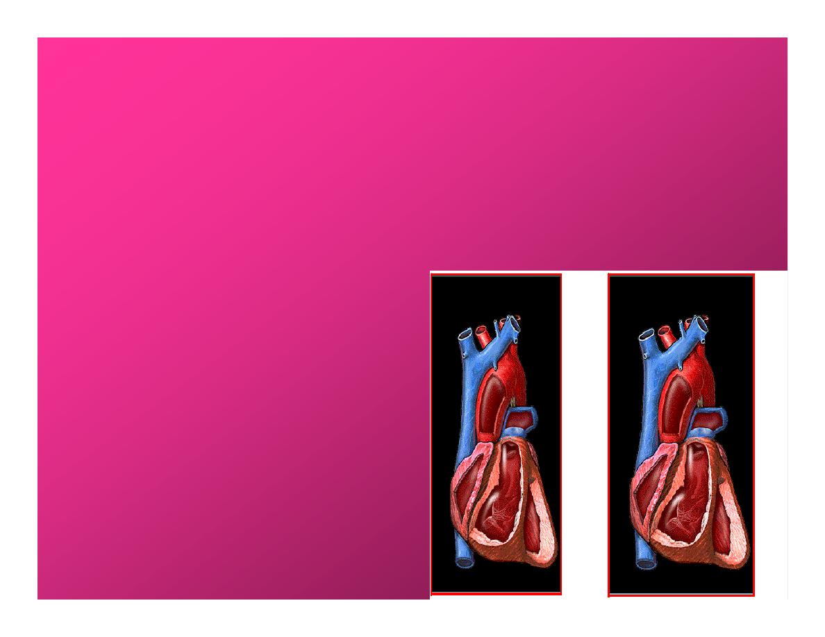
Cardiac Cycle (the mechanical events)
The cardiac cycle
includes all events
related to the flow
of bl. Through the
heart during one
complete heart
beat.
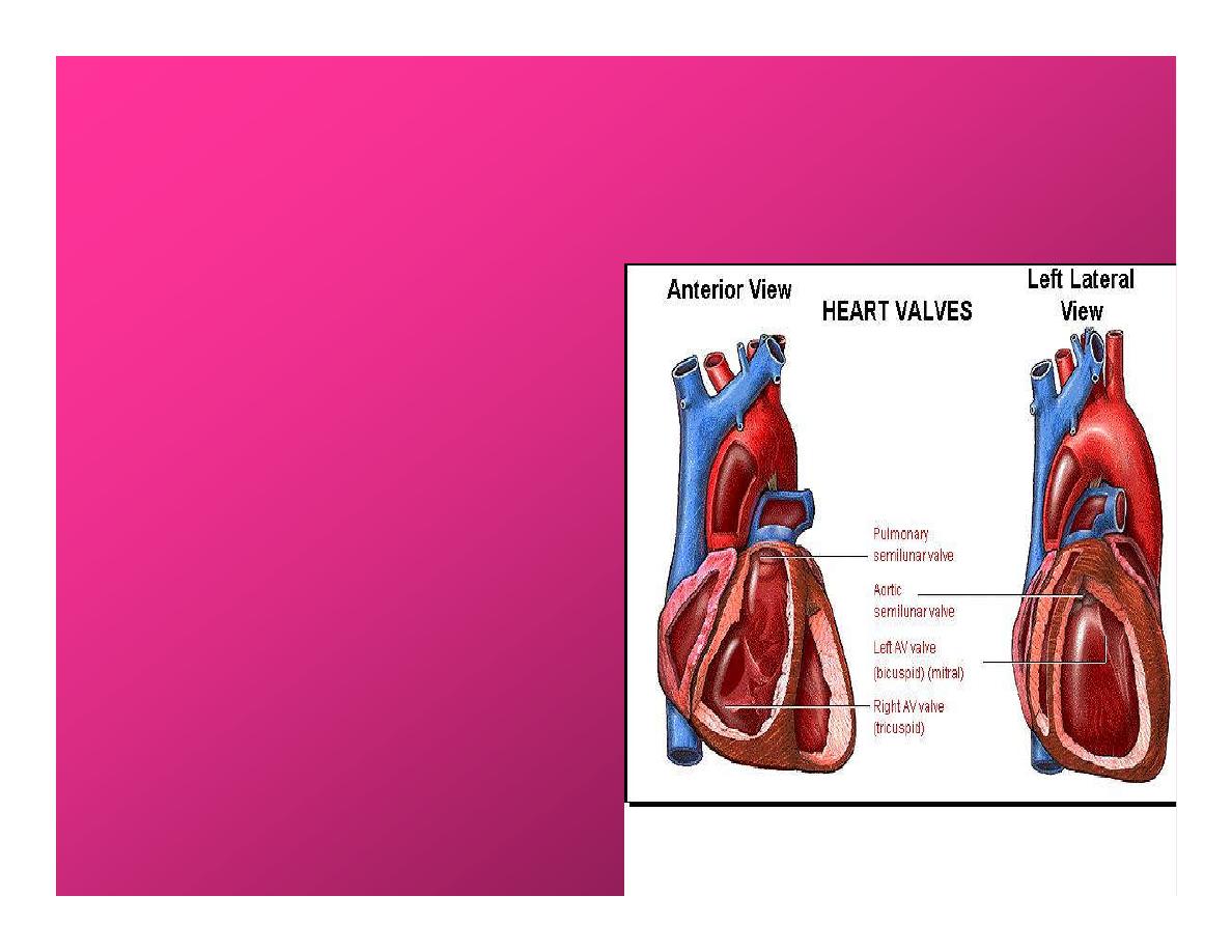
Cardiac Cycle
•
During the cardiac cycle,
heart valves open & close in
response to differences in
bl.p.on their two sides.
•
These figure show
•
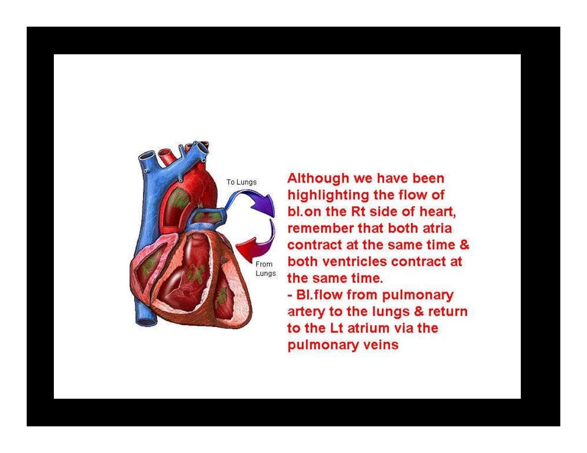
Pulmonary semi lunar V.
•
Aortic semilunar V.
•
Lt. AV valve (mitral v)
•
Rt. AV valve (tricuspid).
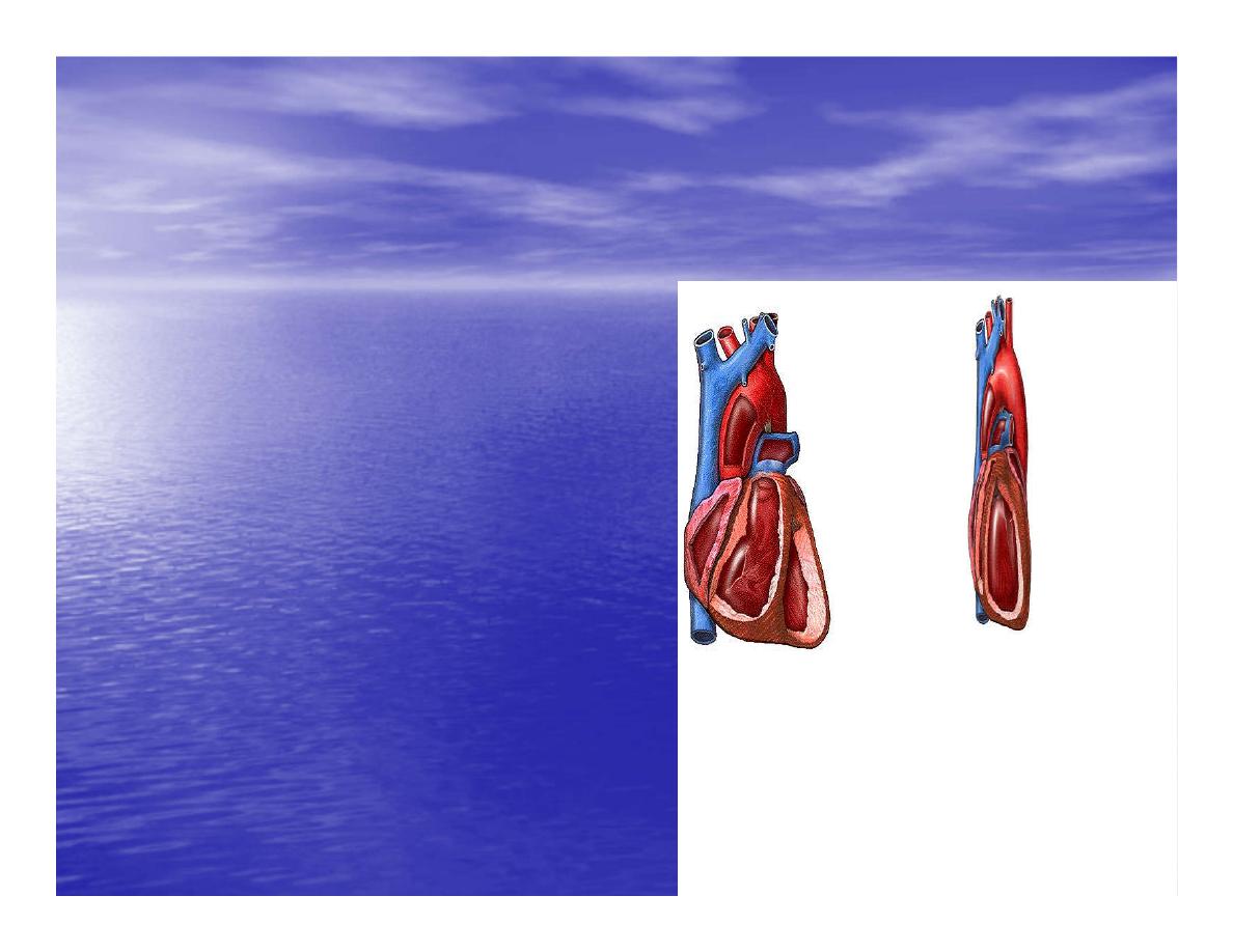
Over View:
Phases of the Cardiac Cycle
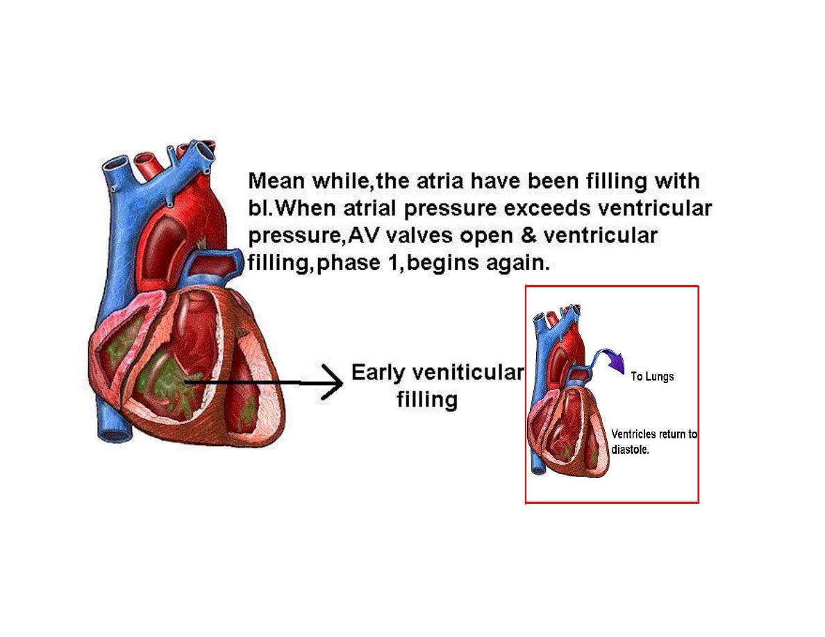
1.Ventricular filling:- occur
during mid-to-late V.diastol
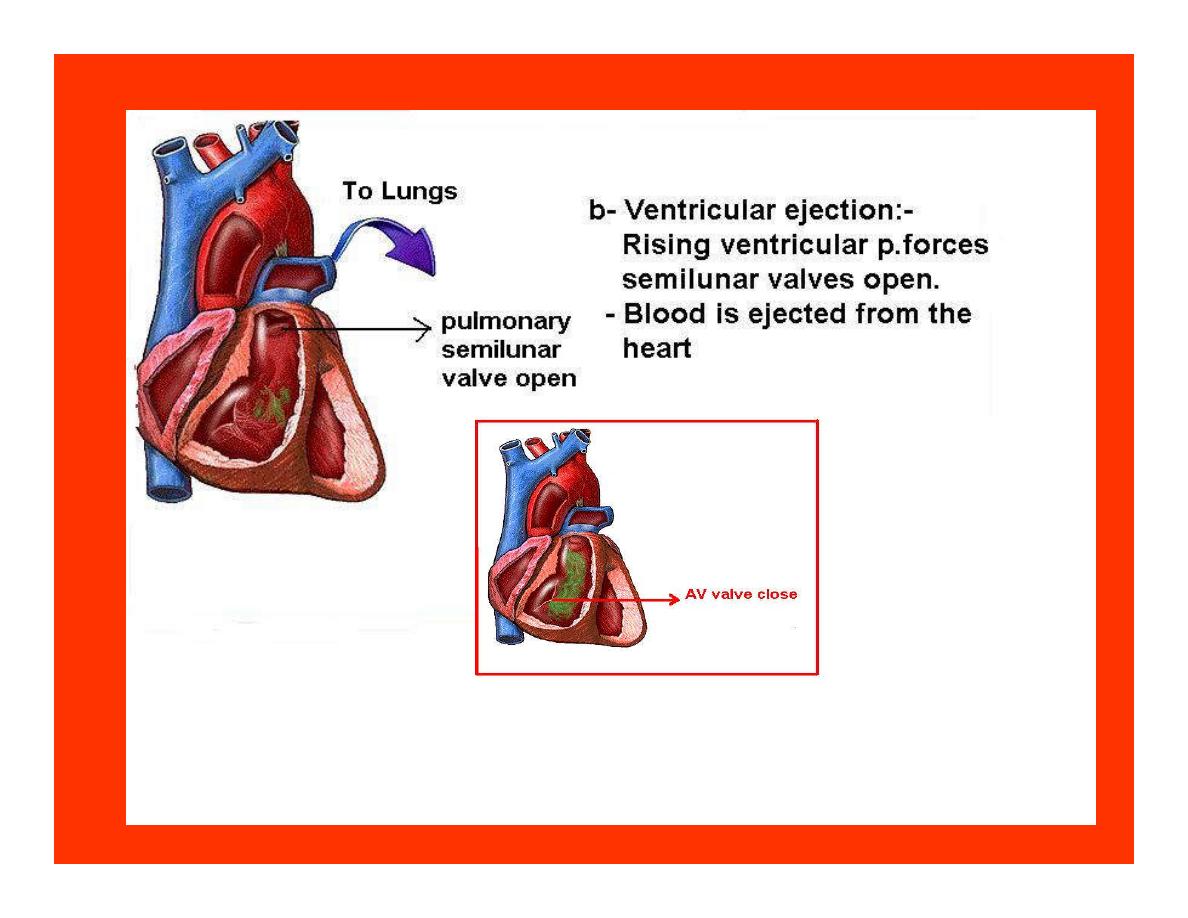
2. Ventricular Emptying
(ventricular systole) which
include:-
-
Isovolumetric contraction
-
Ventricular ejection.
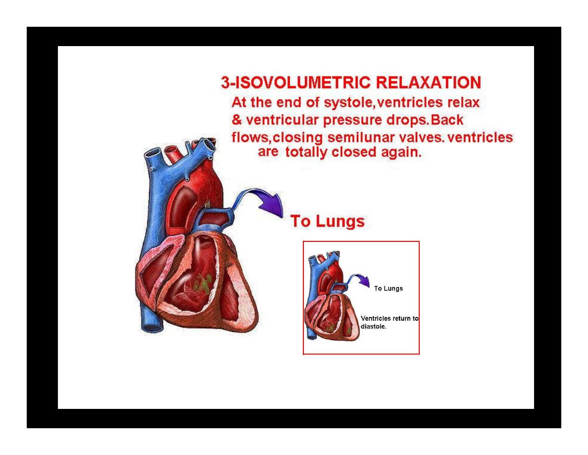
- Isovolumetric relaxation
occur during early diastole.
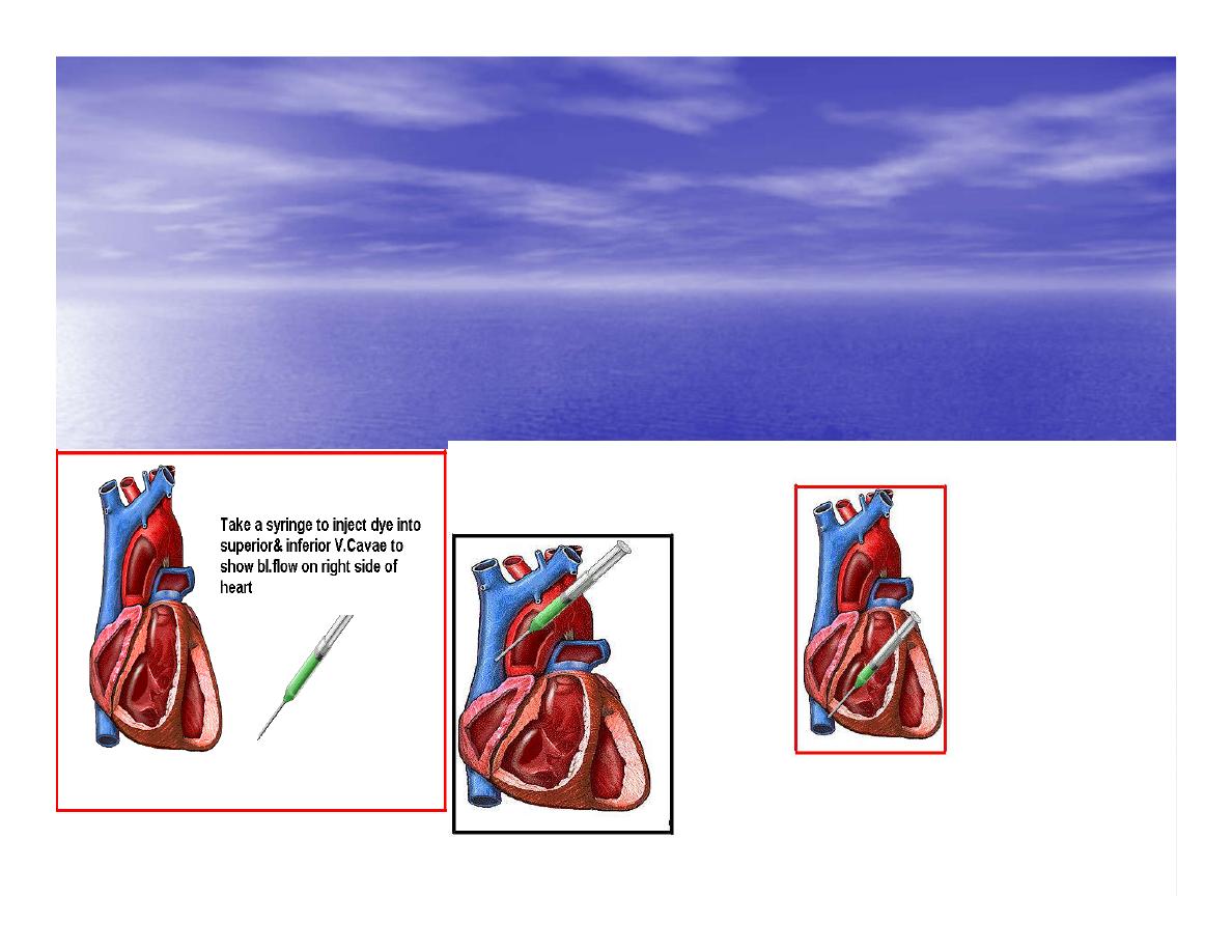
Cardiac Cycle
•
Inject dye into superior & inferior venae
cava to show bl.flow on Rt.side of heart
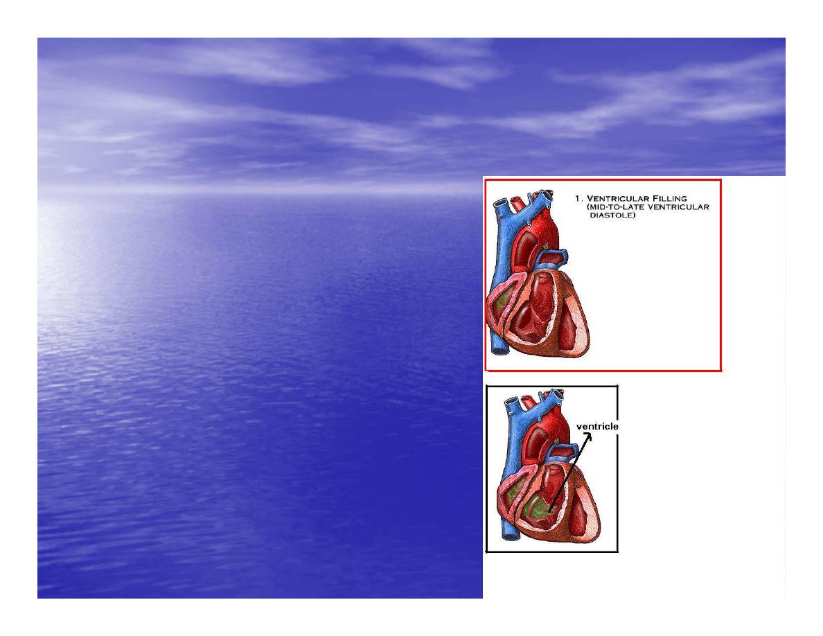
Phases of cardiac cycle
•
1
st
phase:-of cardiac
cycle.
1- Ventricular filling (Mid-
to-Late V.D) when
heart chamber relax
A- Bl.flow passively into
the atria, through open
AV valves,& into
ventricles, where the
pressure is lower.
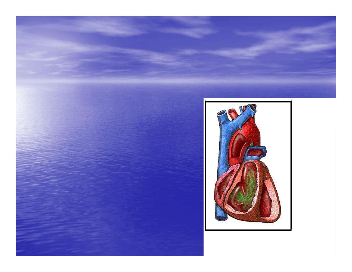
Ventricular filling
B-
Atria contract, forcing the
remaining blood into
ventricles.
C- During the last 3
rd
of
ventricular diastolic time
the atria contract & give
an additional 20%-30%
of the filling of the
ventricle during each
cardiac cycle.
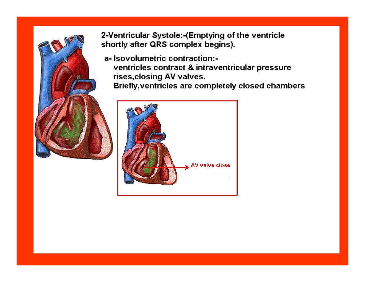


Function of the atria as pumps
•
The propagated action potential which is
represented by p wave on (ECG) precedes
atrial contraction.
•
Bl.normally flows continually from the great
veins into the atria & about 70% of this flows
pass directly through the atria into the
ventricles even before the atria contract.
•
Then, trial contraction causes an additional 20-
30% filling of the ventricles.

Central venous pressure (CVP)
•
The p.in the Rt atrium is called CVP
•
Measured clinically from the level of the column of
bl.in the neck veins (jugular v)
•
While subject is reclining at an angle of about 45
0
where the heart & sternal angle at the same level.
•
Venous pressure can be also measured by inserting a
syringe needle connected to a pressure recorder or to
a water manometer directly into a vein.
•
CVP can be measured accurately by inserting a
catheter through the veins into the Rt.atrium.
•
CVP is decreased during inspiration (negative
pressure breathing) & shock.
•
Increased by expiration&heart failure.

Jugular venous pressure
•
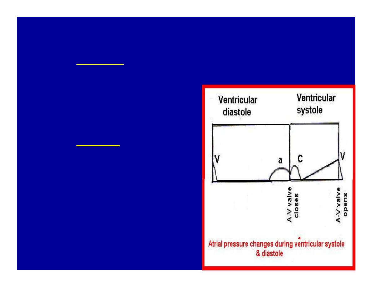
During cardiac cycle ,the atrial pressure
are transmitted to the great veins,
producing 3 characteristic waves in the
jugular pulse (also called jvp) & these
are:
•
1. The a wave
•
2. The c wave
•
3. The v wave.
Jugular venous pressure
•
The a wave : which is
caused by atrial contraction
and the atrial pressure
increases (about 4-6mmHg
in the right atrium and 7-
8mmHg in the left atrium).
•
The c wave: occurs when
the ventricles begin to
contract. It is caused to less
extent by slight back flow of
blood into the atria at the
onset of ventricular
contraction and mainly by
bulging of the AV valves
backward toward the atria
because of increasing
pressure in the ventricles.
JVP
•
The V wave: Occurs toward the end of
ventricular contraction and it results from
slow build up of blood in the atria while the
AV valves are closed during ventricular
contraction.
•
venous pressure falls during inspiration as a
result of the increased negative intrathoracic
pressure and rises again during expiration.

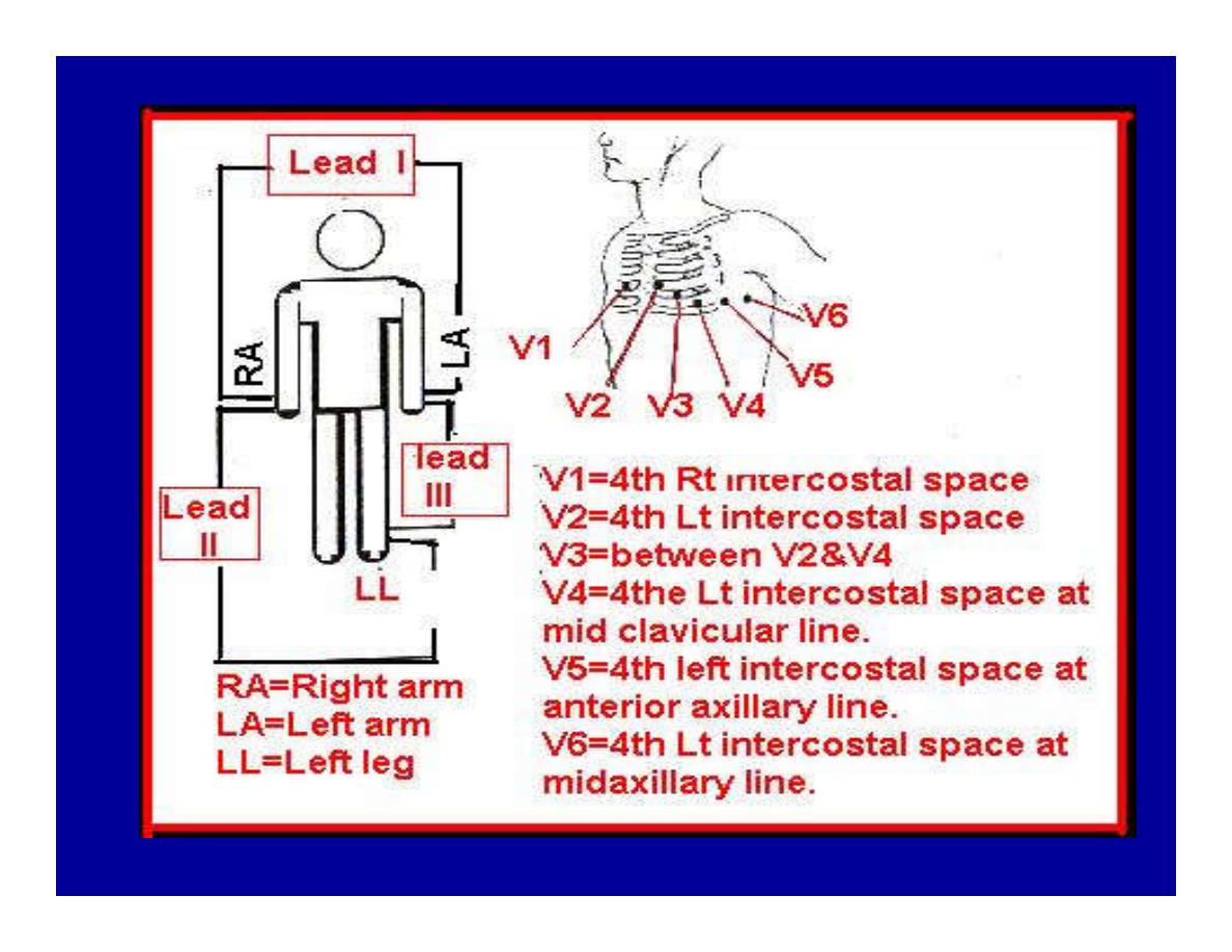
Normal electrocardiogram
•
As the transmission of the depolarization wave
(cardiac impulse) passes through the heart,
electrical currents spread into the surrounding
tissues and to the surface of the body which can
be recorded by E.C.G.
•
Are merely the recordings of differences in
voltage between tow electrodes on the body
surface as a function of time.

Calibration
•
A limited amount of information is provided by the
height of the P,QRS,& T waves, provided the machine
is properly calibrated. A standard signal of 1mV should
move the stylus vertically 1cm.(2 large squares),& this
‘calibration’ signal should be included with every
record.
•
When the machine is properly calibrated,
-tall P waves indicated RT atrial hypertrophy.
-tall R waves in the Lt ventricular leads indicate Lt.V.H
-& tall R waves in the Lt.ventricular leads indicate
Lt.V.H.& tall T waves indicate hyperkalaemia.
•
Small complex indicate a pericardial effusion.

RECORDING ECG
•
1- the patient must lie down & relax.
•
2- Connect up the limb leads’making certain that
they are applied to the correct limb.
•
3- Calibrate the record with the 1ml V signal
•
4- Record the six standard leads-3 or 4 complexes
are sufficient for each.
•
5- Record the six chest leads (V leads).

The 12 -lead ECG
•
E.C.G.interpretation is easy if you remember
the directions from which the various leads
look at the heart.
•
Standard ECG uses 9 electrodes which are
used to form 12 ‘lead's recording axis.
•
Six of these recording axis are in the frontal
plane,6 in the horizontal plane.

The limb leads-frontal planes axes
•
The most common set of connections is with
electrodes connected to both arms & the left leg give
3 limb leads (AVL,AVR,AVF).
•
Alternate connections(Einthoven’s triangle)
produces the 3 limb leads(1,11,111).
•
The 6 limb leads give electrical views of the heart in
the frontal plane.
•
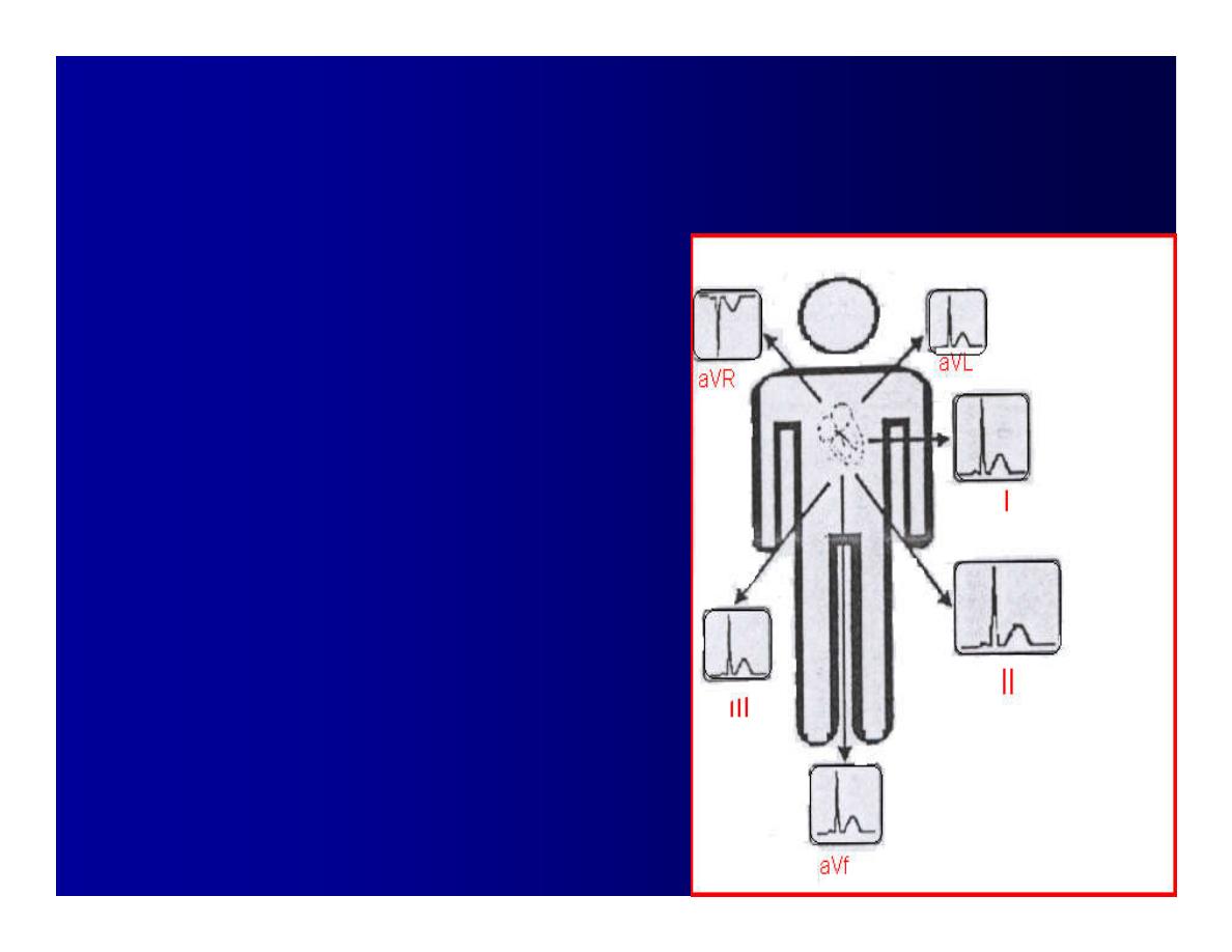
The six ‘standard 'leads can be through of as looking
at the heart in a vertical plane (that is,from the sides
or the feet). Thus
Standard leads
•
Leads 1, AVL,V5,V6 look at the
Lt.lateral surface of the heart.
•
Leads 111 & AVF at the inferior
surface
•
Lead AVR looks at the atria.
The septal leads, V1 and V2
septal wall of the left ventricle
The anterior leads, V3 and V4
anterior wall of the left ventricle
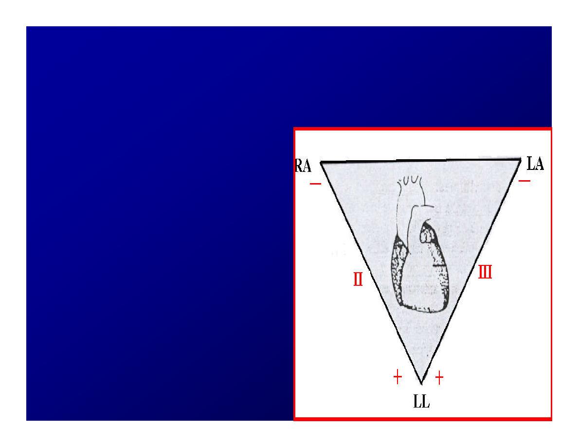
Standard Leads
•
Lead I- horizontal axis
+ electrode on Lt.arm
negative on Rt.
•
Lead II- axis at 60
0,
+
electrode on Lt.leg,
negative on Rt.arm
•
Lead III-axis at 120
0,
+
electrode on Lt leg,
negative on Lt.arm.

Chest leads- horizontal plane axes
•
The V lead (chest leads) is attached to the
chest wall by means of a suction electrode, &
recordings are made from 6 position overlying
the 4th & 5th rib spaces.
•
The 6 V leads (V1,V2,V3,V4,V5,V6) look at the
heart in a horizontal plane from the front &
the Lt side.
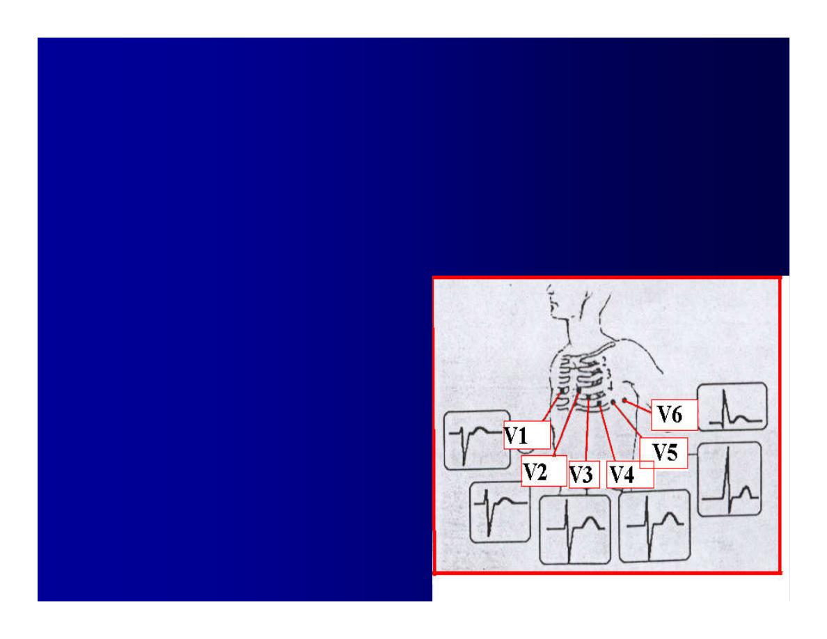
Thus:
•
Leads V1 & V2 look at
the Rt.ventricle
•
Leads V3 & V4 look at
the septum between
the ventricles (the
anterior wall of the Lt
ventricle)
•
Leads V5 & V6 at the
lateral walls of the Lt.V

The Shape of the QRS complex
in leads I,II,III
•
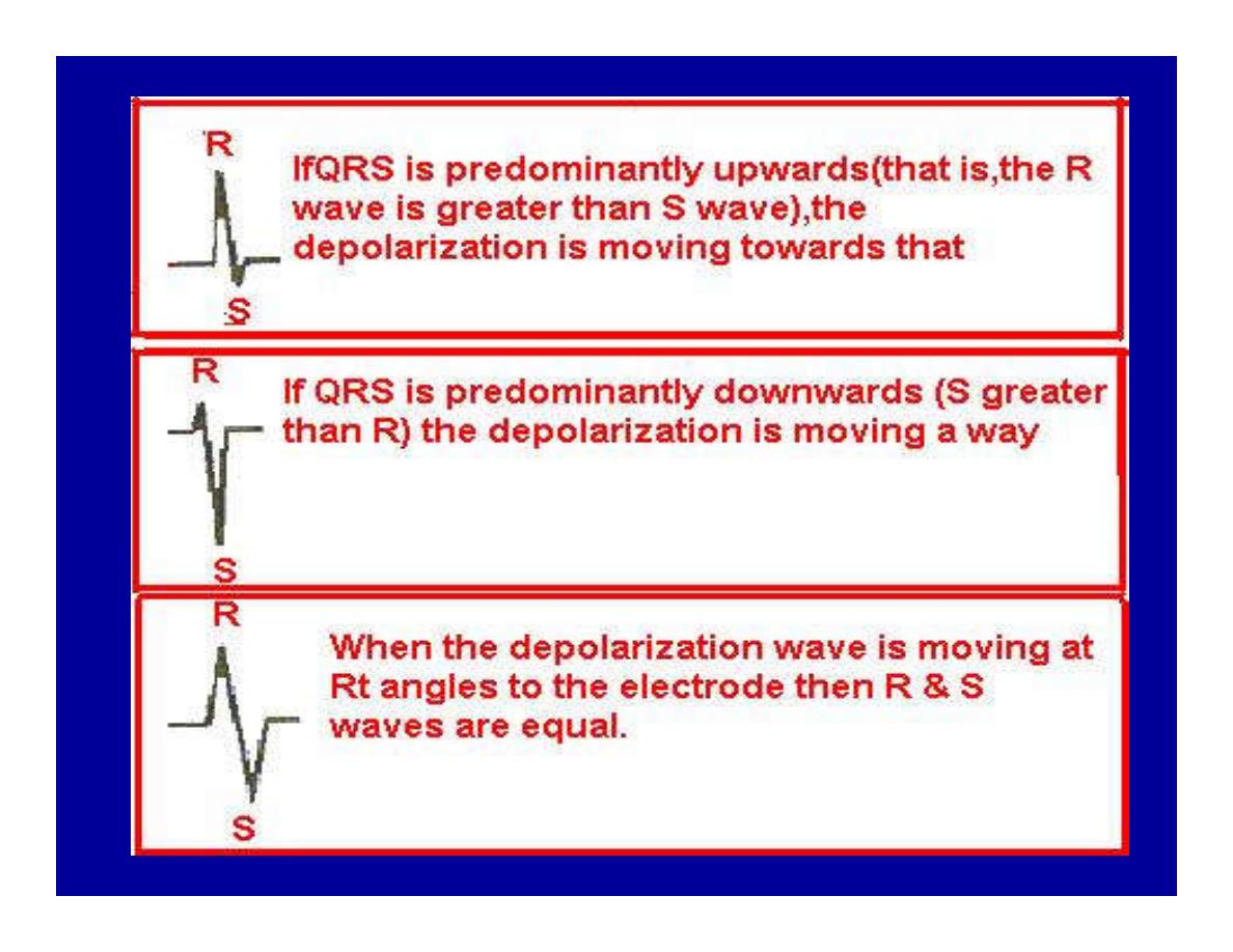
The E.C.G. machine is arranged so that when
depolarization wave spreads towards a lead the
needle moves upwards & when it spreads a
way from the lead the needle moves
downwards.
•
The deflection of the QRS complex thus shows
the direction in which the wave of
depolarization is spreading.
NORMAL ECG
•
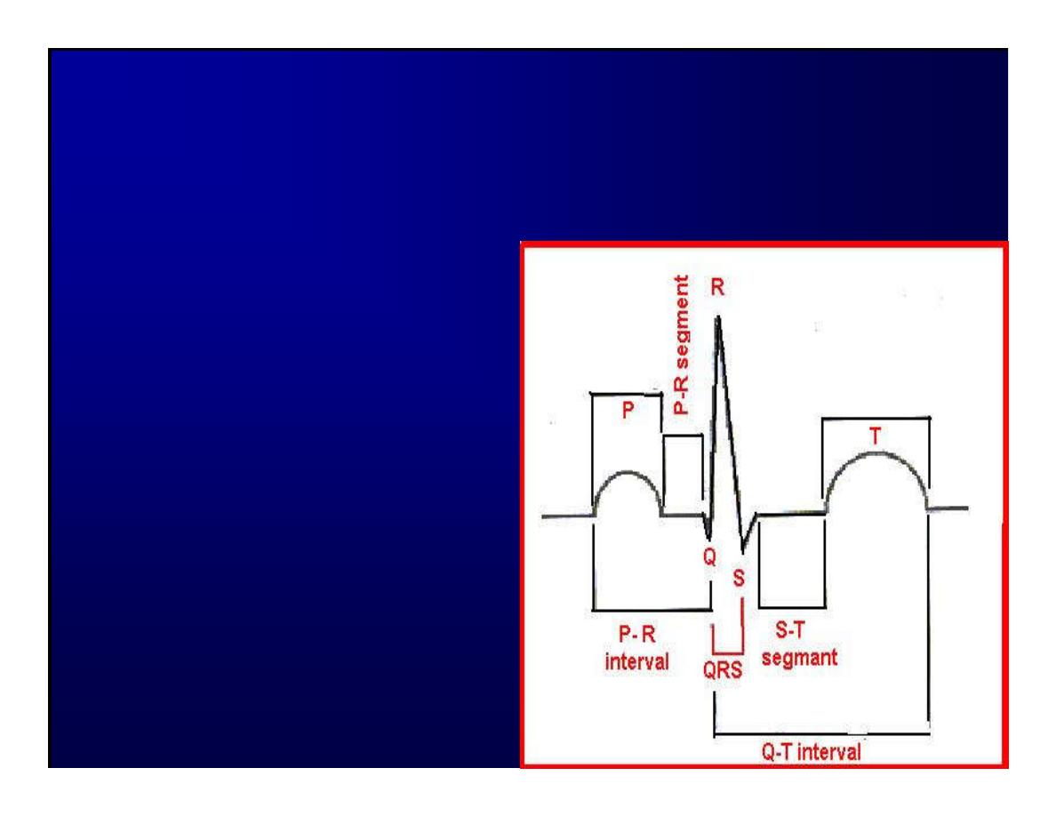
The normal E.C.G is
composed of a P wave,
•
a QRS complex and
•
a T wave.
•
The QRS complex is
often three separated
waves the Q wave, the R
wave and the S wave.
ECG
•
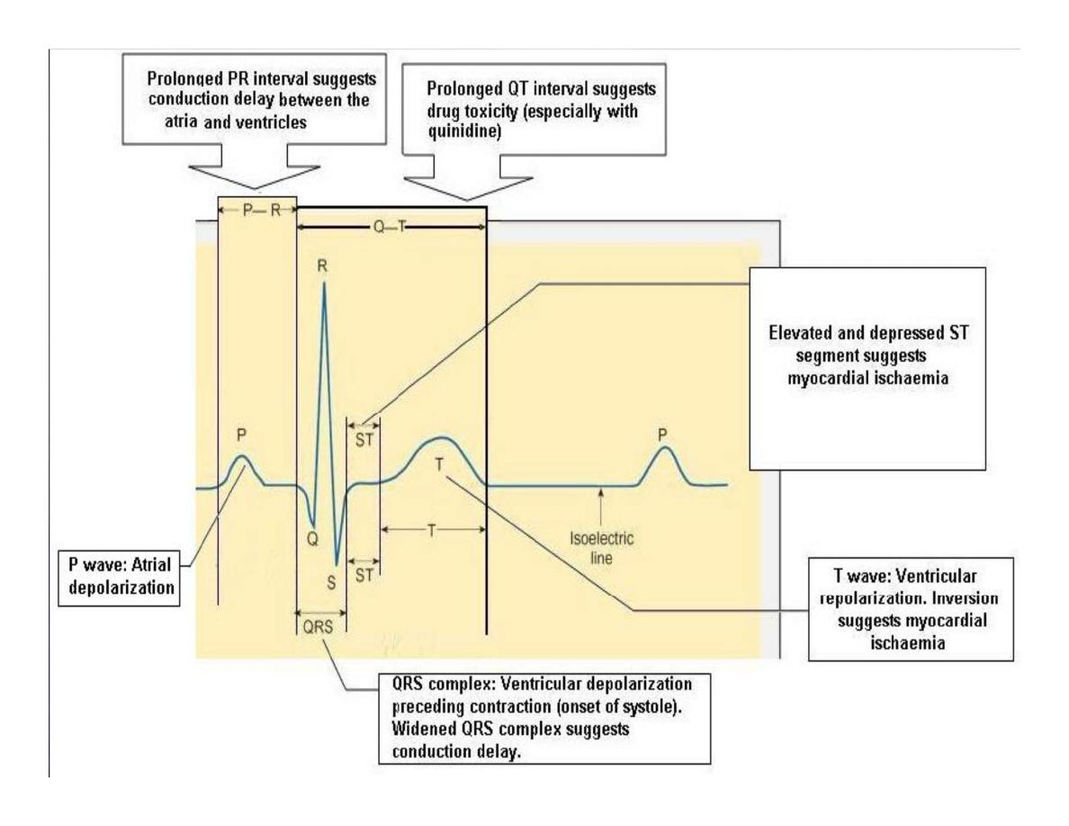
The P wave is caused by electrical currents
generated as the atria depolarize prior to
contraction.
•
QRS complex is caused by current generated
when the ventricles depolarize prior to
contraction.
•
The T wave is caused by currents generated as
the ventricle recover (repolarize) from the
state of depolarization.
ECG
•
The atria repolarize a approximately 0.15 to
0.20 sec after the depolarization wave which is
just at the moment that the QRS wave is being
recorded in the E.C.G.
•
The
PR
interval is the period between the
beginning of the
atrial depolarization
and the
beginning of the
ventricular depolarization
.It
is a measure of atrio-ventricular conduction
time.
ECG
•
The QT interval
is the time from the
beginning of the ventricular depolarization to
the end of repolarization (about o.41 sec at 50
beats/min in normal male subjects).
•
The R-R interval
is the time between tow
successive complexes and is usually used to
measure the cardiac cycle length.
•
The regular heart rat can be measured using
R-R interval
according to the following
equation:-
Heart rate= 60/R-R interval (in sec)
Duration (sec)
Type of ECG wave
&interval
0.08
P wave duration
0.05-0.10
QRS duration
0.15-o.25
T wave duration
0.12-0.20
PR interval
Heart rate-dependent
Q-T interval
It is imp to be
remembered
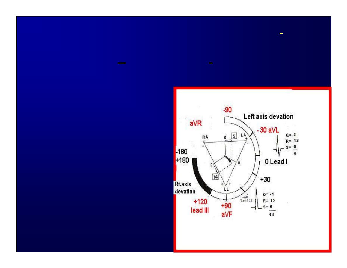
The normal electrical axis lies between -30º and+ 120º .
Right axis deviation is present when the mean electrical
axis lies between+ 120º and +180º. as occurs in
right
ventricular hypertrophy &Rt bundle branch block
•
Left axis deviation is
present when the
mean electrical axis
lies between -30º and
-90ºas occurs in
obesity,
Left
ventricular
hypertrophy and left
bundle branch block .

Systematic approach to read E.C.G
1. Rate
2. Rhythm
3. Axis deviation
4. Presence of ventricular hypertrophy
5. P wave
6. P-R interval
7. Q wave
8. QRS complex
9. S-T segment
10. T wave

Heart rate
How to calculate heart rate:-
When the rhythm is regular
When the rhythm is irregular
Rhythm
If the interval between all R waves is equal then
it is regular rhythm
If the interval between all R waves is unequal
then it is irregular rhythm (arrhythmia).
There is a normal arrhythmia called sinus
arrhythmia which occurs with respiration

When the rhythm is regular
Heart Rate = 1500 / No. of small squares between two
consecutive waves or
Heart Rate = 300 / No. of large squares between two
consecutive waves
NOTE: The above formulas are correct when the paper
speed is running at the
standard rate of 25 mm/s.
All ECG machine run at a standard rate and use paper with
standard size sequres.Each large square (5mm) represents
0.2 seconds and one small square represents 0.04 seconds.
So there are 5 large squares per second and 300 per
minute. Thus the ECG event such as QRS complex
occurring once per large squares is occurring at a rate of
300 per minute.
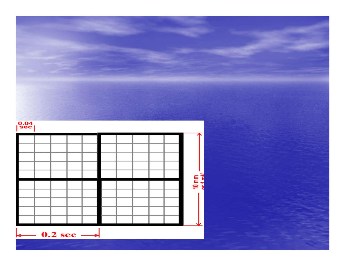
ECG graph paper
A typical electrocardiograph runs at a paper speed of 25 mm/s, although faster paper
speeds are occasionally used. Each small block of ECG paper is 1 mm². At a paper
speed of 25 mm/s, one small block of ECG paper translates into 0.04 s (or 40 ms).
Five small blocks make up 1 large block, which translates into 0.20 s (or 200 ms).
Hence, there are 5 large blocks per second. A diagnostic quality 12 lead ECG is
calibrated at 10 mm/mV, so 1 mm translates into 0.1 mV.

When the rhythm is irregular
Heart rate=number of R-R interval in
30 large squars × 10

Rate
If the rate is < 60 beat / min (sinus bradycardia) (or < 50
beats/min during sleep).
If the rate is >100 beat / min (sinus tachycardia)
Sinus arrhythmia
Heart Rate increases during inspiration
And Heart Rate decreases during expiration
The above is normal and is called sinus arrhythmia "sinus”
from (SA node) and
arrhythmia (irregular rhythm)".
Axis deviation
The normal range for the cardiac axis is between - 30° and 90°.
An axis lying beyond
- 30° is termed left axis deviation, whereas an axis > 90° is
termed right axis
deviation.

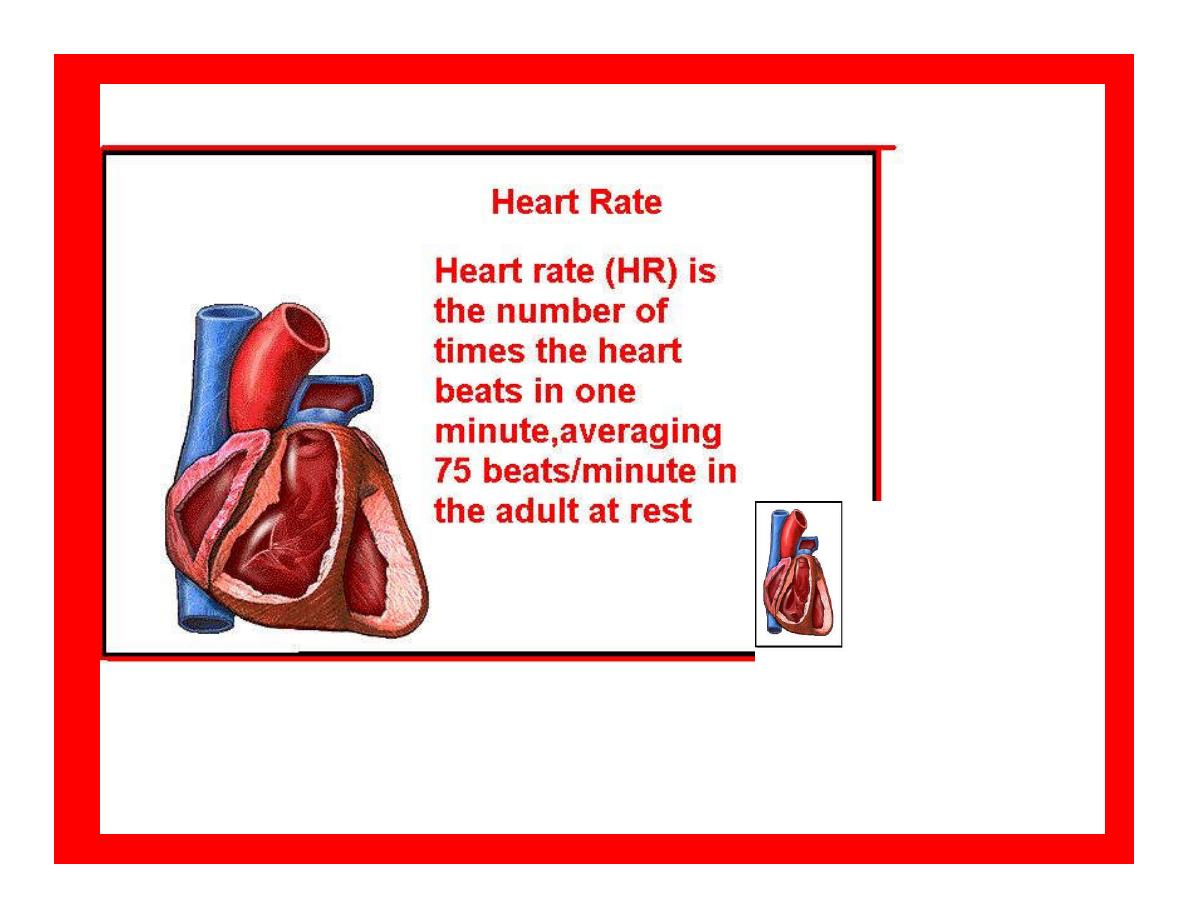
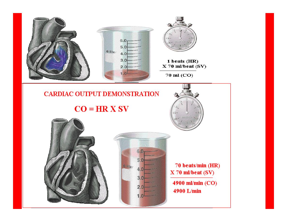
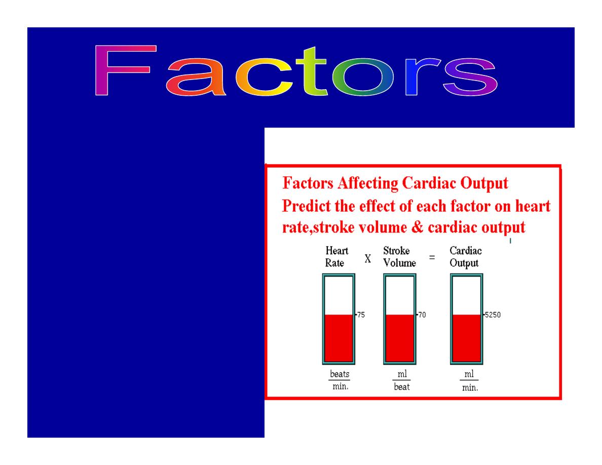
Cardiac out put (C.O)
•
Is the volume of blood pumped by
each ventricle per minute.
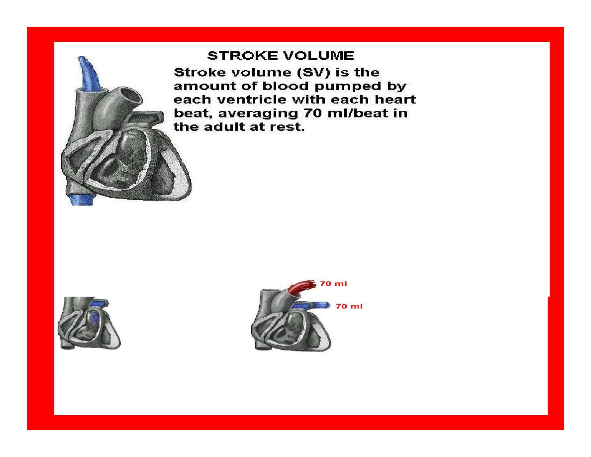
•
This stroke volume (SV) is the
volume of blood pumped by each
ventricle /beat.
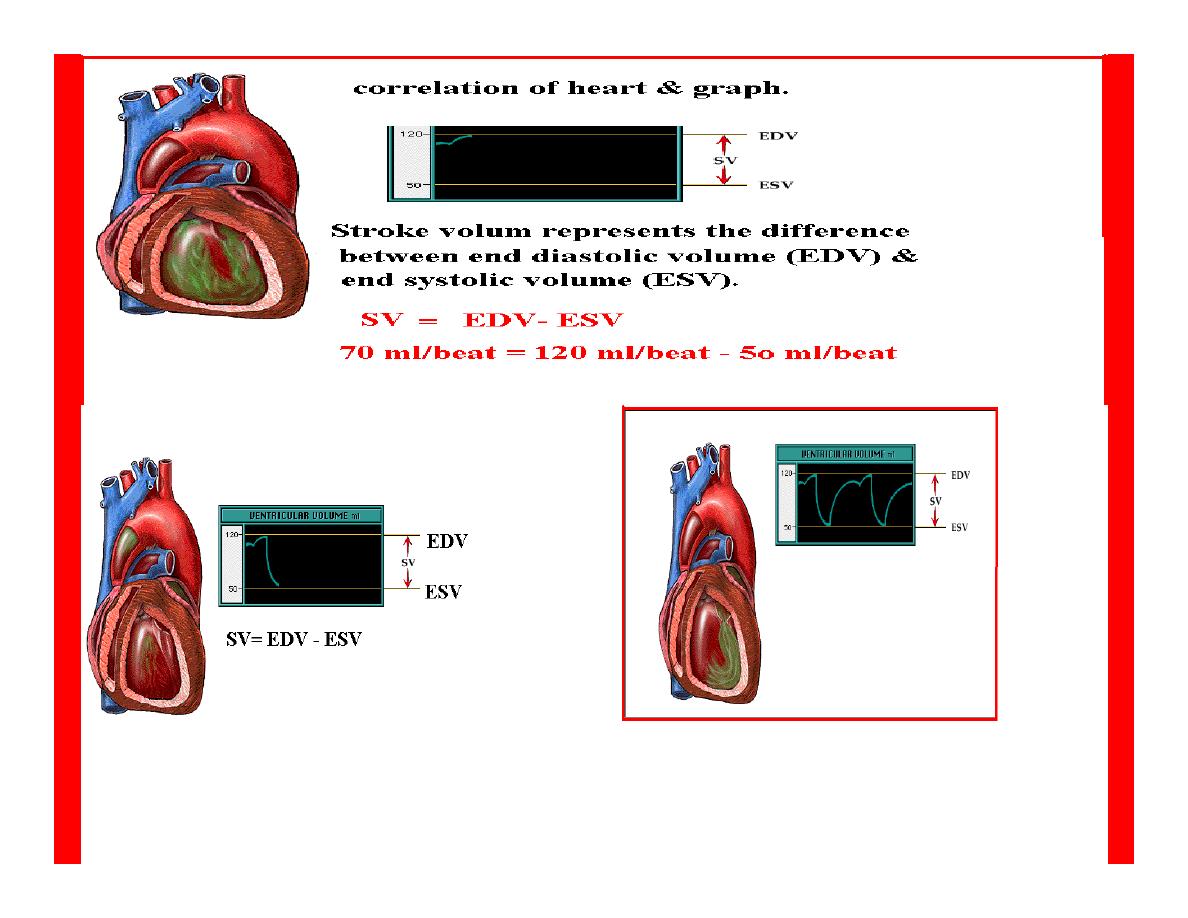
•
Therefore:-
•
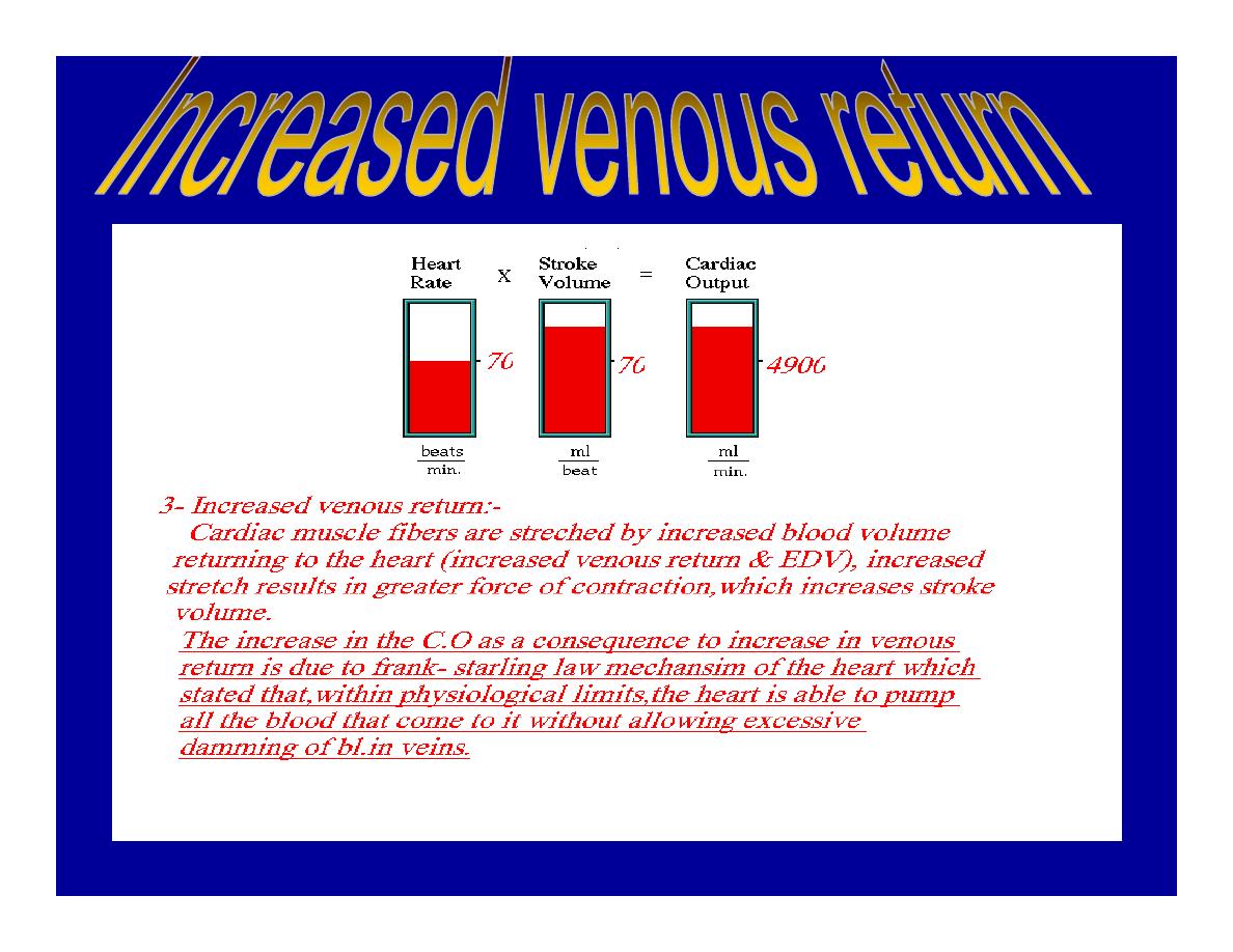
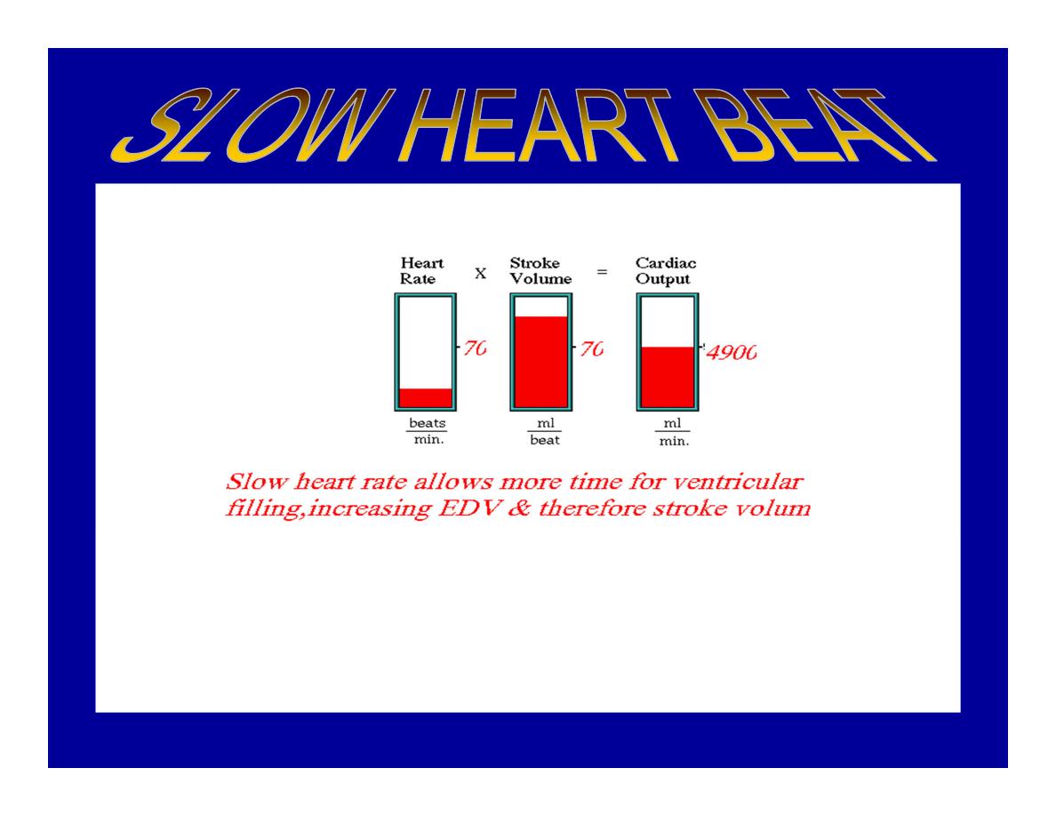
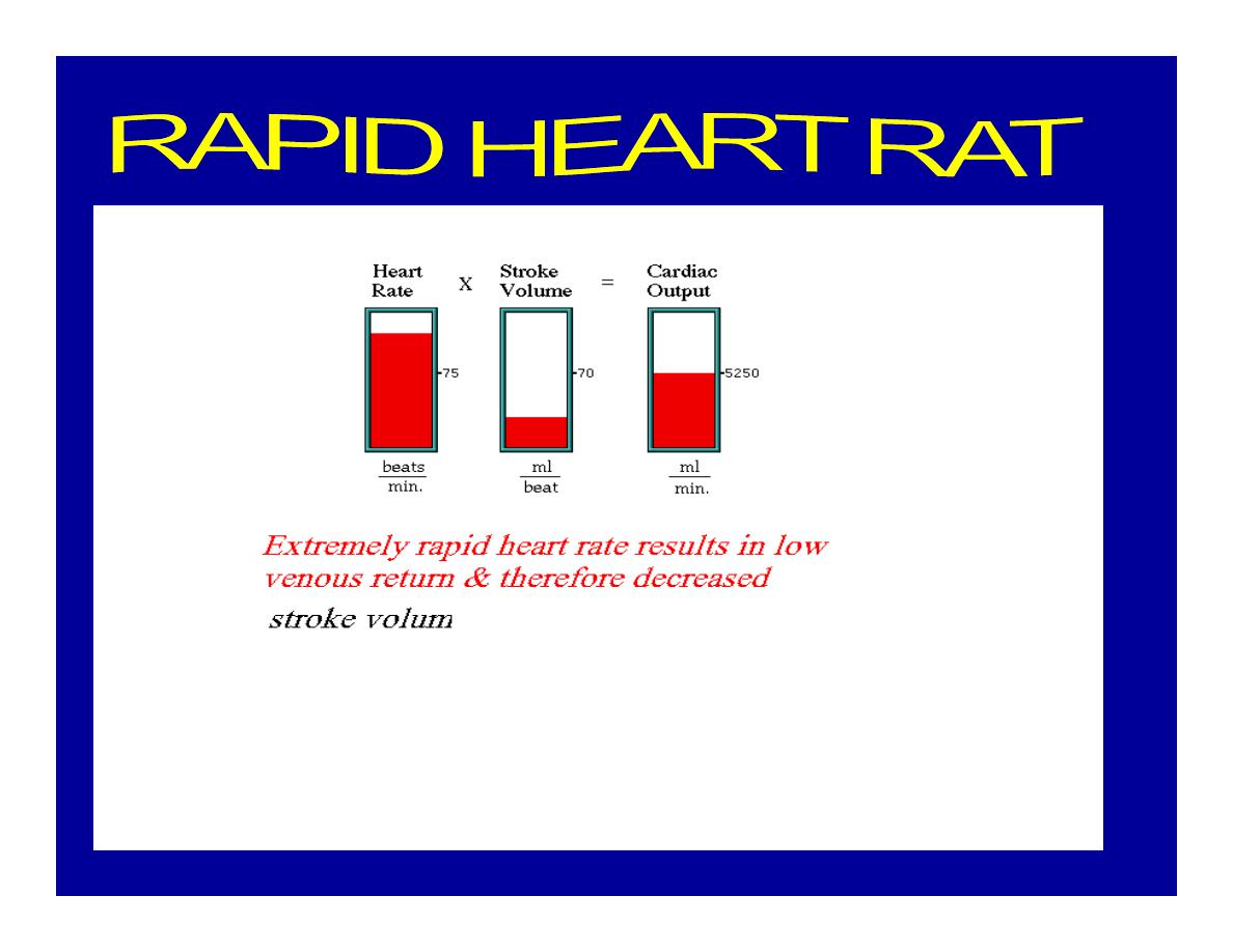
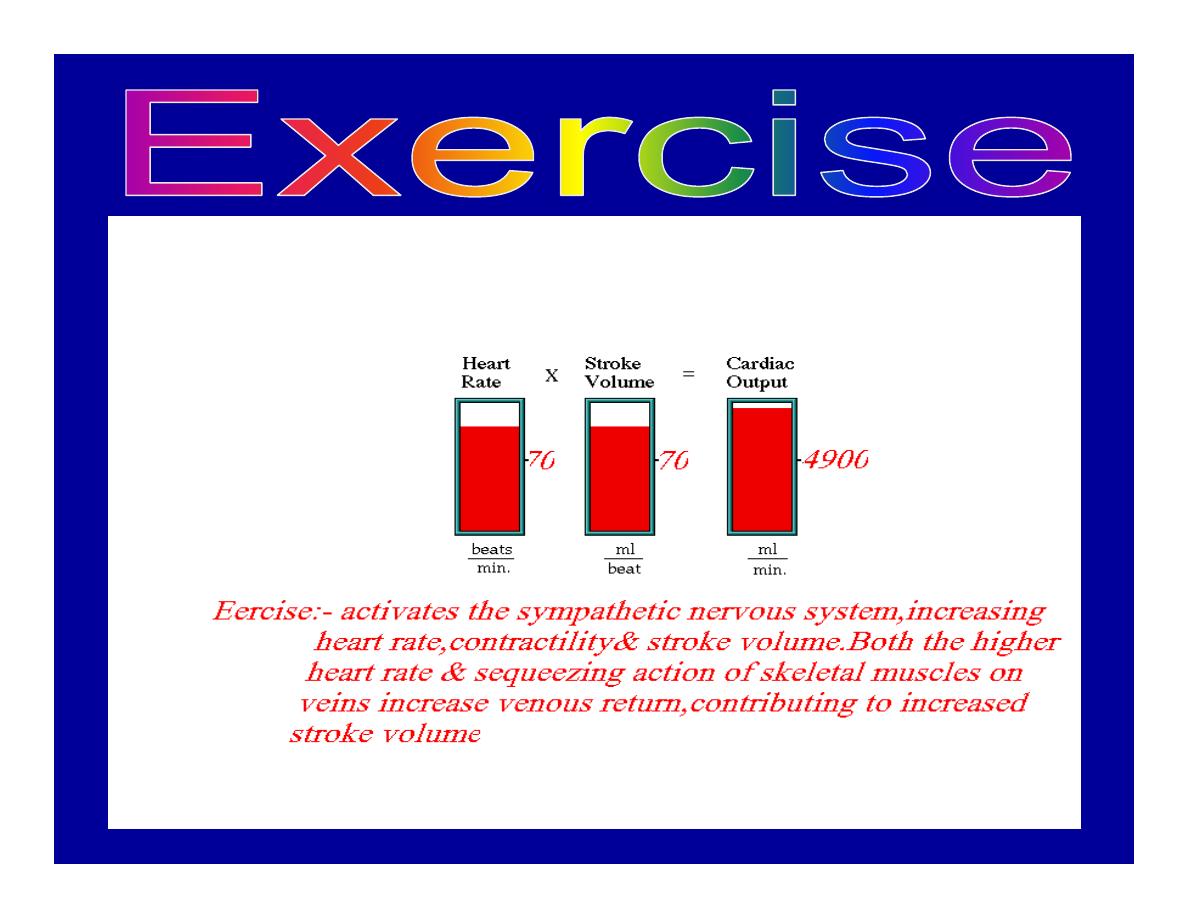
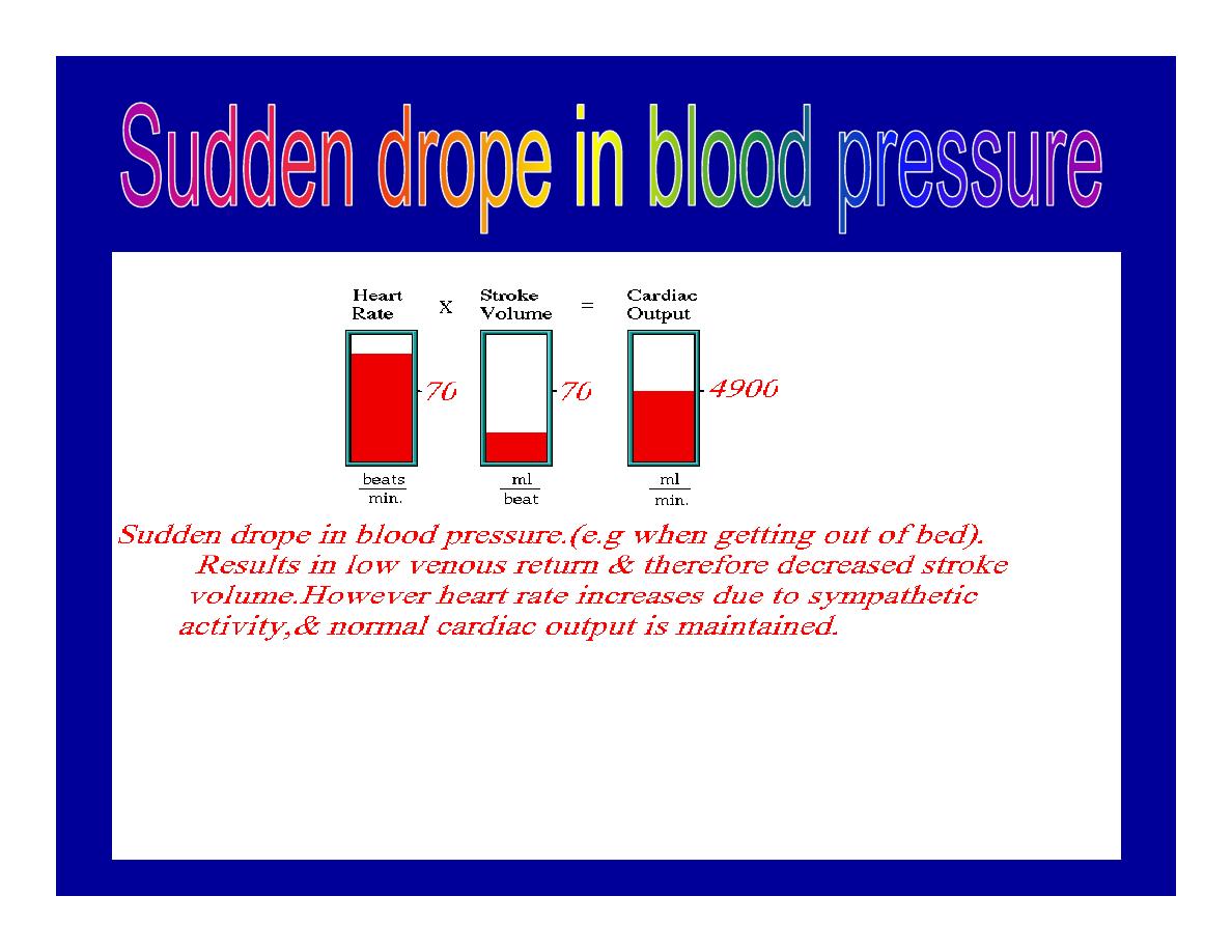
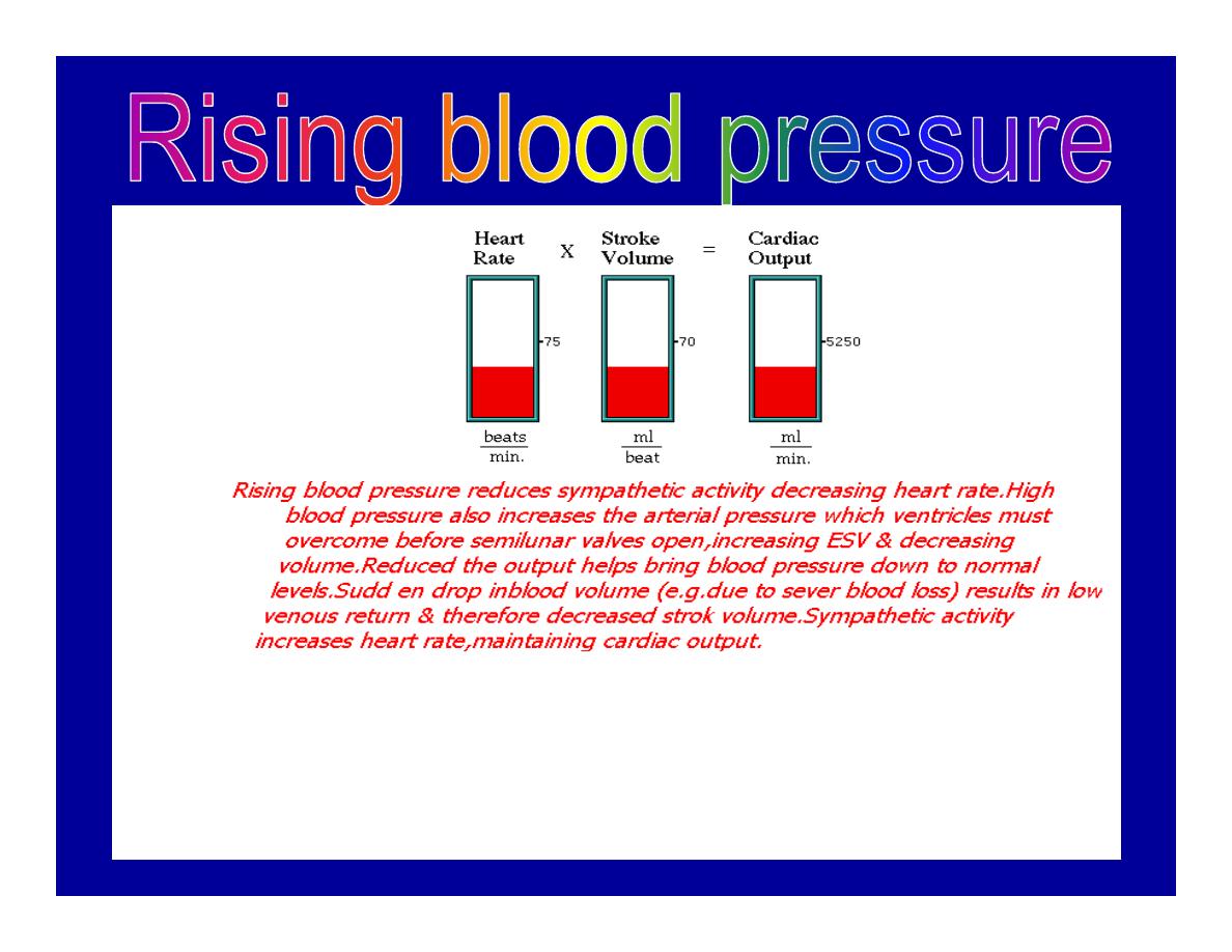
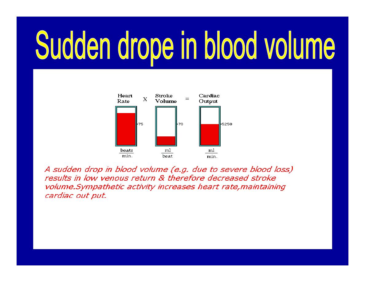
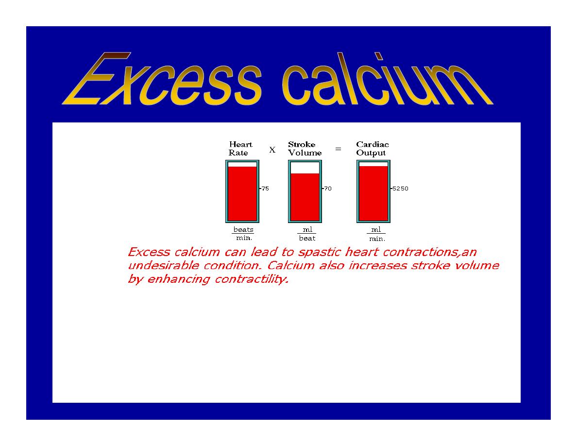
C.O = Stroke volume χ Heart rate
= 70 χ 70=4900 ml/min.

STROKE VOLUME
•
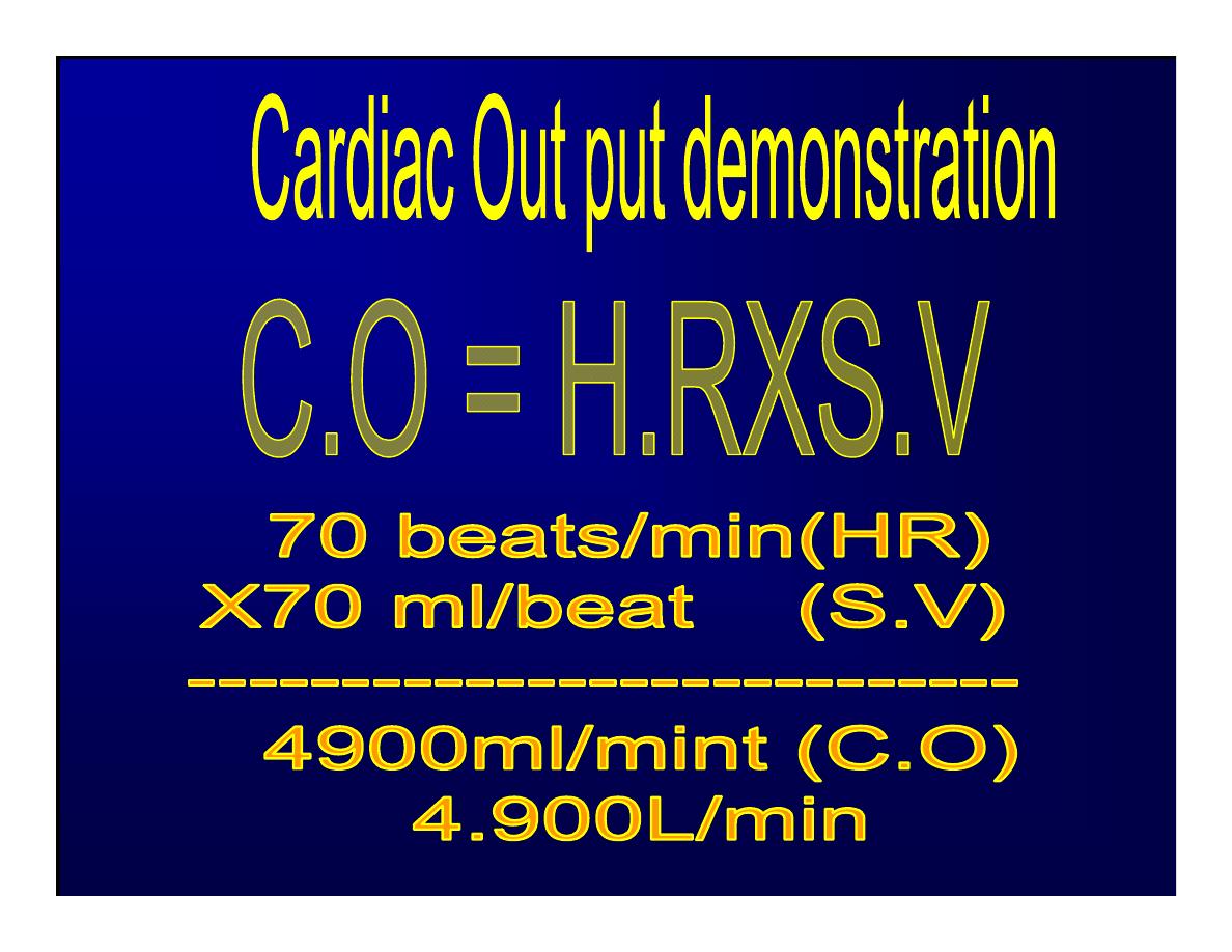
By the time diastole ends each ventricle has filled by
bl. This amount of bl.is the EDV.
•
The amount of bl.ejecte during systole is stroke
volume at the end of systole the volume of
bl.remaining in each ventricle is ESV.
•
For example:-
•
each ventricle normally contain a bout 120ml of bl.
By the end of diastole.
•
By end of systole about 50mll of bl. This mean
about 70ml/beat is pump out by each ventrical
during systole.
•
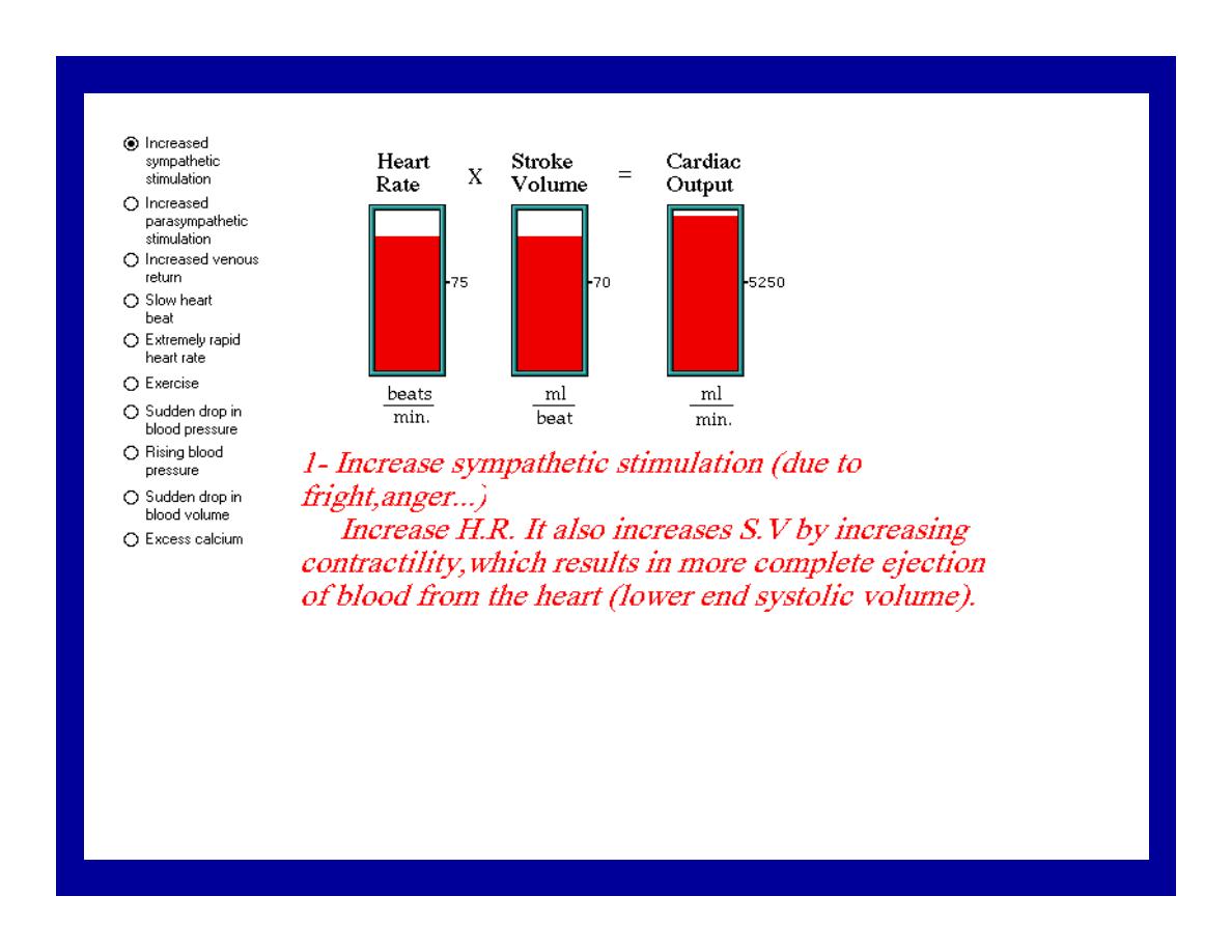
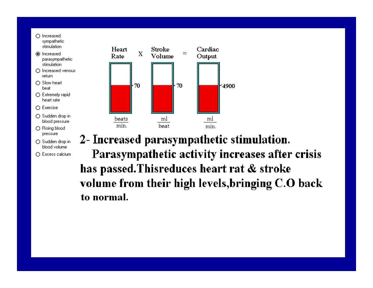
The key factor
regulating s.v is the
amount of stretching
that occur to
ventricular cardiac
m.prior to the
v.contraction.
•
The more cardiac
stretching the more
forcefully to contract
these contraction
increase s.v
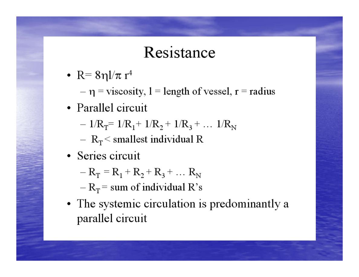
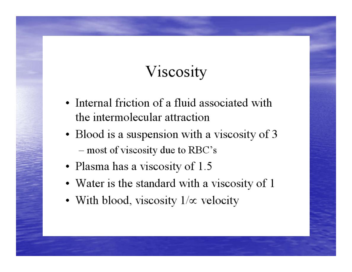
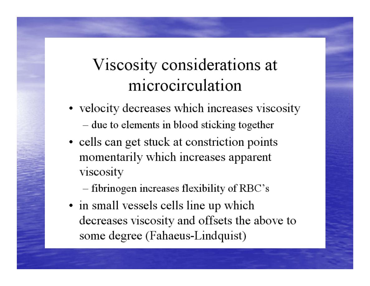
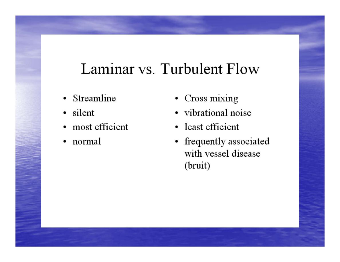
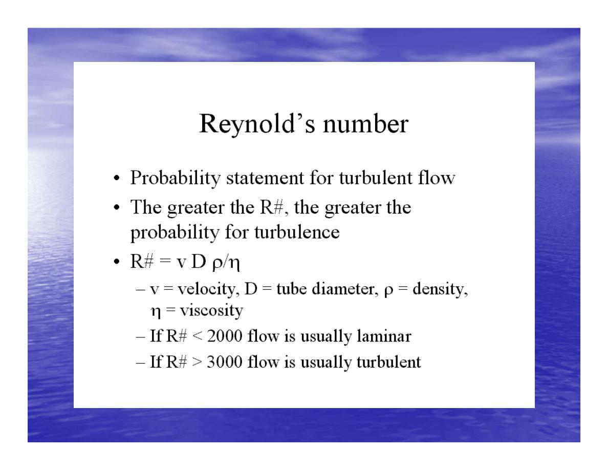

Reynolds increased
•
Decreased bl.viscosity(ex.decrease Hct,anemia)
•
Increased bl.velocity (ex.narrawing of avessel).
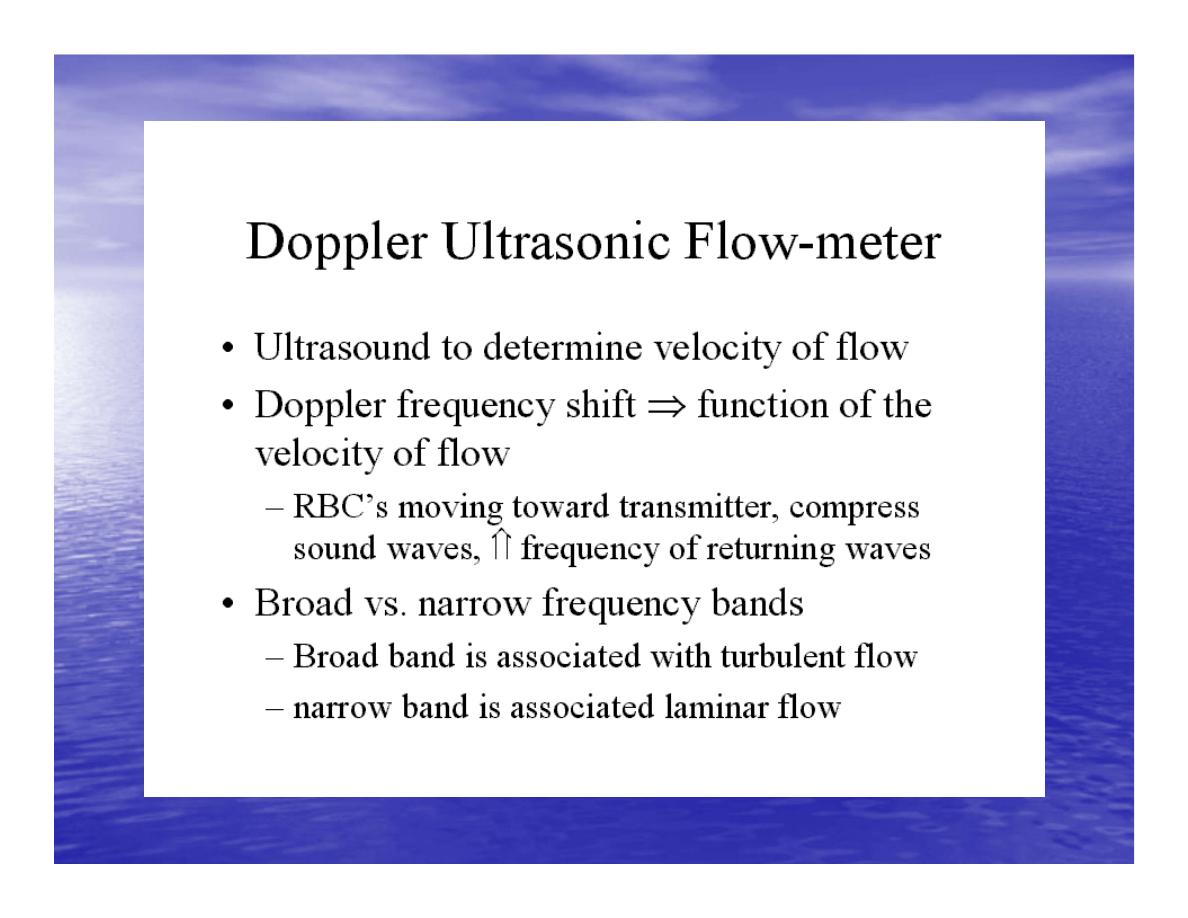
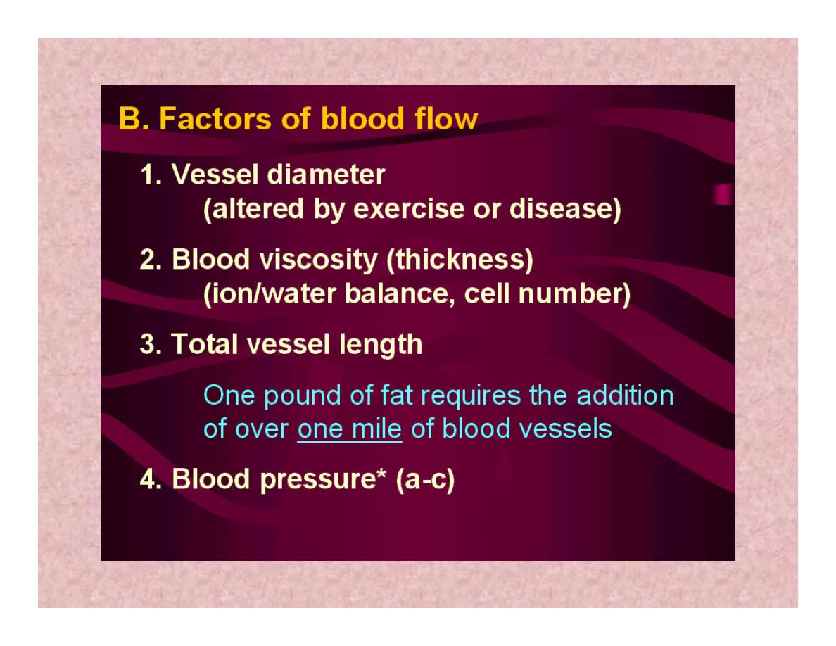
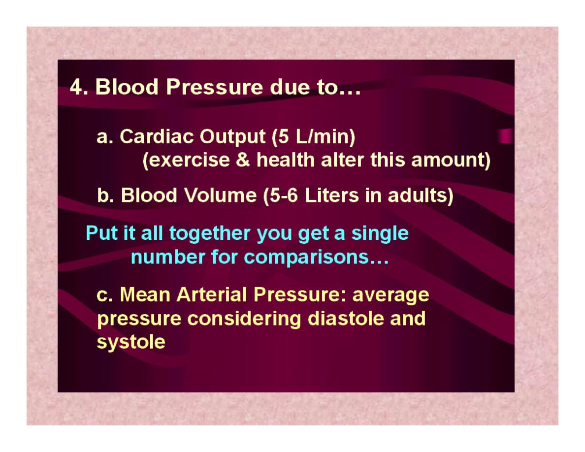
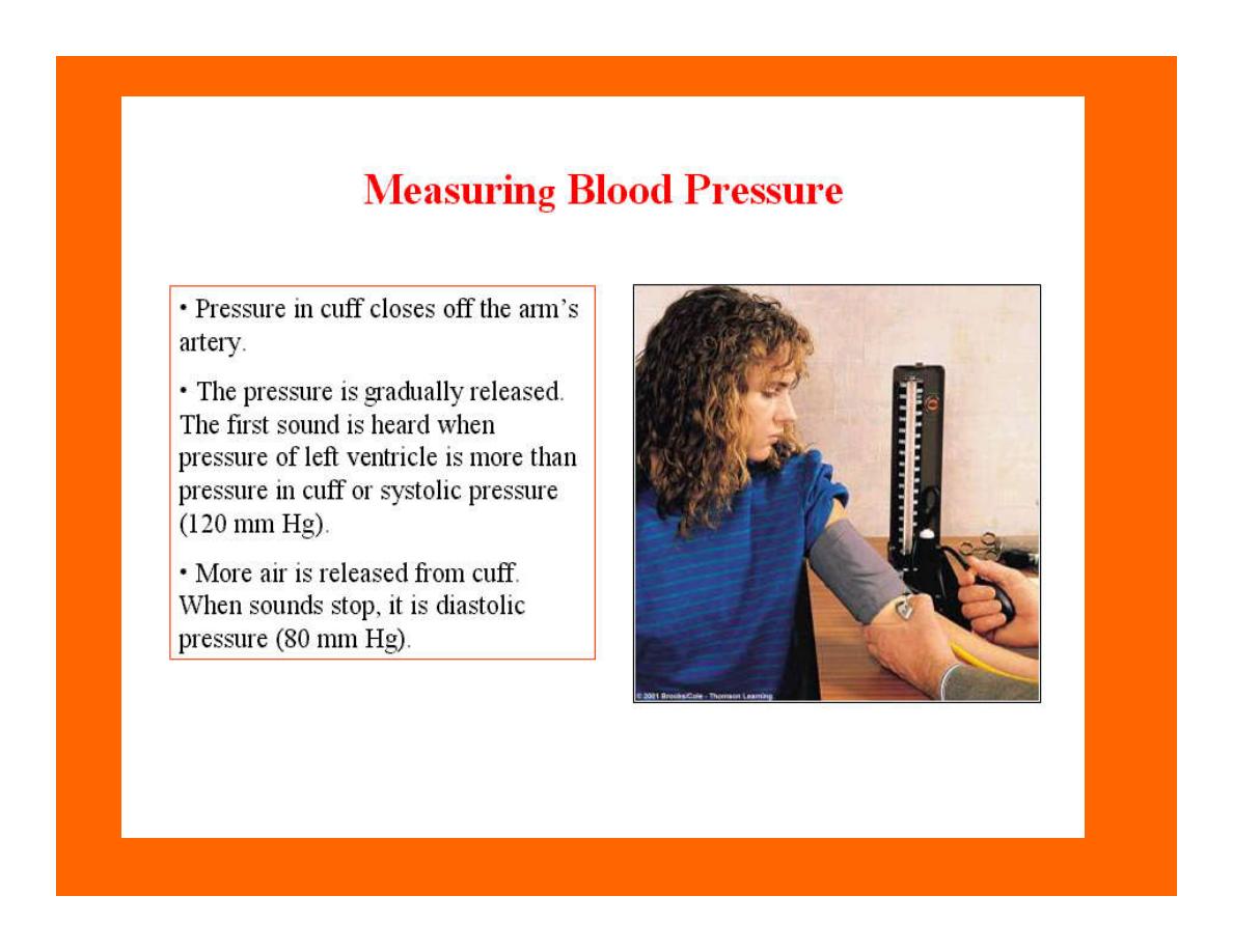
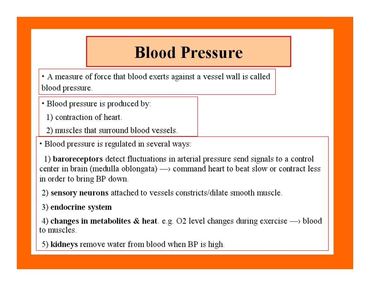
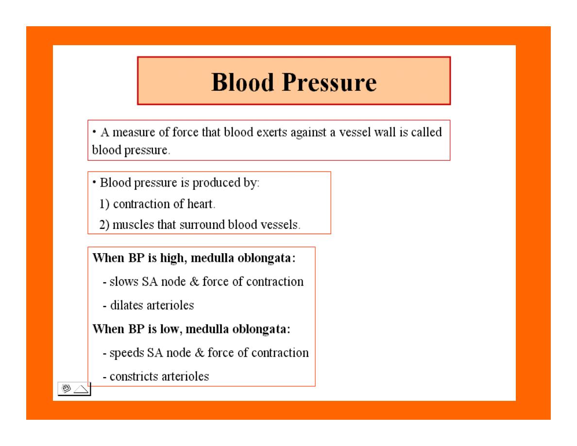
Measuring of Bl.pressure
•
Methods of measuring blood
pressure:
•
Blood pressure can be recorded
direct by using mercury manometer with
electronic pressure transducers or indirect
by mercury sphygmomanometer
(palpatory and auscultatory methods)
[refere to the practical Manual].
•Palpatory method:
Feel the radial pulse of the subject by
placing the three middle fingers on the radial
artery against the radius bone with mild pressure
of the distal finger, you can feel the radial pulse by
index finger. Raise the pressure in the cuff to 200
mm Hg. Start to lower the pressure slowly while
you are feeling the radial pulse. Once you feel the
pulsation of the radial artery, record the reading on
the manometer. This reading gives you the systolic
pressure. Repeat this procedure many times and
record your result.
•Auscultatory method:
From your surface anatomy
knowledge of the antecubital fossa, put
the diaphragm of the stethoscope at
the position of the brachial artery just
below the lower edge of the cuff (not
underneath the cuff !). Raise the
pressure to 200 mm Hg, then lower the
pressure slowly and steadily

Phase measure Bl.pressure
The following phases of the character of the sound (Korotkow or Korotkoff
sounds) will be heard by the stethoscope:
Phase 1: If the pressure is above systolic no sound can be heard. When
the systolic pressure is reached a clear loud sound is heard with each heart
beat.
Phase 2: When you lower the pressure more the sound becomes softer.
Phase 3: As the pressure is lowered, the sound becomes louder and
banging in character.
Phase 4: The sound will disapper completely (sometimes this taken as the
diastolic pressure).
The exact cause of Korotkoff sounds is still debated but they are
believed to be caused mainly by blood jetting through the partly occluded
vessel. The jet causes turbulence in the open vessel beyond the cuff, and
this sets up the vibrations heard throug the stethoscope
Regulation of arterial blood
pressure
•
There are tow main mechanisms for regulation
of BP and these are:
•
[A] Rapidly acting mechanism (short –
term regulation
•
[B] Slow acting mechanism (long-term
regulation
)

Rapidly acting mechanism
•
which are rapid to begin acting, they
begin to act within seconds and
become fully active within a minute
or so, and lose their capability for
pressure control after few hours or a
few days (adaptating mechanisms).
Rapidly acting mechanism
•
These mechanisms include:
•
(1) Nervous pressure control
mechanisms which include:
•
[I] The baroreceptors feedback
mechanism,
•
[II] The central nervous system
ischemic mechanism,
•
[III] The chemoreceptor
mechanism.
•

•
(2) Other pressure control mechanisms which
begin to react within minutes and become fully
active within 30 minutes to several hours and
include:
•
[I] The hormonal mechanism which
include norepinephrine
epinephrine,vasopressin,and renin-angiotensin
vasoconstrictor mechanisms.
•
[II] Stress relaxation mechanism,
•
[III] Capillary fluid shift.

B] Slow acting mechanism
(long-term regulation)
•
, which are slow to begin acting and
control the arterial BP over a period of
days,weeks, months, and years.Their
effectuveness becomes steadily greater
with time (non adapting mechanisms),
and able to bring the pressure all the way
back to normal.These mechanisms include
the renal-body fliud –pressure control
mechanisms.

[A] Rapidly acting mechanism (Short-
term regulation):
•
The barorecepttors feedback
mechanism: This reflex is mediated by
baroreceptors located in the carotid
sinus at the bifurcation of the common
carotid arteries, and their afferent nerve
fibers pass to the glossopharyngeal
nerves, and baroreceptors which are
located along the arch of the aorta, and
their afferent nerve fibers pass through
the vagi.

Increase in Bl.pressure
•
A rise in pressure stretches the baroreceptors and causes them to
transmit signals into the nucleus of tractus solitarius which is the
sensory termination of both the vagal and glossopharyngeal nerves
at central nervous system.
•
From this nucleus an inhibitory signals pass to vasoconstrictor area
of the VMC and an excitotry signals pass to the vagal
(parasympathetic) center.
•
The feedback signals are then sent back through autonomic nervous
circulation cause vasodilatation (decrease peripheral resistance), a
decrease in heart and contractility.All will reduce arterial blood
pressure downward toward the normal level.
•

Sudden decrease in bl.pressure
•
when a person sits or stand after having
been lying down. Immediately upon
standing, the arterial pressure in the head
and upper part of the body obviously
tends to fall,
•
the falling pressure at the baroreceptors elicits
an immediate reflex,resulting in strong
sympathetic discharge throughout the body, and
this minimizes the decrease in the head and
upper body.
•

sudden decrease in bl.pressure
•
Strong pressure to the neck over the
bhfurcations of the carotid arteries in
human being can excite the baroreceptors
of the carotid sinus,causing the arterial
pressure to fall as much as 20 mm Hg in
normal person and even a more marked
reduction in BP and even stop the heart
completely in older atherosclerotic
patients which lead to fainting (carotid
sinus syncope).

The chemoreceptor
mechanism
•
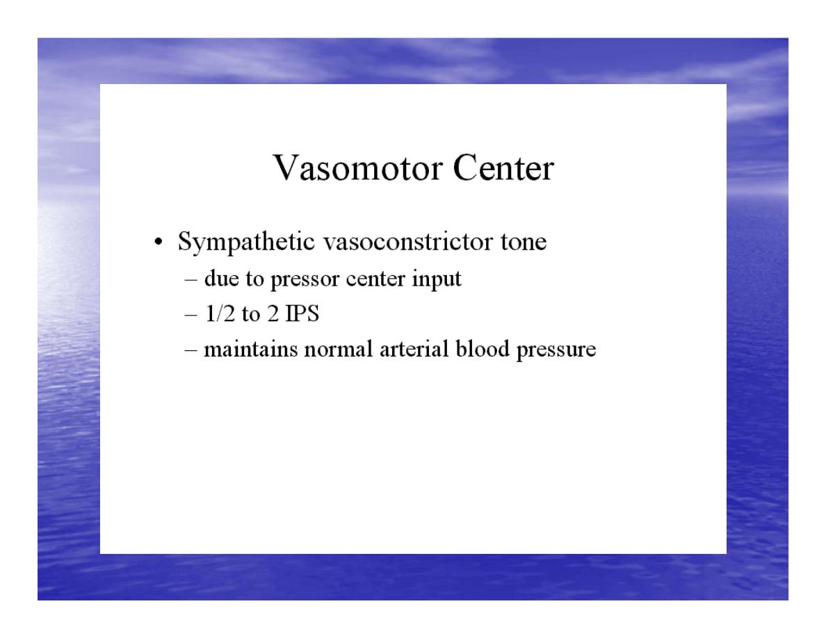
Whenever the arterial pressure falls below critical level,
the chemoreceptors become stimulated because of
diminished blood flow to the aortic and carotid bodies
and excess
2
and therefore diminished availability of O
ions are not removed by the slow flow of
and H+
2
CO
blood. The signals from the chemoreceptors are
transmitted to VMC to excite it, and this elevate the BP
back toward normal level whenever it falls too
low.Chemoreceptors mechanism start to operat when
the blood pressure falls below 80 mm H
g.

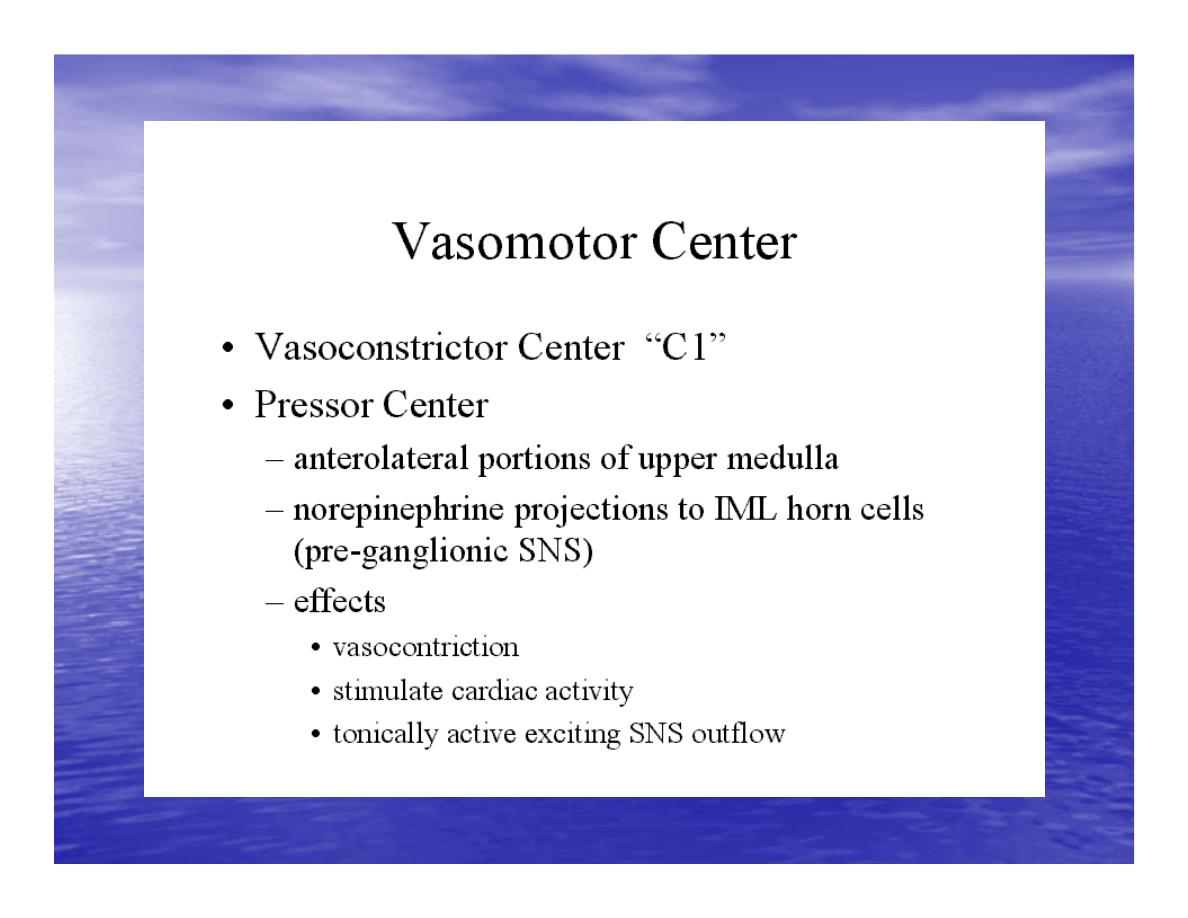
Short-Term Regulation of Rising Blood Pressure
•
When rising Pressure
•
• Rising blood pressure
•
• Stretching of arterial walls
•
• Stimulation of baroreceptors in carotid
•
sinus, aortic arch, and other large
•
arteries of the neck and thorax
•
• Increased impulses to the brain

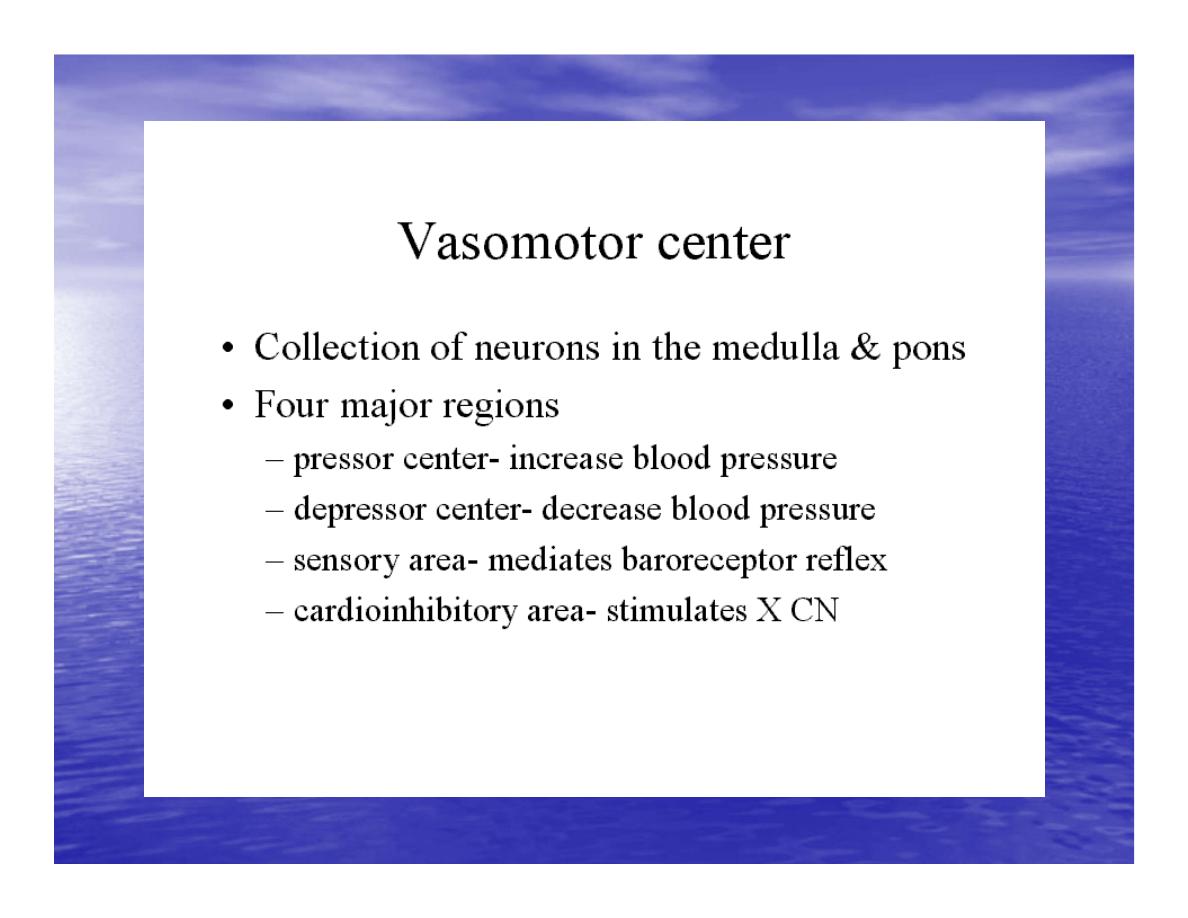
So the effect of baroreceptors
ٍ
•
Increased impulses to brain from
baroreceptors
•
• Increased parasympathetic activity and
decreased sympathetic activity
•
• Reduction of heart rate and increase in
arterial diameter
•
• Lower blood pressure
•
Page 5. Increased Parasympathetic
Activity

Increased Parasympathetic
Activity
•
• Effect of Increased Parasympathetic and
•
Decreased Sympathetic Activity on Heart
•
and Blood Pressure:
•
• Increased activity of vagus
•
(parasympathetic) nerve
•
• Decreased activity of sympathetic cardiac
•
nerves
•
• Reduction of heart rate
•
• Lower cardiac output
•
• Lower blood pressure

Decreased Sympathetic
Activity
•
Effect of Decreased Sympathetic Activity
on Arteries and Blood Pressure:
•
• Decreased activity of vasomotor fibers
(sympathetic nerve fibers)
•
• Relaxation of vascular smooth muscle
•
• Increased arterial diameter
•
• Lower blood pressure
Regulation of Rising Blood
Pressure
•
Short-term regulation of Rising Blood Pressure
•
• Rising blood pressure
•
• Stretching of baroreceptors
•
• Increased impulses to the brain
•
• Increased parasympathetic activity
•
• Decreased sympathetic activity
•
• Slowing of heart rate
•
• Increased arterial pressure
•
• Reduction of blood pressure
Diameter

Short-term Regulation of
Falling Blood Pressure
•
Falling blood pressure
•
• Baroreceptors inhibited
•
• Decreased impulses to the brain
•
• Decreased parasympathetic activity, increased sympathetic activity
•
• Three effects:
•
1. Heart: increased heart rate and increased contractility
•
2. Vessels: increased vasoconstriction
•
3. Adrenal gland: release of epinephrine and norepinephrine which
enhance heart rate,
•
contractility, and vasoconstriction
•
• Increased blood pressure

Sympathetic Activity on
Heart and Blood Pressure
•
Increased activity of sympathetic cardiac
nerves
•
• Decreased activity of vagus
(parasympathetic) nerve
•
• Increased heart rate and contractility
•
• Higher cardiac output
•
• Increased blood pressure

Vasomotor Fibers
•
Effect of Increased Sympathetic Activity
on Arteries and Blood Pressure:
•
• Increased activity of vasomotor fibers
(sympathetic nerve fibers)
•
• Constriction of vascular smooth muscle
•
• Decreased arterial diameter
•
• Increased blood pressure

Sympathetic Activity on
Adrenal Gland and Blood Pressure
•
• Increased sympathetic impulses to
•
adrenal glands
•
• Release of epinephrine and
•
norepinephrine to bloodstream
•
• Hormones increase heart rate,
•
contractility and vasoconstriction.
•
Effect is slower-acting and more
•
prolonged than nervous system control.
•
• Increased blood pressure

Regulation of Falling Blood
Pressur
•
Regulation of Falling Blood Pressure
•
• Falling blood pressure
•
• Baroreceptors inhibited
•
• Decreased impulses to the brain
•
• Decreased parasympathetic activity
•
• Increased sympathetic activity
•
• Increased heart rate and contractility
•
• Increased vasoconstriction
•
• Release of epinephrine and
•
norepinephrine from adrenal gland
•
• Increased blood pressure
Hormonal mechanisms:
•
Hormonal
:
Hormonal mechanisms
regulation of the circulation means
regulation of tissue blood flow by
substances in the body fluids, such as by
hormones, ions, or so forth. Among the
most important of the humoral factors
that affect circulatory function are the
following:

Vasocontrictor agents:
Nor epinephrine has vasoconstrictor effects
in almost all vascular beds of the body,
and epinephrine has similar effects except
it has a mild vasodilator effects in both
skeletal, cardiac muscle and liver.

Long-Term Regulation of
Low BP
•
• Long-term regulation of blood pressure is primarily
accomplished by altering blood volume.
•
• The loss of blood through hemorrhage, accident, or
donating a pint of blood will lower blood pressure
•
and trigger processes to restore blood volume and
therefore blood pressure back to normal.
•
• Long-term regulatory processes promote the
conservation of body fluids via renal mechanisms and
•
stimulate intake of water to normalize blood volume and
blood pressures.

Loss of Blood
•
When there is a loss of blood, blood
pressure and blood volume decrease.
•
So Juxtaglomerular Cells in the kidney
monitor alterations in the blood pressure.
If blood pressure falls
•
too low, these specialized cells release the
enzyme renin into the bloodstream.

Renin-Angiotensin
Mechanism: Step 1
•
The renin/angiotensin mechanism consists
of a series of steps aimed at increasing
blood volume and
•
blood pressure.

Step 1
:
•
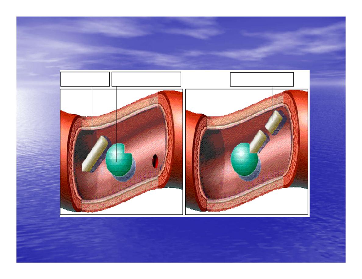
Step 1: Catalyzing Formation of
Angiotensin I: As renin travels through the
bloodstream, it binds to
•
an inactive plasma protein,
angiotensinogen, activating it into
angiotensin I.

Step 2:
•
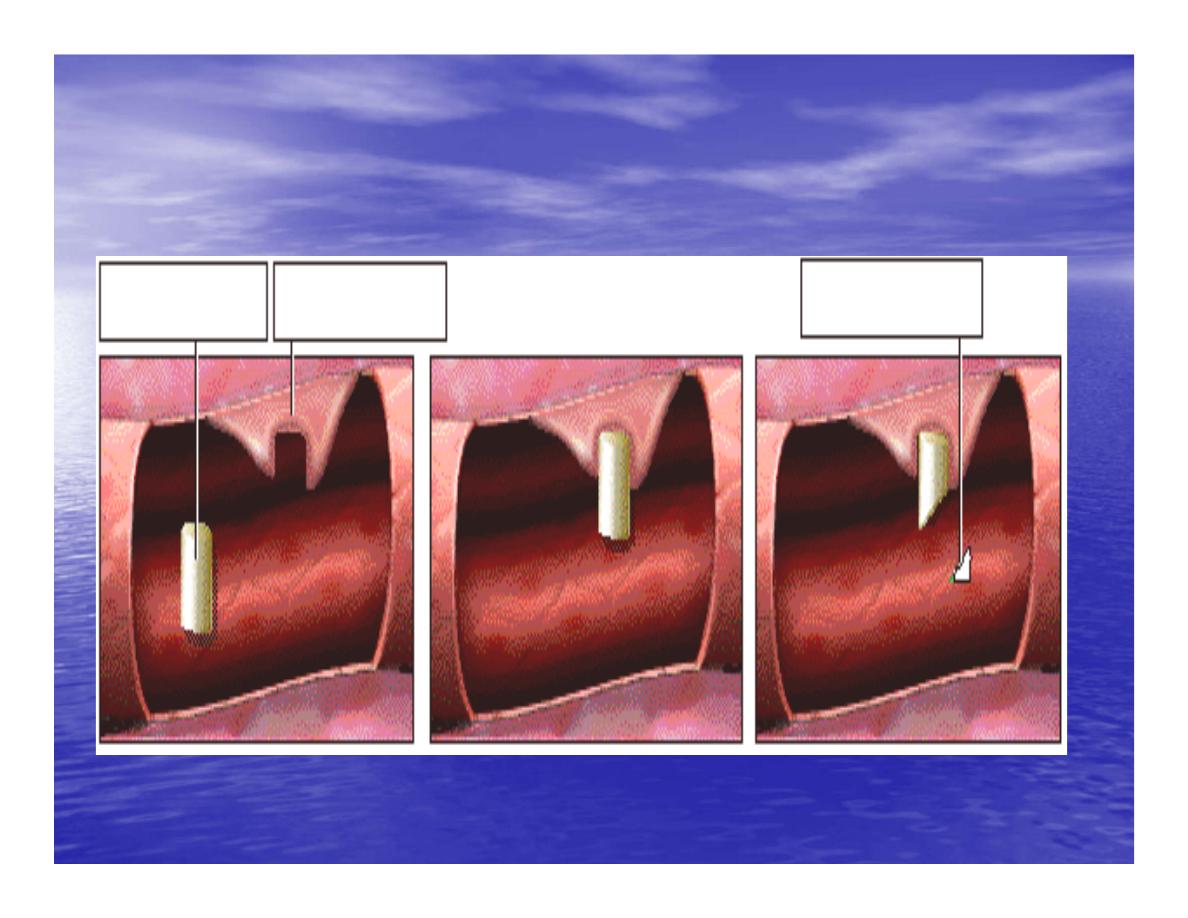
Step 2: Conversion of Angiotensin I
to Angiotensin II: As angiotensin I passes
through the lung
•
capillaries, an enzyme in the lungs
converts angiotensin I to angiotensin II.
Step 2:

Step 3: Angiotensin II in the
Bloodstream
•
Step 3: Angiotensin II Stimulates
Aldosterone Release: Angiotensin II
continues through the
•
Blood stream until it reaches the
adrenal
gland.

Release of Aldosterone
•
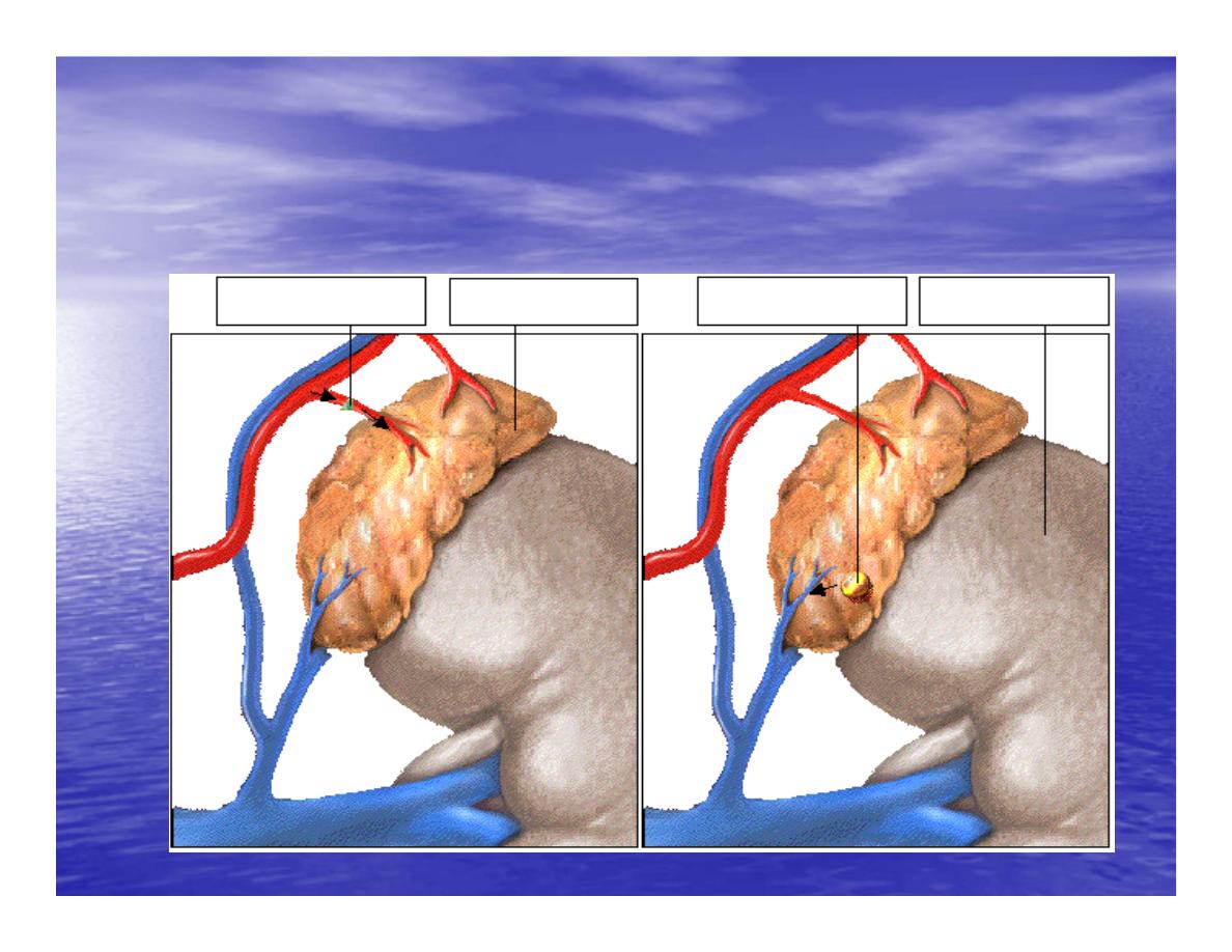
Step 3: Angiotensin II Stimulates
Aldosterone Release: Here it
stimulates the cells of the adrenal
cortex to release the hormone
aldosterone
Release of Aldosterone

•
Angiotensin II as a Vasoconstrictor
•
• A secondary effect is that angiotensin II
is a vasoconstrictor and therefore raises
blood pressure in
•
the body's arterioles.

Aldosterone Mechanism
•
. Aldosterone Mechanism
•
• Long-Term Regulation: Aldosterone
Mechanism: The target organ for
aldosterone is the kidney.
•
Here aldosterone promotes increased
reabsorption of sodium from the kidney
tubules.

• Long-Term Regulation:
Aldosterone Mechanism:
•
Distal Convoluted Tubule
•
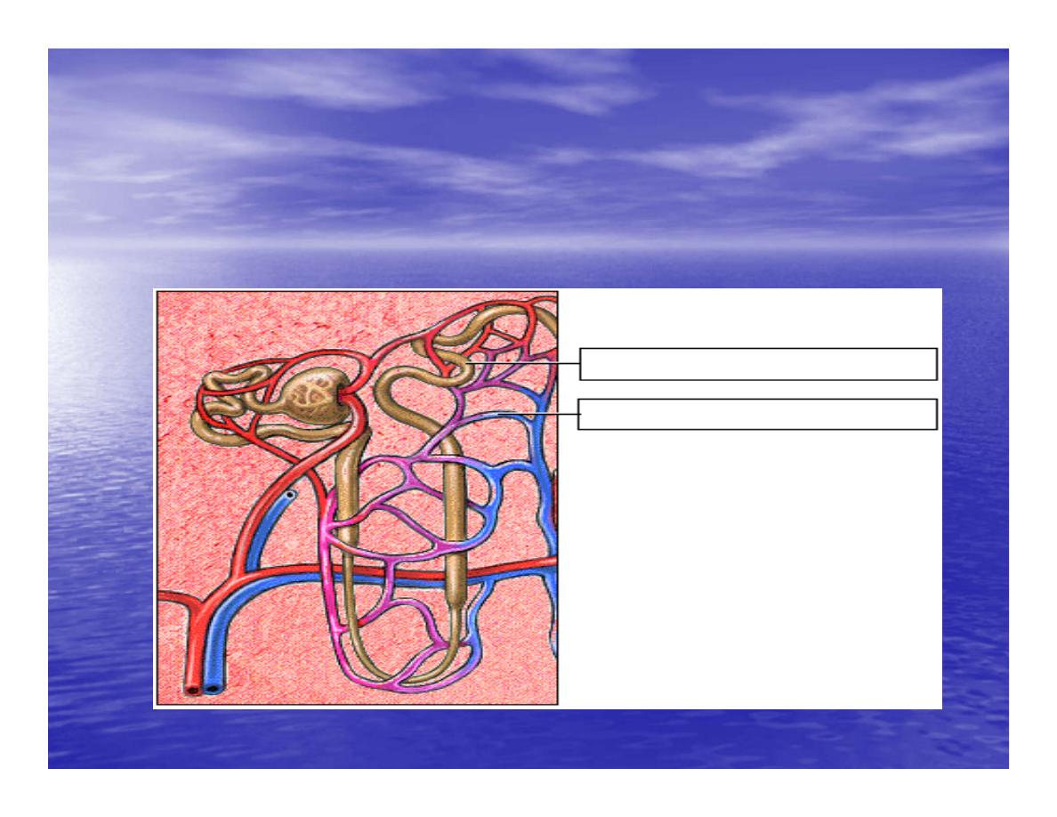
• Each distal convoluted tubule winds
through the kidney and eventually empties
its contents into
•
a urine-collecting duct.
•
• The peritubular capillaries absorb solutes
and water from the tubule cells as these
reclaimed from the filtratesubstances
Distal Convoluted Tubule

Long-Term Regulation:
Aldosterone Mechanism
•
Sodium Reabsorption
•
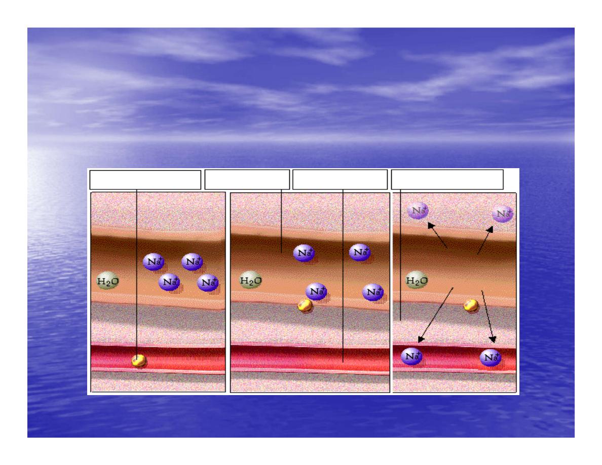
• Aldosterone stimulates the cells of the
distal convoluted tubule to increase the
active transport of
•
sodium ions out of the tubule into the
interstitial fluid, accelerating sodium
reabsorption.
Long-Term Regulation:
Aldosterone Mechanism

Long-Term Regulation:
Aldosterone Mechanism
•
Water Reabsorption
•
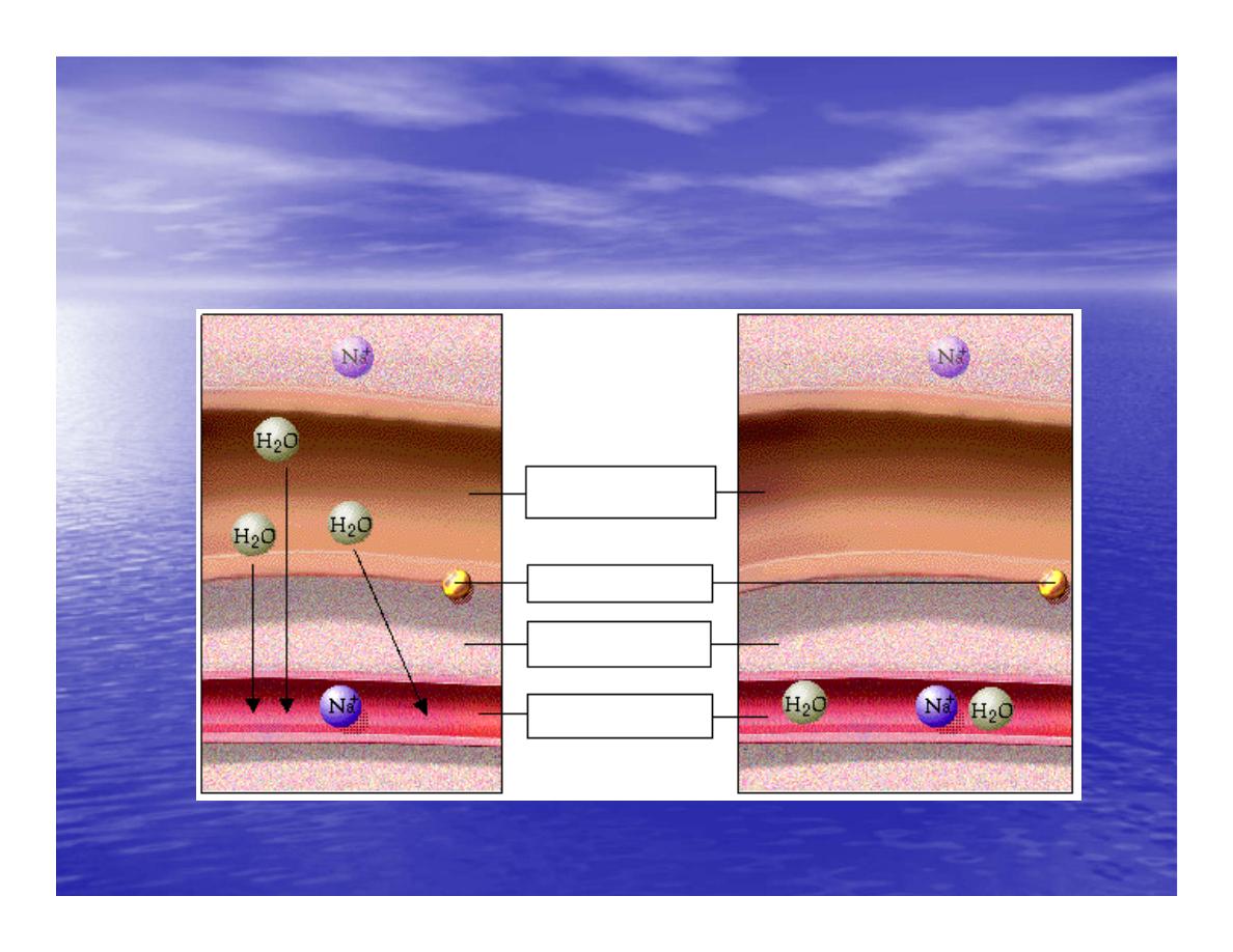
• As sodium moves into the bloodstream,
water follows. The reabsorbed water
increases the blood
•
volume and therefore the blood pressure.
Long-Term Regulation:
Aldosterone Mechanism

Increase in Osmolarity
•
Increase in Osmolarity
•
• Dehydration due to sweating, diarrhea, or excessive
urine flow will cause an increase in osmolarity
•
of the blood and a decrease in blood volume and blood
pressure.
•
Long-Term Effect of Osmolarity on BP
•
• As increased osmolarity is detected there is both a
short and long-term effect. For the long-term
•
effect, the hypothalamus sends a signal to the posterior
pituitary to release antidiuretic hormone

Antidiuretic Hormone
•
Antidiuretic Hormone
•
• ADH increases water reabsorption in the kidney.
•
ADH in Distal Convoluted Tubule
•
• ADH promotes the reabsorption of water from the
kidney by stimulating an increase in the number
•
of water channels in the distal convoluted tubules and
collecting tubules (ducts).
•
• These channels aid in the movement of water back
into the capillaries, decreasing the osmolarity of
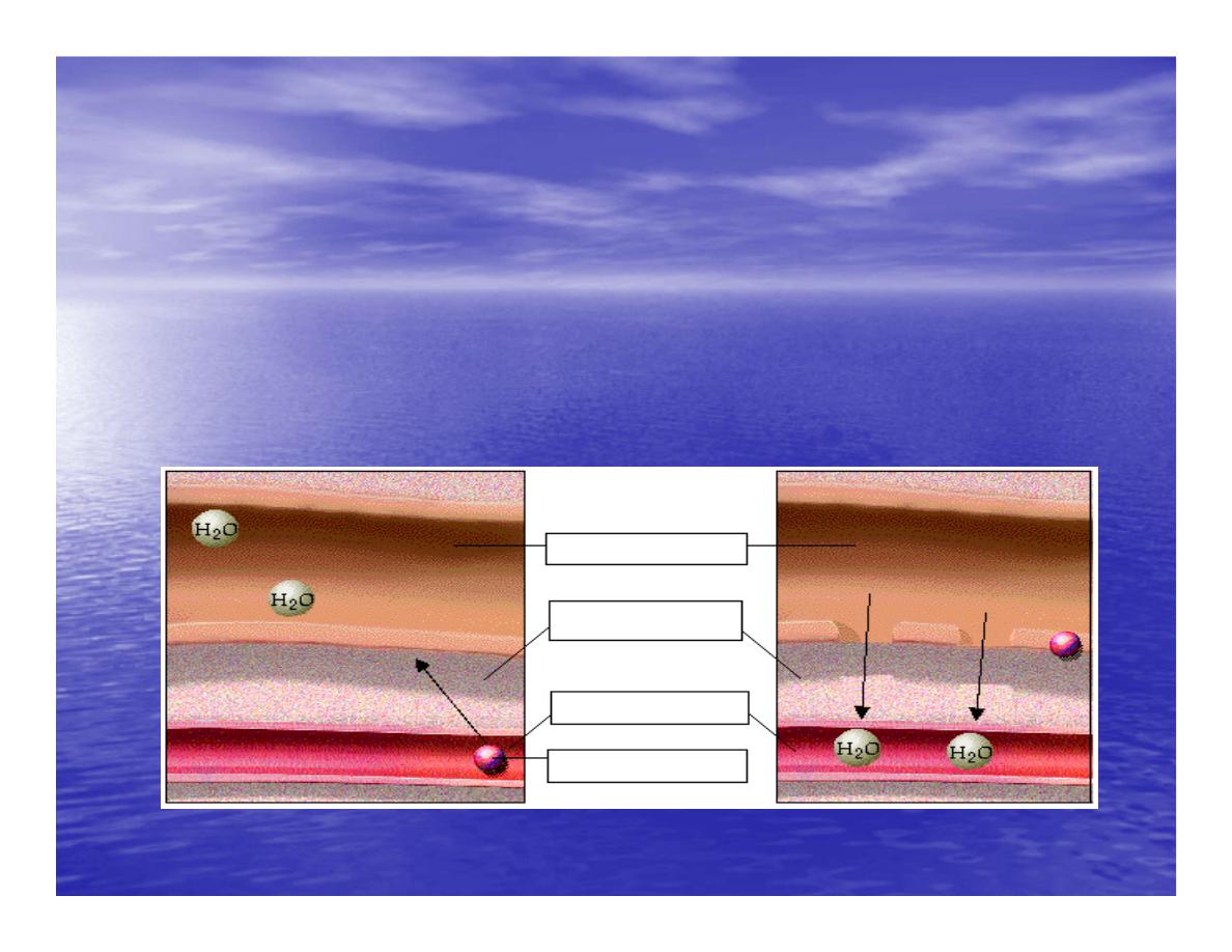
the blood volume and therefore blood pressure.

Short-Term Effect of
Osmolarity on BP
•
• A short-term effect of increased
osmolarity is the excitation of the thirst
center in the
•
hypothalamus. The thirst center stimulates
the individual to drink more water and
thus
•
rehydrate the blood and extracellular fluid,
restoring blood volume and therefore
blood pressure.

Summary
•
•
In the short-term, rising blood pressure
stimulates increased parasympathetic activity,
which leads
•
to reduced heart rate, vasodilation and lower
blood pressure.
•
• Falling blood pressure stimulates increased
sympathetic activity, which leads to increased
heart
•
rate, contractility, vasoconstriction, and blood
pressure
.

Summary
•
•
Long-term blood pressure regulation involves
renal regulation of blood volume via the
reninangiotensin
•
mechanism and aldosterone mechanism.
•
• Increased blood osmolarity stimulates release
of antidiuretic hormone (ADH), which promotes
•
reabsorption of water, and excites the thirst
center, resulting in increased blood volume and
bloo
d
