Cells of the Nervous System
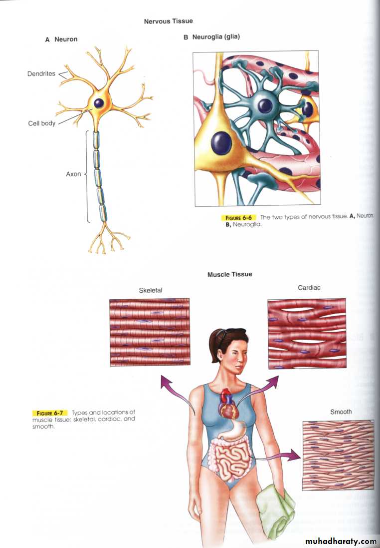
The human nervous system consists of billions of nerve cells (neurons) plus supporting cells (neuroglial).
1. Neurons—basic functional units (nerve cells), able to respond to stimuli, conduct impulses, and communicate with each other and with other types of cells like muscle cells
2. Neuroglia or glial cells
Neurons
Types of Neurons (according to function) ;Afferent (sensory), Interneurons, Efferent (motor)Neurons
Sensory or afferent neuron
Interneuron
Motor or efferent Neuron
Length of Fibers
Long dendrites and short axon
Short dendrites and short or long axon
Short dendrites and long axons
Location
Cell body and dendrite are outside of the spinal cord; the cell body is located in a dorsal root ganglion
Entirely within the spinal cord or CNS
Dendrites and the cell body are located in the spinal cord; the axon is outside of the spinal cord
Function
Conduct impulse to the spinal cord
Interconnect the sensory neuron with appropriate motor neuron
Conduct impulse to an effecter (muscle or gland)
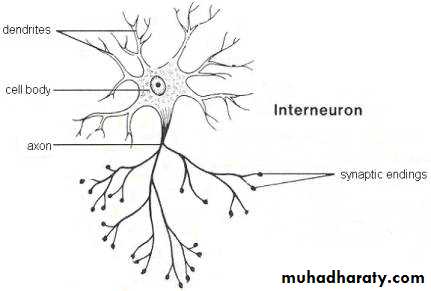
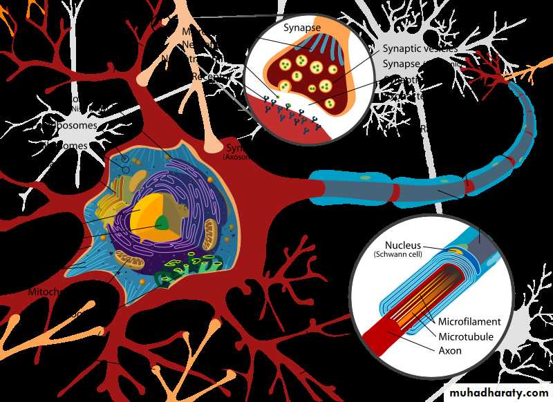
Neurons consist of: Cell Body, Dendrite, Axons.
A. Cell Body
Contains nucleus, control center of cell
Nutritional center where macromolecules produce
Nucleus surrounded by cytoplasm that contains typical organelles as well as some specialized ones
Dendrite
Highly branched unmyelinated processes emerging from cell bodyReceiving portion of neuron
Number per neuron varies from 1-hundreds
Receives information from other neurons in form of neurotransmitters (chemicals that stimulate other neurons)
Axons (nerve fiber)
Only one axon
Long process that may be myelinated and transmit nerve impulses away from cell body
Important features:
Joins cell body at axon hillock; expanded region
First portion called initial segment
Junction of axon hillock and initial segment is trigger zone (production of Action Potential)
Axon collaterals—may branch off axon
Axon and its axon collaterals end at many fine processes called axon terminals
Tips of axon terminals have synaptic end bulbs or synaptic terminals
Synaptic end bulbs; swellings that contain synaptic vesicles that store neurotransmitters
Cell membrane of axon; axolemma. Same general structure as other cell membranes (Phospholipid bilayer, has Na+-K+ pumps)
Axons
DendritesTake information away from the cell body
Smooth surface
Generally only 1 axon per cell
No ribosomes
Can have myelin
Branch further from the cell body
Bring information to the cell body
Rough surface(dendritic spines)
Usually many dendrites per cell
Have ribosomes
No myelin insulation
Branch near the cell body
Synapse; The junction between a nerve cell and another cell .
The space between two cells is known as the synaptic cleft. To cross the synaptic cleft requires the actions of neurotransmitters (small molecules, some are even hormones).
Neurotransmitters are stored in small synaptic vesicles clustered at the tip of the axon.
Messages travel within the neuron as an electrical action potential.
Arrival of the action potential causes some of the vesicles to move to the end of the axon and discharge their contents into the synaptic cleft. Released neurotransmitters diffuse across the cleft, and bind to receptors on the other cell's membrane, causing ion channels on that cell to open.
Some neurotransmitters cause an action potential, others are inhibitory.
The time for neurotransmitter action is between 0,5 and 1 millisecond.
Neurotransmitters are either;
destroyed by specific enzymes in the synaptic cleft.diffuse out of the cleft.
reabsorbed by the cell.
Transmitters in the Nervous System:
More than 30 organic molecules are thought to act as neurotransmitters;Acetylcholine.
Catecholamines; epinephrine, norepinephrine.
Serotonin, dopamine, glutamate, secretin, endorphins, Gamma aminobutyric acid (GABA), Oxytocin, Angiotensin II.
Even gas molecules such as nitric oxide (NO) can act as local transmitters (The gas types are not stored, but are made on demand).
Neuroglia or Glial Cells:
About 90% of cells in human nervous system, outnumber neurons by 5XDo not transmit nerve impulses and can divide in mature nervous system.
Regulating the internal environment of neurons in the central nervous system.
Types of Neuroglia
Schwann cells; Forming myelin sheaths only in peripheral nervous system (PNS)
Oligodendrogliocytes; Forming myelin sheaths only in central nervous system (CNS)
Microglia; Protecting neurons (via phagocytosis)
Astrocytes; which are found throughout the brain;
send processes to blood vessels, form the blood–brain barrier
send processes that envelop synapses and the surface of nerve cells.
produce substances that are tropic to neurons, and they help to maintain the appropriate concentration of ions and neurotransmitters.
Astroglia has receptors for many neurotransmitters.
Myelination:
Myelin; lipid rich sheath that covers some axons at regular intervals & acts as an insulator. Ions responsible for carrying current across membrane cannot permeate this thick barrier.
Internodes
Nodes of Ranvier; between myelinated regions where axonal membrane is naked and exposed to ECF.
These gaps in myelination, play an important role in the transmission of electrical signals along the axon.
Electrophysiology of Neurons:
Nerve impulse is result of movement of ions across the axon plasma membrane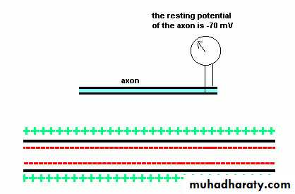
Concentrations of specific ions on the 2 sides of axon plasma membrane of a resting neuron (one not conducting an impulse) are very different.
The concentrations of chloride ions (Cl-) and calcium ions (Ca2+) are also maintained at greater levels outside the cell
Concentration of K+ ions is approximately 30 times greater inside the cell than outside
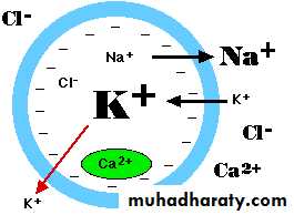
Concentration of Na+ is approximately 10-15 times greater outside the cell than inside.
In a resting neuron, K+ ions diffuse out of a cell through k+ leak channels
There are no Na+ leak channels making the plasma membrane essentially impermeable to Na+ ions.
K+ are positively charged and their movement out of cell through leak channels leaves inside of the membrane slightly more negatively charged than outside
This separation of positive and negative charge is called potential and measured in millivolts(mV)
Transmembrane potential; is the voltage difference across the cell membrane between the ICF and ECF
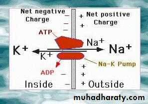
A resting cell has potential difference of about -70 (mV), called resting membrane potential (RMP). Negative sign indicates negativity of inside of cell relative to the outside of cell.
What factors contribute to membrane potential?
An unequal distribution of (Na+) and (K+) ions, occurs on the two sides of a nerve cell membrane because carriers actively transport these two ions: sodium from the inside to the outside and potassium from the outside to the inside by SODIUM - POTASSIUM PUMP.
Two types of signals produced by membrane changes:
1. Graded potential (incoming signals)Occurs in dendrites and cell bodies
Are proportional to stimulus intensity
Short-lived, ineffective beyond a short distance.
Critical to body’s function; synaptic potentials,
receptor potentials, endplate potentials, pacemaker potentials
Summation; small changes can be added to each other and if enough of them might reach point called threshold potential
Threshold potential necessary for action potential to be generated
2. Action Potential
Action Potential; Brief, rapid change in membrane voltage that is propagated along the axon to the synaptic terminals, also called nerve impulse (nerve impulse is series of action potentials)Potential reverses so that inside of cell transiently becomes more positive than outside
Long-distance; do not diminish in strength as they travel throughout neuron
Occurs in a few thousandths of second
Involves channels for Na+ and K+
All-or-none (occur as a maximal depolarization if stimulus reaches threshold. Or do not occur at all if stimulus is below threshold);
If the stimulus is too low there is no action potential (this is the "none" part)
If the stimulus is above a threshold the action potential is always the same size- it does not get larger for stronger stimuli
As the action potential travels along the axon it does not die out, but stays the same size
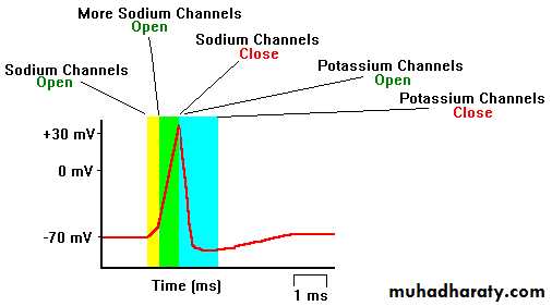
Process of one action potential:
After stimulation an interval called (latent period) that ends with the start of the action potential and corresponds to the time it takes the impulse to travel along the axon from the site of stimulation to the recording electrodes
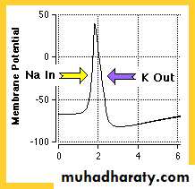
Begins when a graded potential reaching trigger zone, depolarizes membrane (becomes less negative) to threshold (usually around -60mV)
The first manifestation of the approaching action potential is a beginning depolarization of the membrane.
Voltage-gated Na+ channels open in response to stimulus and Na+ rushes in and voltage-gated K+ channels begin to open slowly (triggered by same stimulus)
After an initial 15 mV of depolarization, the rate of depolarization increases, to form firing level or threshold.
Rapid Na+ entry causes depolarization to +30mV or so
K+ moves from cell to ECF following gradient and at same time Na+ gates are closing so membrane potential rapidly becomes more negative, sending the cell towards resting potential, repolarization.
When membrane potential reaches -70mV K+ channels have not yet closed so K+ continues to leave cell (after hyperpolarization).
Voltage-gated K+ channels close, K+ leakage into cell exceeds movement out, bringing membrane potential back to RMP of -70mV.
Summary:
Refractory Period:
Time when cell membrane is insensitive to additional stimulation=refractory.Refers to fact that once an action potential as begun, for about 1msec, second action potential cannot be triggered, no matter how large the stimulus
Refers to patches of membrane, not whole neuron
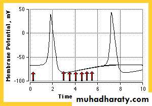
Action potentials cannot overlap and cannot travel backward because of refractory periods
Two types of refractory periods:
Absolute refractory period; corresponding to the period from the time the firing level is reached until repolarization is about one-third complete
Relative refractory period; lasting from this point to the start of after-depolarization.
Impulse Conduction (propagation of action potentials)
Impulses typically travel along neurons at a speed of anywhere from 1 to 120 meters per second
The speed of conduction can be influenced by:
The diameter of a fiber
Temperature
The presence or absence of myelin. Neurons with myelin conduct impulses much faster than those without myelin.
Conduction velocity for non-myelinated nerve = ~1meter/sec (depends upon diameter)
Conduction velocity for myelinated nerve = ~100 meters/sec
In non-myelinated nerve fiber an impulse travels by continuous conduction.
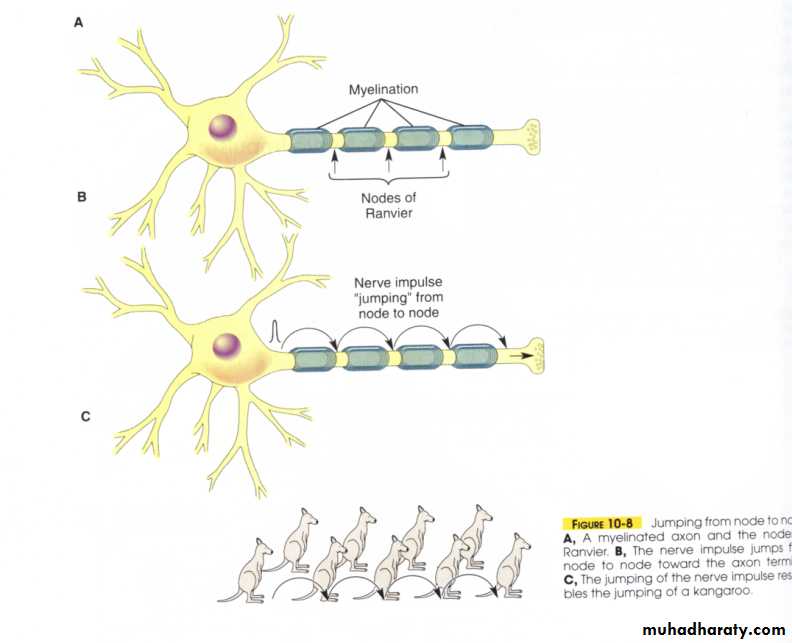
In myelinated neuron, Between areas of myelin are non-myelinated areas called the nodes of Ranvier. So, action potentials only occur along the nodes and, therefore, impulses 'jump' over the areas of myelin - going from node to node in a process called saltatory conduction (salutatory meaning 'jumping'): Because the impulse 'jumps' over areas of myelin, an impulse travels much faster along a myelinated neuron than along a non-myelinated neuron.
(Demyelinating diseases causes severe nerve defects. In Autoimmune diseases, immune system attacks nerves)
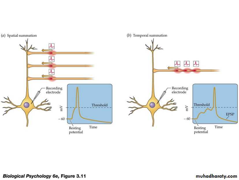
Summation:
1 - Temporal summation ; A second means for transmitting signals of increasing strength is by increasing the frequency of nerve impulses in each fiber.2- Spatial summation ; whereby increasing signal strength is transmitted by using progressively greater numbers of fibers.
Spatial summation
PROPERTIES OF MIXED NERVES
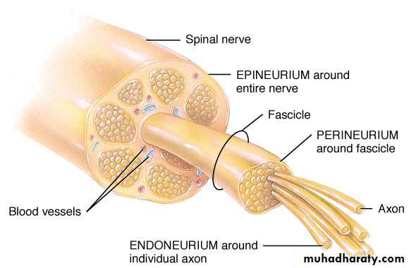
Peripheral nerves in mammals are made up of many axons bound together in a fibrous envelope called the epineurium.
Potential changes recorded from extracellular, such nerves therefore represent an algebraic summation of the all-or-none action potentials of many axons.
Subthreshold stimuli; Electrical potential level which don’t produce action potential (none of the axons are stimulated and no response occurs).
Threshold stimuli; Electrical potential level (voltage) at which an action potential or nerve impulse is produced (axons with low thresholds fire and a small potential observed).
As the intensity of the stimulating current is increased, the axons with higher thresholds are also discharged. The electrical response increases proportionately until the stimulus is strong enough to excite all of the axons in the nerve.
The stimulus that produces excitation of all the axons is the maximal stimulus, and application of greater, supramaximal stimuli produces no further increase in the size of the observed potential
Compound Action Potentials:
Mixed nerve is made up of families of fibers with various speeds of conduction.Another property of mixed nerves, as opposed to single axons, is the appearance of multiple peaks in the action potential, called compound action potential.
When all the fibers are stimulated, the activity in fast-conducting fibers arrives sooner than the activity in slower fibers.
The number and size of the peaks vary and depends on the number and type of fibers stimulated.
NEUROTROPHINS (Trophic Support of Neurons):
A number of proteins necessary for survival and growth of neurons.Some of these neurotrophins are products of the muscles or other structures that the neurons innervate, but others are produced by astrocytes.
neurotropins needs high affinity receptors on neurons to act.
Nerve growth factor (NGF); a protein growth factor that is necessary for the growth and maintenance of sympathetic neurons and some sensory neurons.
Another neurotrophins; Brain-derived neurotrophic factor (BDNF), Neurotrophin 3 (NT-3), Neurotrophin4/5(NT-4/5).
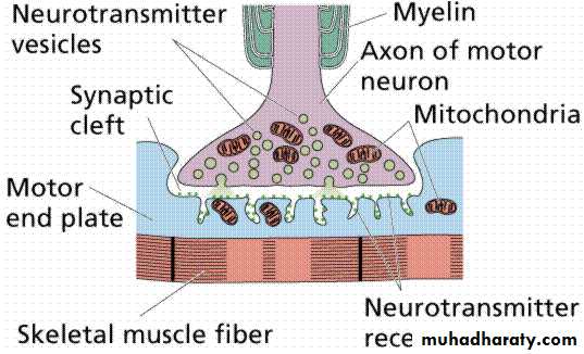
Synapses are specialized cell-to-cell contacts that allow the information encoded by action potentials to pass to another cell.
Synapses occur at the junction between the processes of two neurons or between a neuron and an effectors cell (e.g., a muscle or gland). neuromuscular junction (NMJ); is a contact between a nerve and a muscle.
There are two types of synapses:
1. Electrical synapses; occur where two cells are joined by gap junctions, which conduct current from cell-to-cell via nonselective pores. Cardiac muscle is an example of cells that are electrically coupled via gap junctions.
2. Chemical synapses; involve the release of a chemical transmitter by one cell that acts upon another cell.
Action potentials in a presynaptic cell cause the release of the chemical transmitter, which crosses a narrow cleft to interact with specific receptors on a postsynaptic cell.
Chemical Transmitters Carry the Signal Across Synapses & Neuromuscular Junctions:
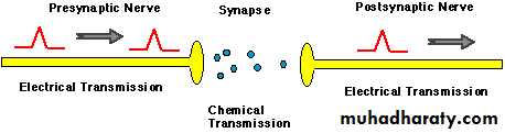
At the synapse there is a break in electrical transmission (the action potential cannot cross), instead chemicals are released that carry the signal to the next nerve.
In neuromuscular junction (NMJ); the action potential stops and the signal is carried by a chemical
There is a delay at synapses, chemical transmission is slower than electrical transmission
Chemical Transmitters Are Made and Stored in the Presynaptic Terminal:
The end of the nerve enlarges into an axon terminal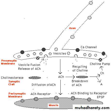
Transmitters are made in the terminal and are stored in tiny vesicles so that they can be released whenever an action potential comes along
Transmitters are made only by the incoming (presynaptic) nerve
Because the transmitter is only on one side the impulse can go in only one direction
Calcium is Required for Transmitter Release:
Transmitter release requires Ca2+ ions
The action potential coming in to the terminal opens Ca channelsCa comes rushing in
Transmitter Diffuses Across the Synaptic Gap and Binds to a Receptor:
The synaptic gap is short and the transmitter travels across it by simple diffusion
When Transmitter Binds to a Receptor it Produces an EPSP or an IPSP:
When the transmitter binds to the receptor ion channels are opened Ions rush into the postsynaptic cellIf the ions depolarize the postsynaptic cell they produce an excitatory postsynaptic potential (EPSP)
Most transmitters produce EPSPs (acetylcholine, epinephrine, norepinephrine)
If the ions make the postsynaptic membrane more negative they produce an inhibitory postsynaptic potential (IPSP)
The major transmitters producing IPSPs are glycine and GABA (gamma amino-butyric acid)
There are both excitatory and inhibitory nerves coming into most synapses
If There Are Enough EPSPs an Action Potential Will be Produced in the Postsynaptic Membrane:
If there are enough EPSPs the postsynaptic membrane will be depolarized to the threshold level and an action potential will be produced, then the signal will travel along the second nerve or muscle cell
IPSPs make the membrane potential more negative and cancel out EPSPs
Integrating Signals:
A single neuron, especially one in the central nervous system, may have thousands of other neurons synapsing on it.
Some of these release activating (depolarizing) neurotransmitters; others release inhibitory (hyperpolarizing) neurotransmitters.
The receiving cell is able to integrate these signals. The diagram shows how this works in a motor neuron.
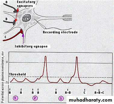
The EPSP created by a single excitatory synapse is insufficient to reach the threshold of the neuron.
EPSPs created in quick series, will add together ("summation"). If they reach threshold, an action potential is generated.
The EPSPs created by separate excitatory synapses (A + B) can also be added together to reach threshold.
Activation of inhibitory synapses (C) makes the resting potential of the neuron more negative. The resulting IPSP may also prevent what would otherwise have been effective EPSPs from triggering an action potential.
Axon hillock( has no synapses and has a lower threshold than elsewhere on the cell), it has the ability to regulate action potential (EPSPs & IPSPs)