THE CENTRAL NERVOUS SYSTEM
The brain is covered by tough meninges and protected by boneThe Meninges:
Both the spinal cord and brain are covered in three continuous sheets of connective tissue, the meninges. From outside in, these are the;
dura mater; pressed against the bony surface of the interior of the vertebrae and the cranium
the arachnoid
the pia mater
The region between the arachnoid and pia mater is filled with cerebrospinal fluid (CSF).
The Brain is Immersed in Cerebrospinal Fluid
The brain is a hollow tube with bulges: has 4 interconnected fluid-filled reservoirs (ventricles) filled with CSF
Total volume ~150 mL
CSF circulates: secreted into ventricles by the choroid plexus & absorbed into veins draining the brain.
Some CSF passes into central canal of spinal cord
Protection: acts as a cushion and reduces injury
Brain floats in CSF; therefore, pressure at the base of the brain is reduced.
CSF excrete waste products (harmful metabolites, drugs and other substances) away from the brain.
There is a Special Barrier Between the Brain and the Blood
There is a "blood brain barrier"; brain capillaries are tighter and less permeable than those in the rest of the body, protects brain from many chemicals and bacteria
Hydrophobic compounds cross the blood brain barrier more readily than hydrophilic ones
The Extracellular Fluid (ECF) of the Central Nervous System
The cells of the central nervous system are bathed in a fluid that differs from that serving as the ECF of the cells in the rest of the body.
The fluid that leaves the capillaries in the brain contains far less protein than "normal" because of the blood-brain barrier.
Homeostatic mechanisms
For proper function, the brain must maintain and regulate pressure inside the skull (intracranial pressure) as it also maintains the flow of oxygen and nutrients to its tissues. Both of these are accomplished by balancing changes in blood flow and CSF volume.
White Matter vs. Gray Matter
Both the spinal cord and the brain consist of
white matter = bundles of axons each coated with a sheath of myelin
gray matter = masses of the cell bodies and dendrites, each covered with synapses.
In the spinal cord, the white matter is at the surface, the gray matter inside.
The brain receives nerve impulses from
the spinal cord and
12 pairs of cranial nerves
Some of the cranial nerves are "mixed", containing both sensory and motor axons
Some, e.g., the optic and olfactory nerves (numbers I and II) contain sensory axons only
Some, e.g. number III that controls eyeball muscles, contain motor axons only.
THE CENTRAL NERVOUS SYSTEM
The central nervous system (CNS) includes the brain and spinal cord.The brain;
All sensation and consciousness originates in the brain.
The brain consists of many regions.
Brain is 2% of body weight(~1400 gm) but uses 12% of body energy
14% of the blood flow goes to the brain
The blood flow per kilogram is equal to that of a muscle doing heavy exercise
The spinal cord is the primary pathway for messages between peripheral areas of the body and the brain. It also mediates reflexes.
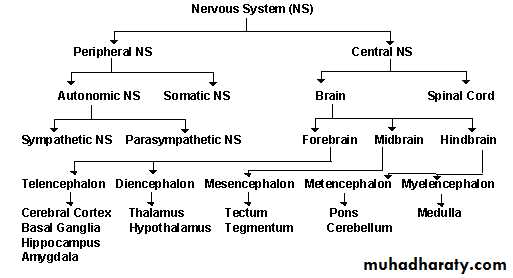
I-The Forebrain
CEREBRUMThe cerebrum is the largest part of the brain, and is the seat of thoughts, perceptions, and voluntary actions. “cortex” is often used interchangeably with cerebrum, but it really refers to the surface layer of gray matter.
The surface area of the brain is increased by its many folds (gyri), which are separated by fissures (sulci).
The left and right cerebral hemispheres are joined by the corpus callosum; amass of nerve fibers that allows communication between corresponding centers in the right and left hemispheres.
Each hemisphere is divided into four lobes, based on anatomic landmarks and functional differences. The lobes are named for the cranial bones that lie over them
frontal lobe
temporal lobe
parietal lobe
occipital lobe
PRIMITIVE STRUCTURES
A group of unpaired structures located deep within the cerebrum, called the diencephalon.The Thalamus relays all sensory stimuli (except olfactory) as they ascend to the cerebral cortex (Sensory processing).
Thalamic functions include primitive awareness of pain, screening of incoming stimuli, and focusing of attention.
The Hypothalamus
Functions:One important function of the hypothalamus is the control of body temperature
Emotions
Hunger
Thirst
Circadian Rhythms, including sleep and wake cycles.
The hypothalamus also controls the pituitary, It's the source of 8 hormones, two of which pass into the posterior lobe of the pituitary gland.
Lateral geniculate nucleus (LGN): All signals entering the brain from the optic nerves enter the LGN and undergo some processing before moving on the various visual areas of the cerebral cortex.
Posterior lobe of the pituitary:
Secretes:
antidiuretic hormone (ADH)
oxytocin
Hidden beneath these regions of cerebral cortex the;
olfactory bulbs; they receive input from the olfactory epithelia.
striatum; it receives input from the frontal lobes and also from the limbic system.
limbic system; It initiates primitive drives (hunger, aggression, and sexual and emotional arousal) The limbic system is made up of the:
hippocampus; It is essential for the formation of long-term memories.
amygdala; The amygdala appears to be a center of emotions (e.g. fear). It sends signals to the hypothalamus and medulla which can activate the fight or flight or response of the autonomic nervous system.
II- The Midbrain (mesencephalon)
Midbrain; occupies only a small region in humans (it is relatively much larger in "lower" vertebrates).
We shall look at only three features: mediates the auditory and visual reflexes:
The Reticular Formation; is involved in hearing, sleep, arousal and vomiting.
Substantia Nigra: helps "smooth" out body movements; damage to the substantia nigra causes Parkinson's disease.
Ventral Tegmental Area (VTA): packed with dopamine-releasing neurons that are activated by nicotinic acetylcholine receptors.
The VTA seems to be involved in pleasure: nicotine, amphetamines and cocaine bind to and activate its dopamine-releasing neurons and this may account for their addictive qualities.
III-The hind brain:
CEREBELLUM
The cerebellum "little brain" also has two hemispheres.
Required for:
posture
balance
smooth, coordinated movements (especially simultaneous movements of different body parts)
The cerebellum appears to be a center for learning motor skills
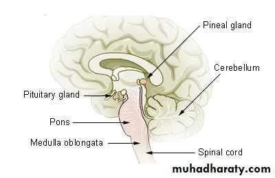
Pons ;
Connects the cerebellum with the cerebrum and the midbrain to the medulla oblongata, and contains one of the respiratory centers, involved in control of breathing.Medulla Oblongata ;
The medulla looks like a swollen tip to the spinal cord.Nerve impulses arising here;
rhythmically stimulate the intercostal muscles and diaphragm, making breathing possible
regulate heartbeat
regulate the diameter of arterioles thus adjusting blood flow.
BRAIN STEM
Composed of the pons, midbrain, and medulla oblongata.
Brain stem relays messages between upper and lower levels of the nervous system.
The cranial nerves originate from the brain stem.
RETICULAR ACTIVATING SYSTEM (RAS)
The RAS, a diffuse network of hyperexcitable neurons fanning out from the brain stem through the cerebral cortex, screens all incoming sensory information and channels it to appropriate areas of the brain for interpretation. RAS activity also stimulates wakefulness.THE SPINAL CORD
The spinal cord joins the brain stem at the level of the foramen magnum and terminates near the second lumbar vertebra.The function of the spinal cord as the major channel of sensory and motor information between the brain and the periphery.
The spinal cord is connected to the periphery via spinal nerves, which are part of the peripheral nervous system (PNS).
Each spinal nerve attaches to the spinal cord by two branches, a dorsal root and a ventral root.
A cross section of the spinal cord reveals;
Gray matter; a central H-shaped mass divided into dorsal (posterior) and ventral (anterior) horns. Gray matter in the dorsal horns relays sensory (afferent) impulses; in the ventral horns, motor (efferent) impulses.
White matter; (myelinated axons of sensory and motor nerves) surrounds these horns and forms the ascending and descending tracts.
THE PERIPHERAL NERVOUS SYSTEM (PNS)
The peripheral nervous system consists of: the cranial nerves (CN), the spinal nerves, and the autonomic nervous system.
CRANIAL NERVES
The 12 pairs of cranial nerves transmit motor or sensory messages, or both, primarily between the brain or brain stem and the head and neck.All cranial nerves, except for the olfactory and optic nerves, originate from the midbrain, pons, or medulla oblongata.
The cranial nerves are sensory, motor, or mixed (both sensory and motor) as follows:
olfactory (CN I) — Sensory: smell
optic (CN II) — Sensory: vision
oculomotor (CN III) — Motor: extraocular eye movement (superior, medial, and inferior lateral), papillary constriction, and upper eyelid elevation
trochlear (CN IV) — Motor: extraocular eye movement (inferior medial)
trigeminal (CN V) — Sensory: transmitting stimuli from face and head, corneal reflex; Motor: chewing, biting, and lateral jaw movements
abducens (CN VI) — Motor: extraocular eye movement (lateral)
facial (CN VII) — Sensory: taste receptors (anterior two-thirds of tongue); Motor: Facial muscle movement, including muscles of expression (those in the forehead and around the eyes and mouth)
acoustic (CN VIII) — Sensory: hearing, sense of balance
glossopharyngeal (CN IX) — Motor: swallowing movements; Sensory: sensations of throat; taste receptors (posterior one-third of tongue)
vagus (CN X). Motor — movement of palate, swallowing, gag reflex; activity of the thoracic and abdominal viscera, such as heart rate and peristalsis.
Sensory: sensations of throat, larynx, and thoracic and abdominal viscera (heart, lungs, bronchi, and GI tract)
spinal accessory (CN XI) — Motor: shoulder movement, head rotation
hypoglossal (CN XII) — Motor: tongue movement.
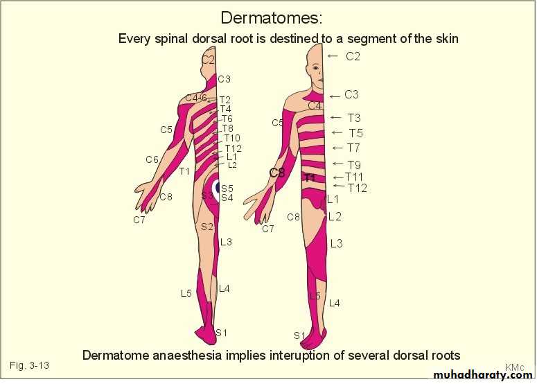
SPINAL NERVES
The 31 pairs of spinal nerves are named according to the vertebra immediately below their exit point from the spinal cord.Each spinal nerve contains of; afferent (sensory) and efferent (motor) neurons, which carry messages to and from particular body regions, called dermatomes.
Peripheral Nervous System:
Divided to :1. Somatic nervous system (voluntary)
a. tissues innervated: skeletal muscle
b. action: always excitatory (cause muscle contraction)
c. CNS control: voluntary (but has involuntary components; e.g. reflexes)
d. no peripheral efferent ganglia or synapses
e. effect of denervation: complete loss of function, paralysis also atrophy
2. Autonomic nervous system (ANS, involuntary)
a. tissues innervated: cardiac muscle, smooth muscle (e.g. viscera, blood vessels), glands (most, but not all)b. action: may be excitatory or inhibitory
c. CNS control: involuntary in general
d. peripheral efferent ganglion and synapse
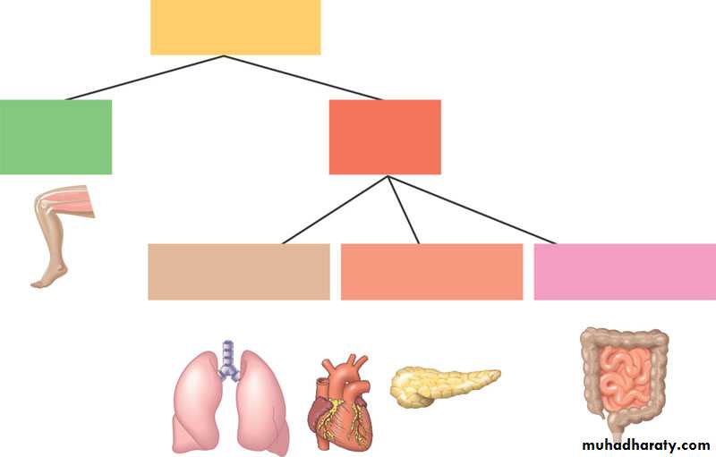
Peripheralnervous systemSomaticnervoussystemAutonomicnervoussystemSympatheticdivisionParasympatheticdivisionEntericdivisione. effect of denervation: generally continues to function, still subject to local and circulating influences, but loss of CNS coordination
f. divisions;
1) sympathetic nervous system
2) parasympathetic nervous system
3. Enteric: innervation contained within the wall of the alimentary tract (sometimes considered as a part of the autonomic system)
AUTONOMIC NERVOUS SYSTEM
The autonomic nervous system (ANS) innervates all internal organs. Sometimes known as the visceral efferent nerves, autonomic nerves carry messages to the viscera from the brain stem and neuroendocrine system.The ANS has two major divisions:
The sympathetic (thoracolumbar) nervous system
The parasympathetic (craniosacral) nervous system.
Sympathetic nervous system
Sympathetic nerves exit the spinal cord between the levels of the 1st thoracic and 2nd lumbar vertebrae; hence the name thoracolumbar.These preganglionic neurons enter small relay stations (ganglia) near the cord.
The ganglia form a chain that transmit the impulse to postganglionic neurons, which reach many organs and glands, and can produce widespread, generalized responses.
The physiologic effects of sympathetic activity include:
pupillary dilation and ciliary muscle relaxationblood vessels vasoconstriction
increased heart rate and contractility
elevated blood pressure
enhanced blood flow to skeletal muscles
heightened respiratory rate
smooth muscle relaxation of the bronchioles
GI tract, and urinary tract sphincter contraction
reduced pancreatic secretion.
increased sweat gland secretion
Parasympathetic nervous system
The fibers of the parasympathetic, or craniosacral, nervous system leave the CNS by way of the cranial nerves from the midbrain and medulla and with the spinal nerves between the 2nd and 4th sacral vertebrae.
After leaving the CNS, the long preganglionic fiber of each parasympathetic nerve travels to a ganglion near a particular organ or gland, and the short postganglionic fiber enters the organ or gland.
Parasympathetic nerves have a specific response involving only one organ or gland.
The physiologic effects of parasympathetic system activity include:
pupillary constrictionincreased pancreatic, salivary, and lacrimal secretions.
reduced heart rate, contractility, and conduction velocity
bronchial smooth muscle constriction
increased GI tract tone and peristalsis with sphincter relaxation
urinary system sphincter relaxation and increased bladder tone
vasodilation of external genitalia, causing erectionThe parasympathetic system has little effect on mental or metabolic activity.
AUTONOMIC TRANSMITTERS
A. Parasympathetic1. preganglionic-postganglionic synapse: acetylcholine (ACh)
2. neuroeffector synapse: acetylcholine (ACh)
B. Sympathetic
1. preganglionic-postganglionic synapse: acetylcholine (ACh)
2.neuroeffector synapse: norepinephrine (NE, noradrenalin)
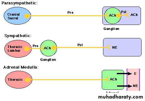
Note exceptions (among several):neurotransmitter released at sweat glands and piloerector muscles is ACh, even though innervation is exclusively sympathetic
C. Adrenal Medulla (endocrine gland, part of sympathetic system);
1. preganglionic-neuroeffector synapse: acetylcholine2. secretion (into circulation): epinephrine E (adrenalin) and norepinephrine NE (noradrenalin)
AnatomicalLocation
PreganglionicFibers
PostganglionicFibers
Transmitter(Ganglia)
Transmitter(Organs)
Sympathetic
Thoracic/Lumbar
Short
Long
ACh
NE
Parasympathetic
Cranial/Sacral
Long
Short
ACh
ACh
AUTONOMIC ACTIONS
A. General Principles
1. Sympathetic system: responses associated with vigorous, often emotional, activity (“fight or flight”)
2. Parasympathetic system: homeostatic (“vegetative”) activities to maintain body internal environment (e.g. digestion and absorption)
3. In organs innervated by both the sympathetic and parasympathetic divisions, their effects are usually (but not always) opposite
Note: often both divisions are active and the state of the organ is determined by the balance between the two.
4. Some organs are innervated by only one division (adrenal medulla, sweat gland and kidney innervated by sympathetic division only, also the blood pressure controlled by sympathetic division only)
5. Sympathetic effects are generally augmented by secretion from the adrenal medulla
6.Sympathetic effects are sometimes general throughout the body, parasympathetic effects are mainly localized.
The Sympathetic is the "Fight or Flight" Branch of the ANS
Emergency situations, where the body needs a sudden burst of energy, are handled by the sympathetic system
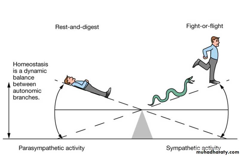
The sympathetic system increases cardiac output and pulmonary ventilation, routes blood to the muscles, raises blood glucose and slows down digestion, kidney filtration and other functions not needed during emergencies
Whole sympathetic system tends to "go off" together
In a controlled environment the sympathetic system is not required for life, but it is essential for any stressful situation
The Parasympathetic is the "Rest and Digest" Branch of the ANS
The parasympathetic system promotes normal maintenance of the body- acquiring building blocks and energy from food and getting rid of the wastes
It promotes secretions and mobility of different parts of the digestive tract.
Also involved in urination, defecation.
Does not "go off" together; activities initiated when appropriate
The vagus nerve (cranial number 10) is the chief parasympathetic nerve
Other cranial parasympathetic nerves are: III (oculomotor), VII (facial) and IX (glossopharyngeal)
Organ
Parasympathetic Response"Rest and Digest"
Sympathetic Response"Fight or Flight"
Eye
Iris constrictsAdjusts for near vision
Iris dilatesAdjusts for far vision
Heart(baro-receptor reflex)
Decreased heart rateCardiac output decreases
Increased rate and strength of contraction Cardiac output increases
Lung Bronchioles
Constriction
Dilation
Liver Glycogen
No effect
Glycogen breakdownBlood glucose increases
Fat Tissue
No effect
Breakdown of fatBlood fatty acids increase
Basal Metabolism
No effect
Increases ~ 2X
Adrenal medulla
No effect

Norepinephrine andepinephrine secreted
Salivary Glands
Saliva production increased
Saliva production reduced
Oral/NasalMucosa
Mucus production increased
Mucus production reduced
Stomach
Increased secretion of HCl & digestive enzymesIncreased motility
Decreased secretionDecreased motility
Intestine
Increased secretion of HCl & digestive enzymesIncreased motility
Decreased secretionDecreased motility
Rectum
Relaxes sphincterContracts wall musclesDefecation promoted
Constricts sphincterRelaxes wall musclesDefecation inhibited
Kidney
Increased urine secretion
Decreased urine secretion
Urinary bladder
Relaxes sphincterDetrusor muscle contractsUrination promoted
Constricts sphincterRelaxes detrusorUrination inhibited
Male Sex Organs
Promotes erection
Promotes ejaculation
B. Sequence of Events
1. Preganglionic cell excited within the CNS
2. Action potiental (AP) travels to ganglion
3 ACh released, exciting postganglionic cell
4. AP travels to nerve endings
5. Transmitter (ACh or NE) released from vesicles in postganglionic ending
6. Transmitter diffuses to end organ (slow compared with neuromuscular transmission); order of seconds
7. Transmitter binds to end organ receptor and induces characteristic effect (e.g. muscle contraction or relaxation, gland secretion or inhibition);
a. cholinergic receptor: binds Ach
b. adrenergic receptor: binds E and/or NE
8. Transmitter is removed from receptor by a combination of the following (note: relatively slow);
a. chemical breakdown (ACh by acetylcholine esterase, NE by COMT or MAO)
b. diffuses away
c. uptake into the postsynaptic cell (to be repackaged into vesicles for later release)
AUTONOMIC RECEPTORS
A. Cholinergic receptorsNormal agonist: acetylcholine
Acetylcholine Receptors; ACh acts on two different types of receptor:
nicotinic receptors; are found at the;
neuromuscular junction of skeletal muscles (only)
post-ganglionic neurons of the parasympathetic nervous system
neurons in the brain(on many, e.g. in the ventral tegmental area).
Nicotine is an agonist , curare is an antagonist (hence its ability to paralyze skeletal muscles)
muscarinic receptors; are found at the;
neuromuscular junctions of cardiac and smooth muscle
glands
post-ganglionic neurons of the sympathetic nervous system.
Muscarine (a toxin produced by certain mushrooms) is an agonist.
Atropine is an antagonist (hence its use in acetylcholinesterase poisoning)
B. Adrenergic receptors
1) Normal agonists: epinephrine and norepinephrine.Norepinephrine is the primary neurotransmitter released by postganglionic sympathetic neurons, with the exception of the cholinergic postganglionic sympathetic neurons serving the sweat glands.
Note: norepinephrine is the same thing as noradrenaline.
The responses of target cells to norepinephrine depend on the specific adrenergic receptor type (i.e., α1, α2, β1, β2, or β3).
The adrenergic receptors are important targets for drugs; it can be agonists or antagonists.
a. alpha-1 (α-1)
1) location: pupil of eye, blood vessel arterioles, veins, alimentary tract sphincters, liver
2) agonist action: excitatory
b. alpha-2 (α-2)
1)location ;Coronary arterioles, skin arterioles, salivary arterioles, intestine & pancreas islet
2) agonist action: excitatory, increases contractility of arterioles. decrease intestine motility & pancrease islet secretions
c. beta-1 (β-1)
1) location: heart
2) agonist action: excitatory, increases heart rate and contractility (force of contraction)
d. beta-2 (β-2)
1) location: lung airways, other places
2) agonist: E, isoproteranol