.
At the end of this lecture the student will be
able to:
Define The Nucleus.
Determine the functions of Nucleus.
Describe the structure of Nucleus.
list the chromosomsal defects.
Recognize to the Nuclear Transport .
Recognize to the Genetics diseases .
Recognize to the clinical features of the
genetic diseases in this lecture.
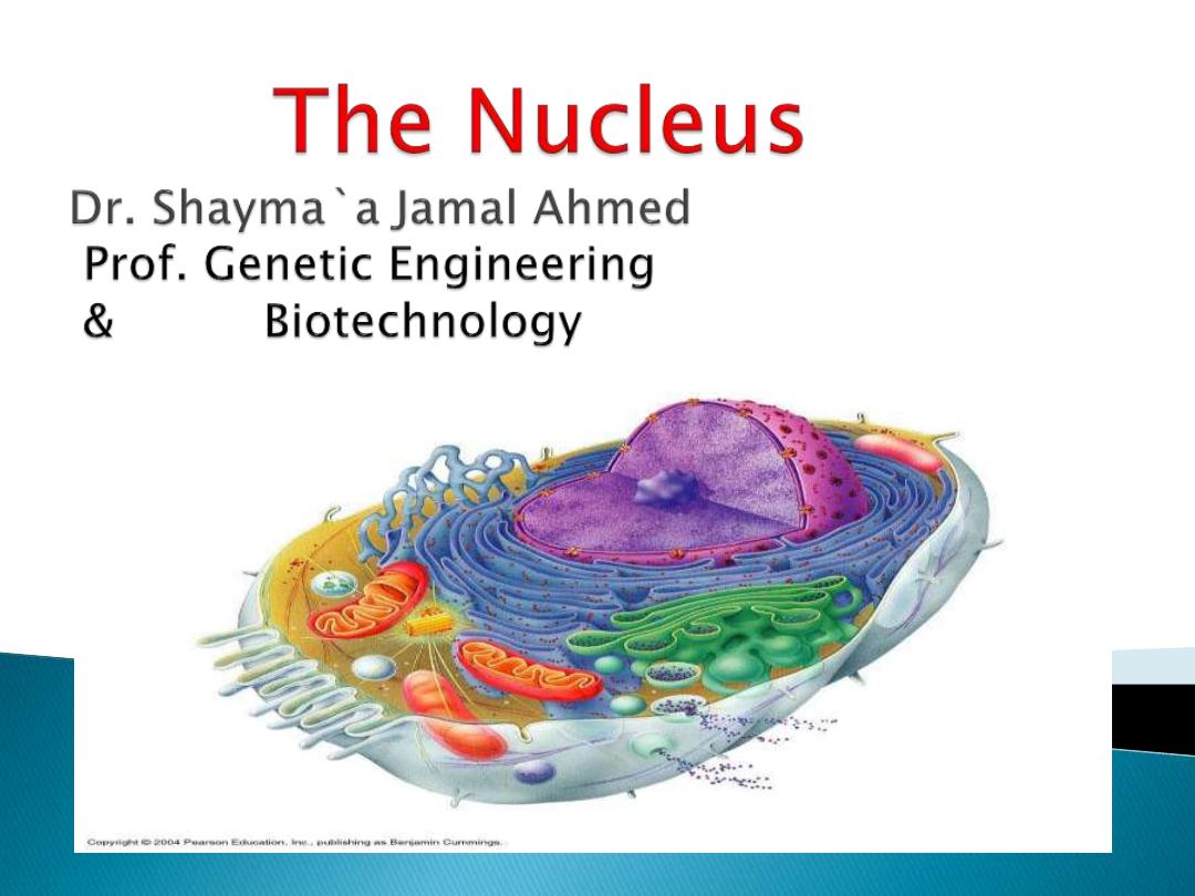
is a highly specialized organelle
that serves as the information
processing and administrative center
of the cell.
A nucleus is the brain of the cell
. It is
the control center where all the
thinking takes place for the cell.
Without a nucleus the cell can not
function.

-
storage and maintenance of the cell’s
genetic information
‐control of gene expression through
transcription
‐synthesis of rRNA and assembly of
ribosomes
1.
Nuclear envelope
2.
Nucleolus
3.
Chromosomes & chromatin
compartment
4.
Other components :
-
nuclear matrix‐protein:
‐
containing febrile network

‐
nucleoplasm:
the fluid substance in which
the solutes of the nucleus are
dissolved
subnuclear particles‐various
discrete specialized structures
within the
nucleoplasm.
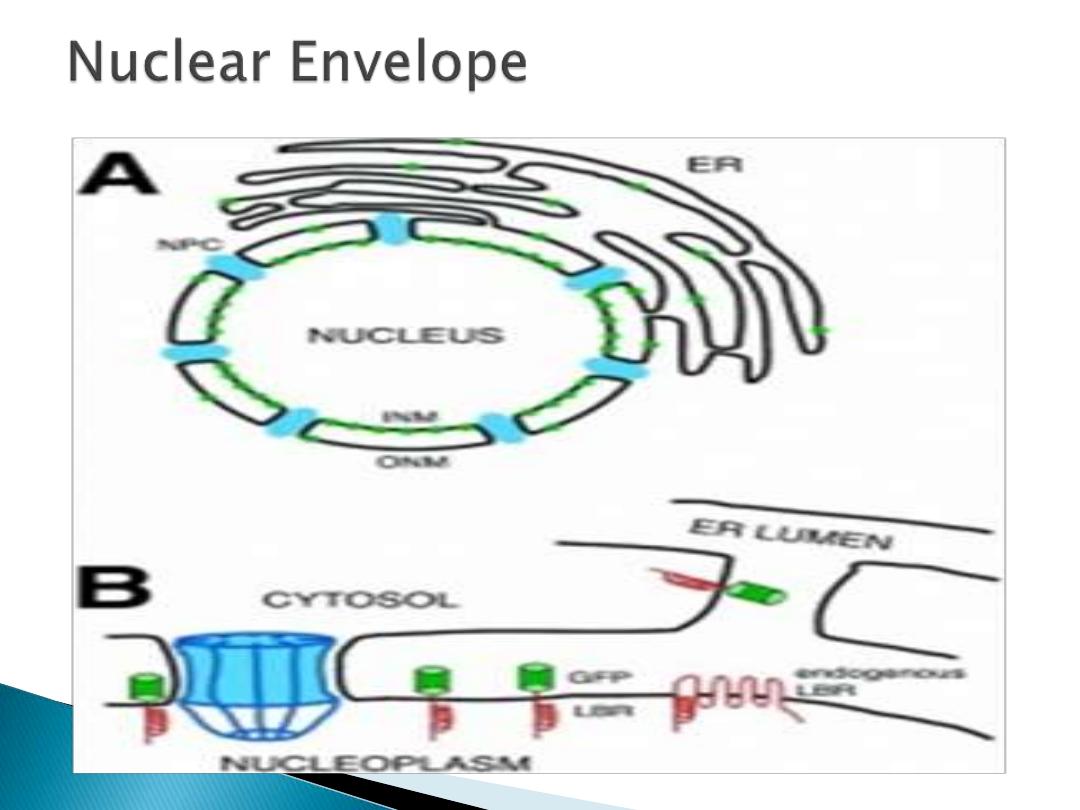
-Nuclear lamina
It is known as
nuclear membrane
, consists of two
cellular membranes , an inner and an outer
membrane, arranged parallel to one another and
separated by 10 to 50 nanometers (nm).
The nuclear envelope completely encloses the
nucleus and separates the cell's genetic material
from the surrounding cytoplasm, serving as a
barrier to prevent macromolecules from diffusing
freely between the nucleoplasm and the
cytoplasm.

The important features are:
1.
Nuclear envelope two
concentric membranes.
2.
Breaks down each mitosis
(recycled)
3.
Outer membrane continuous
with Endoplasmic Reticulum).
4.
Contains holes “
nuclear
pores
”
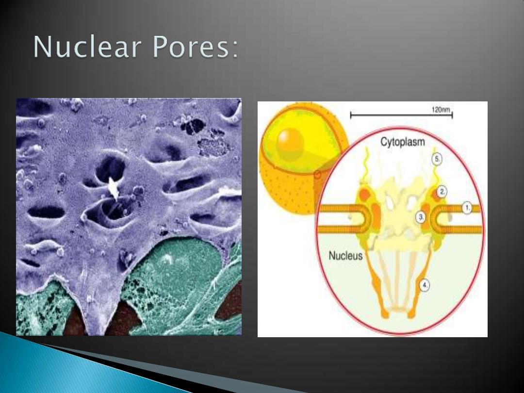
1)
Protein complex
2)
External diameter of about 120 nm (30 times
the size of a ribosome)
3)
Channel diameter 25 nm
4)
channels between nucleus and cytoplasm
(import/export)
5)
passive passage of small polar molecules,
ions,
6)
active (selective/ regulated) passage of
macromolecules, proteins and RNAs
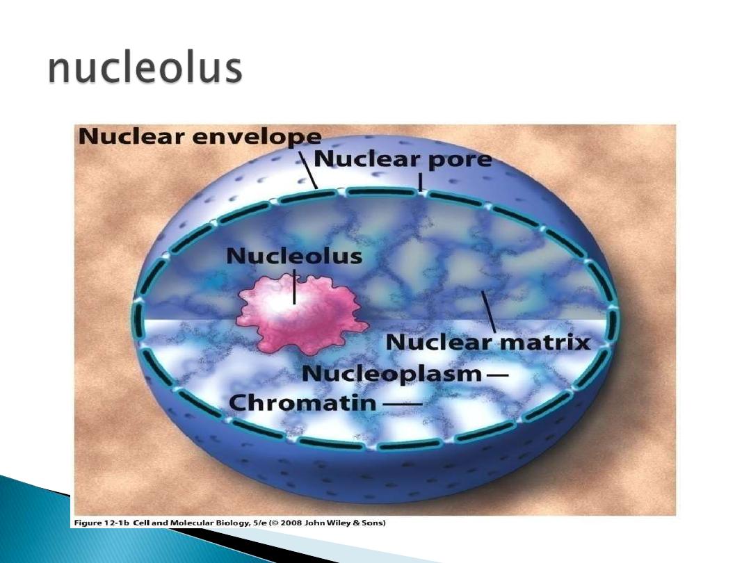
is a dense, spherical-shaped structure present inside
the nucleus. Some of the eukaryotic organisms have
nucleus that contains up to four nucleoli.
The nucleolus plays an indirect role in protein
synthesis by producing ribosomes. These ribosomes
are cell organelles made up of RNA and proteins; they
are transported to the cytoplasm, which are then
attached to the endoplasmic reticulum. Ribosomes
are the protein-producing organelles of a cell.
Nucleolus disappears when a cell undergoes division
and is reformed after the completion of cell division.
1.
The cell nucleus contains the majority of the
cell's genetic material in the form of multiple
linear
DNA
molecules organized into structures
called
chromosomes.
2.
Each human cell contains roughly two meters of
DNA. During most of the cell cycle these are
organized in a DNA-protein complex known as
chromatin
, and during cell division the
chromatin can be seen to form the well-defined
chromosomes familiar from a
karyotype
. A small
fraction of the cell's genes are located instead in
the mitochondria.
1.
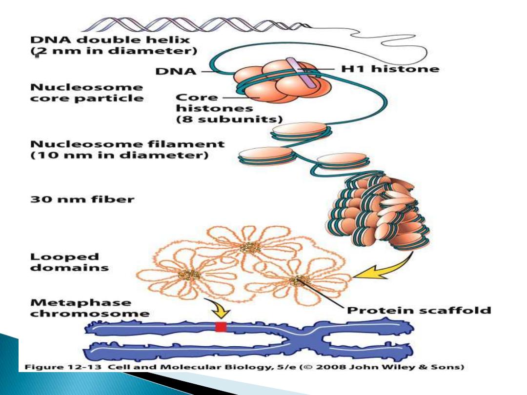
the total length of DNA is ≈ 2m per cell
2.
‐ DNA is combined with histone proteins and
organized into a precise, compact structure ‐a
dense string‐like fibre called chromatin
3.
‐ DNA‐histone units are termed ‘nucleosomes’,
and in the electron microscope, uncondensed
chromatin has a “beads on a string” appearance
4.
‐ nucleosomes are further coiled, compacting
the DNA by a factor of 40
5.
‐ the overall negative charge of the DNA is
neutralised by the positive charge of the
histones.
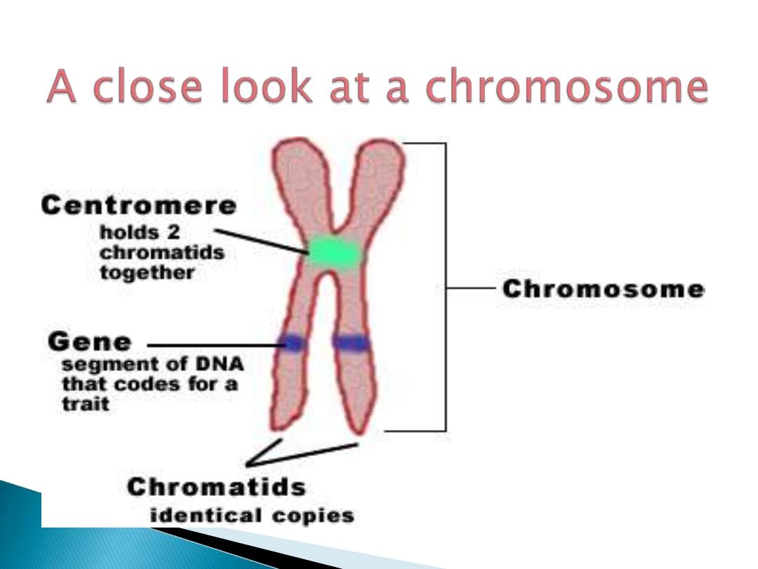
1.
2 telomeres, centromere, replication origins
2.
Telomere- at ends of chromosome (bacterial
DNA circular)
3.
Centromere-
holds duplicated DNA together
4.
Kinetochore
- protein complex forms around
the centromere forms during mitosis
5.
Chromatin
- DNA packed by DNA binding
proteins (histones and non-histones) form
30nm DNA fiber.
6.
2 types of chromatin in interphase nuclei
(based on cytology)
1.
heterochromatin -
highly condensed
(restricted gene transcription)
2.
euchromatin
- less condensed (gene
transcriptio
n)

Any defects occur during cell
division, this will lead to:
The cell may die.
It may develop genetic diseases.
It may escape apoptosis leading
to the progression of cancer.

Chromosomal alterations are two
types:
Numeric :
1-
Aneuploid :
Any number that is not
an exact multiple oh (n).
2-
polyploid:
chromosome numbers
such as 3n &4n (which leads to
spontaneous abortion)

Structural:
structural changes in the
chromosomes result from
chromosomal breakage followed
by loss or rearrangement of
broken part.
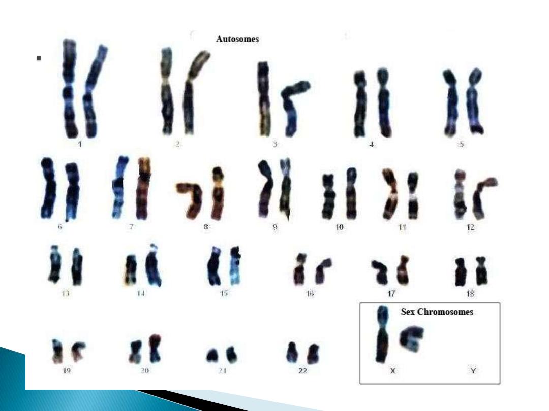
Diagnosis= cytogenetic analysis
By
Chromosome Kayrotyping.
1- translocation:
transfer of a part of one
chromosome to another chromosome,
which are:
I. Balanced reciprocal translocation.
II. Centric fusion translocation
(robertsonian).
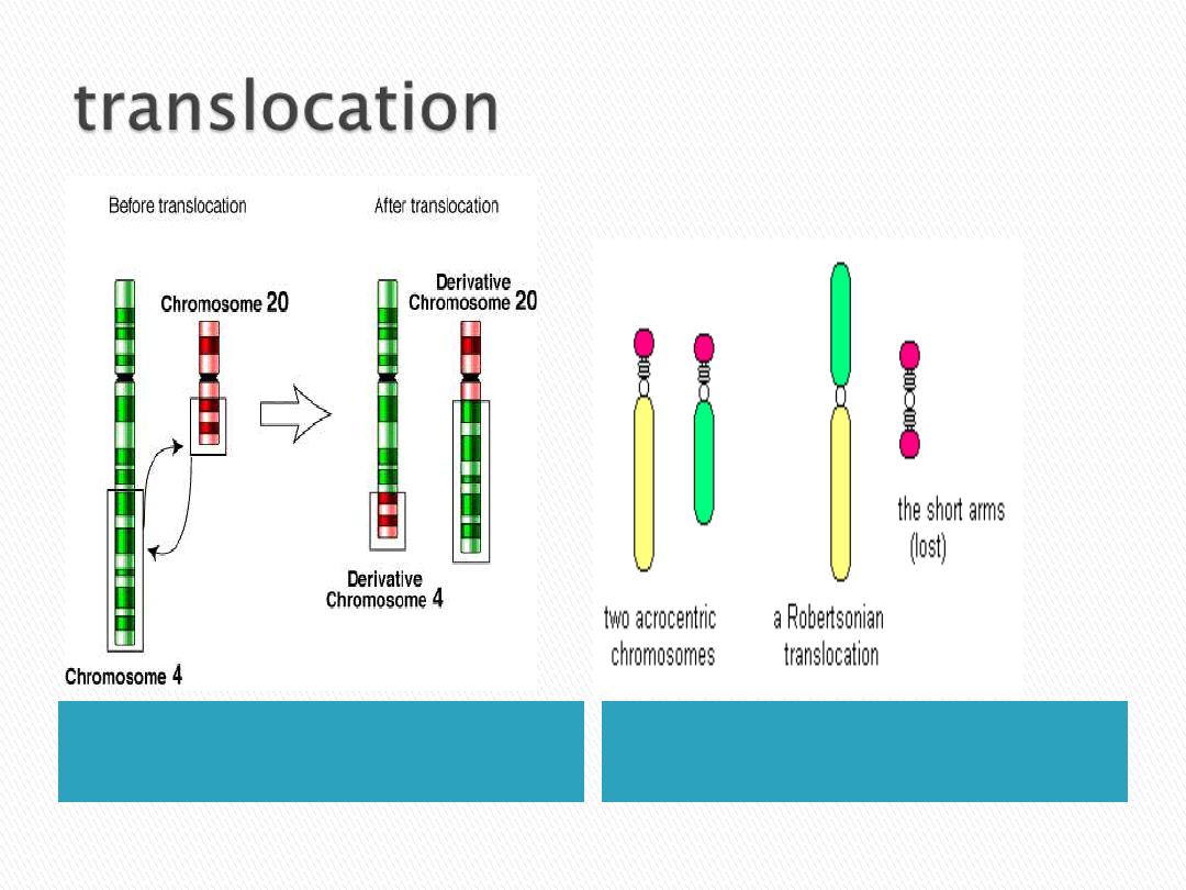
Balance reciprocal
translocation
Centric fusion transloca(
robersonin)

2- isochromosomes :
the centromere divides
horizontally. One or two arms of
the chromosome is then lost, and
the remning arm is duplicated,
resulting in a chromosome with
two short arms only or two long
arms only.
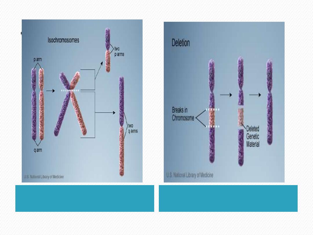
isochromosomes
Deletion

3- Deletion :
loss of a portion of a chromosome
which lead to lost of many genes.
4- inversions:
occur when there are interstitial breaks
in a chromosome, and the segment
reunites after a complete turnaround.
5- Ring chromosome:
It is a variant of a deletion. After loss
of segment from each end of the
chromosome, the unite to form a ring.
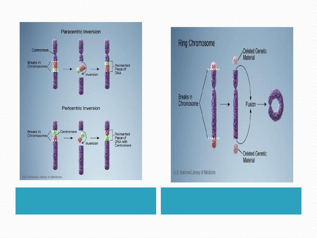
Inversions
Ring chromosome
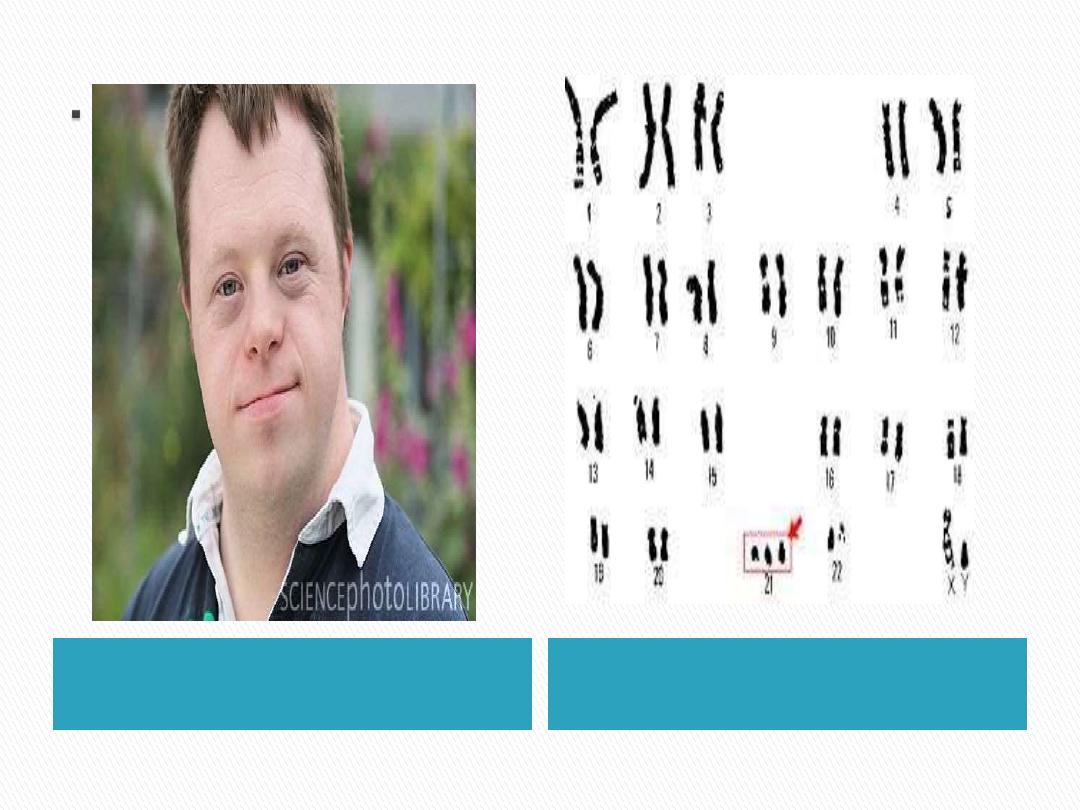
Trisomy 21 (Down Syndrome)
1.
Most common.
2.
Affected persons have trisomy 21(3 chr.
No.21).
3.
Chromosome count is 47.
4.
Cause is meiotic nondisjuntion.
5.
Maternal age has a strong influence on the
incidence of Down Syndrome.
What are the others trisomy?
Phenotype
Genotype
Examples :
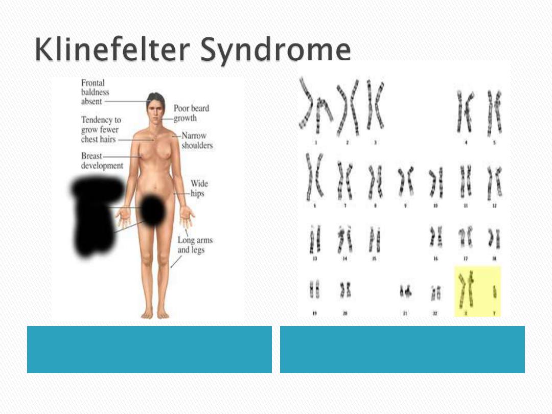
- Klinefelter Syndrome
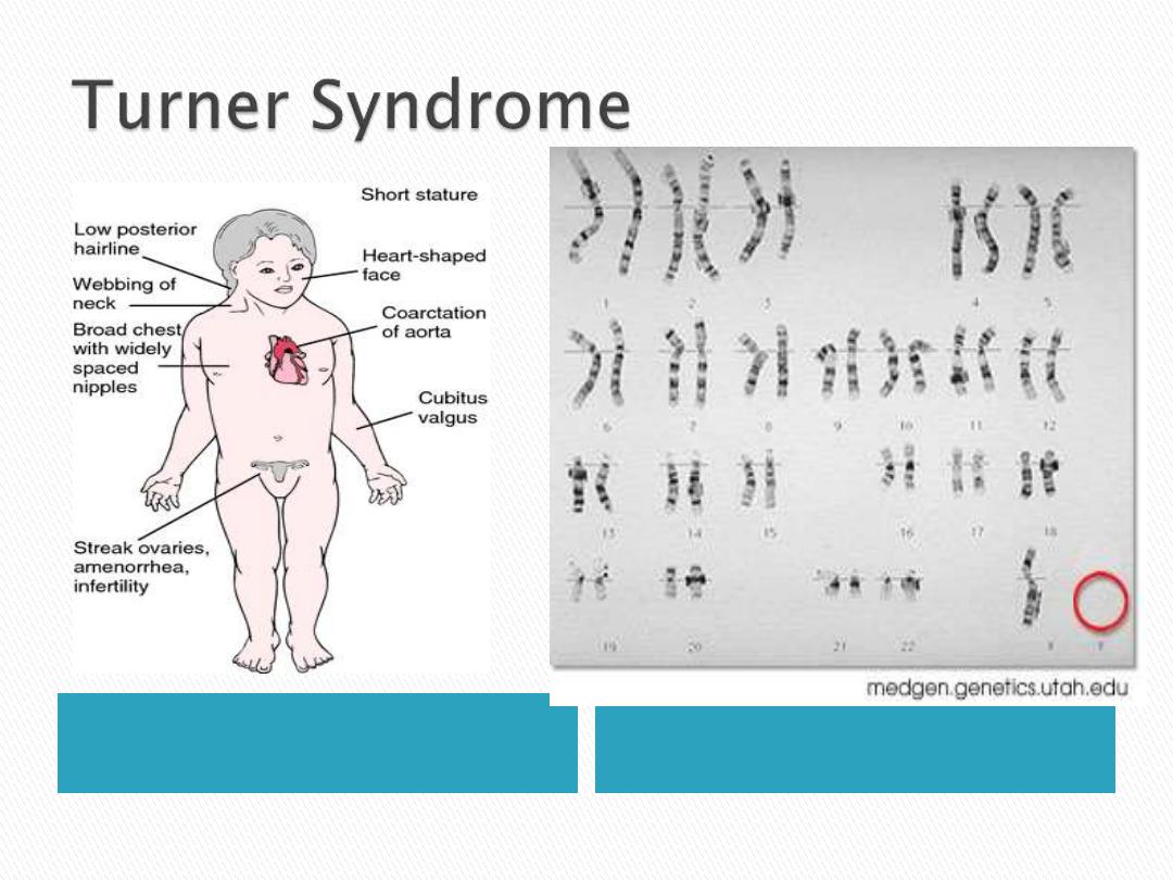
- Turner Syndrome
Compatible with life.
Ranging from 45,x to 49xxxxy
1.
Male hypogonadism.
2.
Most patients are 47,xxy in
karyotype.
3.
Cause nondisjuntion of sex
chromosomes during meiosis.
4.
Advanced maternal age and the
history of irradiation of either may
contribute to the meiotic error.
Phenotype
Genotype
karyotype is 45x.
Cause: Monosomy of x
chromosome.
Hypogonadism in phenotypic
females.
Phenotype
Genotype