Meiosis
Dr. Shayma`a J. Ahmed
Prof. Genetic Engineerin& Biotechnology

Objectives:
At the end of this lecture the student will be able to:
Define the Meiosis.
List the Phases of Meiosis.
Describe the Stages of Meiosis .
Determine the features of Meiosis.
Compare between the mitosis & meiosis.
Describe the Meiosis Sex Differences.
Recognize to the clinical features of the genetic
diseases in this lecture .

Meiosis: making egg and sperm cells
A process called ‘meiosis’ creates new
reproductive cells with half as many
chromosomes as the original cell.
Without meiosis, joining of the egg and
sperm at fertilization would produce
offspring with twice the original number of
chromosomes as it`s parents.

.
starts with one reproductive cell
(containing 46
chromosomes)
that replicates its DNA only once
but divides twice to produce four new cells, each
having
only 23 chromosomes
.
It consists of two nuclear divisions-
Meiosis-I &
Meiosis-II.

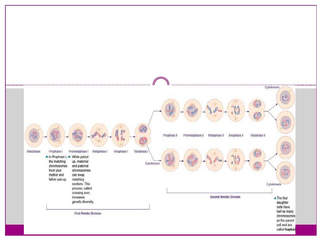
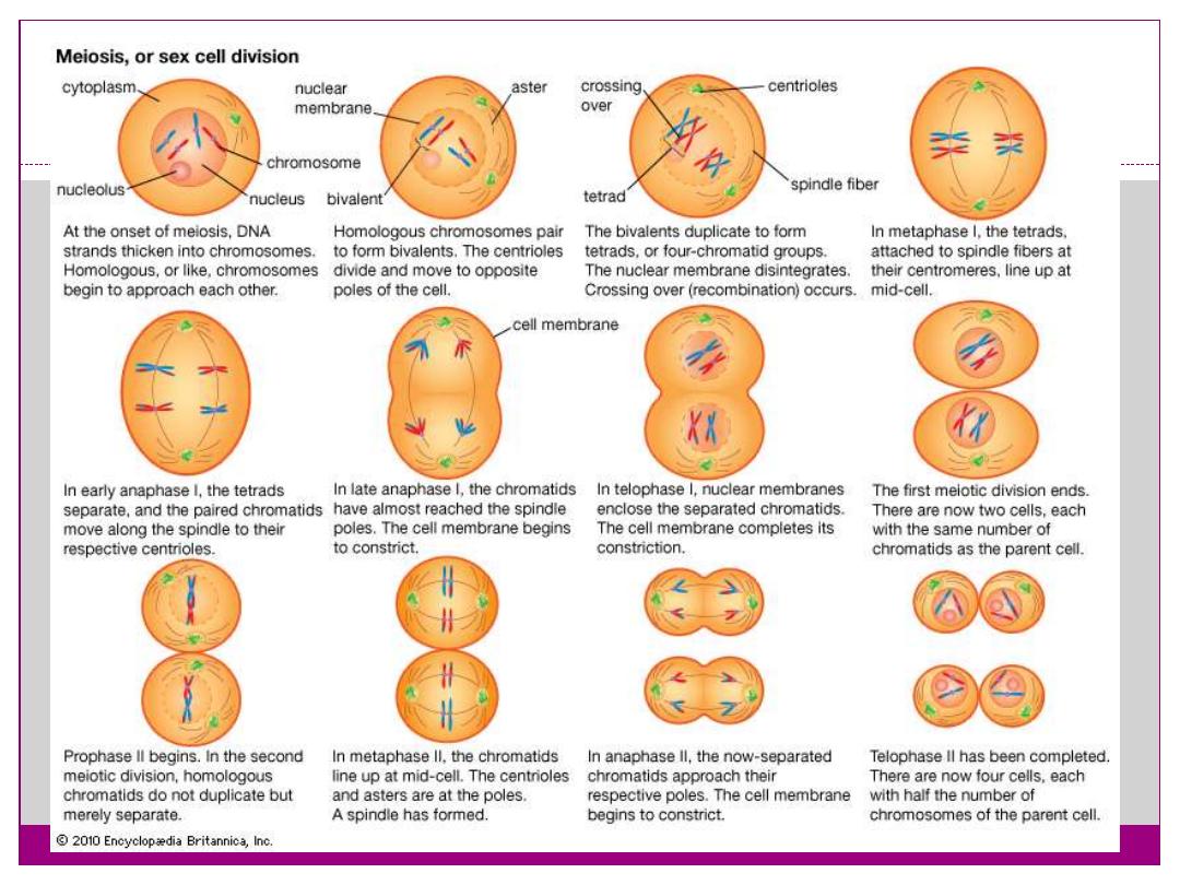
Meiosis-I:
Interphase:
Before meiosis begins, genetic material is
duplicated.
Meiosis- I
has two main purposes:
1- It is the reduction division, so it reduces the
number of chromosomes in half, making the
daughter cells haploid (when the parent cell was
diploid).
2- It is during meiosis I that most of the genetic
recombination occurs.

.
Phases:
Keep in mind that before meiosis begins at all,
the DNA undergoes replication, just like it did
before mitosis started. So, when you first see
chromosomes in meiosis I, they have sister
chromatids, just like in mitosis. It is just that in
meiosis- I, we will be talking about tetrads
becoming visible, lining up, separating, and
decondensing (rather than chromosomes, like in
mitosis).
Finally, cytokinesis occurs, too, any time after
the tetrads have moved out of the equator (just like
in mitosis).

Prophase I:
Prophase I is further divided into five stages (phases):
1- Leptotene:
leptotene phase, leptonema; Greek, leptotene = "thin
threads"
the duplicated paired chromosome homologs condense.
2-Zygotene:
zygotene phase, zygonema, Greek, zygotene = "paired
threads"
homologous chromosomes become closely associated
(synapsis) to form pairs of chromosomes consisting of four
chromatids (tetrads).
the synaptonemal complex begins to form between the two
sets of sister chromatids in each bivalent (the duplicated
chromosome paired with its homologous duplicated
chromosome).

3-Pachytene:
pachytene phase, pachynema; Greek, pachytene =
"thick threads"
crossing over between pairs of homologous
chromosomes to form chiasmata (form between two
nonsister chromatids at points where they have crossed
over)
synaptonemal complex is complete and can be stable
for some time.
Autosomal non-sister chromatids of homologous
chromosomes can now extensively exchange segments
in regions of homology.
Only small regions of non-paired sex chromosomes
interact
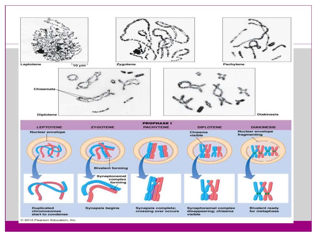
.

4- Diplotene:
diplotene phase, diplonema; Greek, diplonema = "two
threads"
homologous chromosomes begin to separate but
remain attached by chiasmata.
synaptonemal complex degrades and the chromosomes
separate from one another a small amount giving this
appearance.
It is possible that some chromosome uncoiling may
also occur allowing some gene transcription.
In the developing human ovary,
oocytes
remain at the diplotene
stage from fetal life through postnatal childhood, until puberty
when the
lutenizing hormone
(LH) surges stimulate the
resumption of meiosis.

5-Diakinesi:
diakinesis phase; Greek, diakinesis = "moving
through"
homologous chromosomes continue to separate, and
chiasmata move to the ends of the chromosomes.
prophase I ends and chromosomes now recondense,
transcription stops and the transition to metaphase
occurs.

:
Metaphase I
Homologous pairs of chromosomes (bivalents)
arranged as a double row along the metaphase plate.
The arrangement of the paired chromosomes with
respect to the poles of the spindle apparatus is random
along the metaphase plate. (This is a source of genetic
variation through random assortment, as the paternal
and maternal chromosomes in a homologous pair are
similar but not identical.
The number of possible arrangements is 2n, where n is
the number of chromosomes in a haploid set. Human
beings have 23 different chromosomes, so the number
of possible combinations is 223, which is over 8
million.)

.
Anaphase I:
The homologous chromosomes in each bivalent
are separated and move to the opposite poles of the
cell.
Telophase I:
The chromosomes become diffuse and the
nuclear membrane reforms.
Cytokinesis I:
Cellular cytoplasmic division to form two new
cells, followed by Meiosis II.
Note - in oocyte meiosis, the extrusion of the
first polar body (1 PB) indicates completion of the
first meiotic division.
.

Meiosis II:
Prophase II:
Chromosomes begin to condense, nuclear membrane
breaks down and spindle forms.
Metaphase II:
Spindle fibres attach to chromosomes, chromosomes
align in cell centre.
Anaphase II:
Chromosomes separate and move to the opposite
poles of the cell.
Telophase II:
Chromosomes reach spindle pole ends and the
nuclear membrane reforms.
Cytokinesis:
Cellular cytoplasmic division to form new cells.
Compare between the mitosis & meiosis:
Meiosis
Mitosis
1- two division
1- one division.
2- Four daughter cells per cycle.
2- two daughter cells per cycle.
3-Daughter cells genetically are
different.
3- Daughter cells genetically are
identical.
4-4-Chromosome number of daughter
cells is half of parent cell(n).
4-Chromosome number of daughter
cells same as that of parent cells(2n).
5-Occurs in germline cell.
5-Occurs in somatic cells.
6- In human, completes after sexual
maturity.
6-Occurs through out life cycle.
7-Used for sexual reproduction,
producing new gene combinations.
7-Used for growth ,repair & a sexual
reproduction.

:
Meiosis Sex Differences
Female (oogenesis):
Meiosis initiated once in a finite population of cells
1 gamete produced / meiosis
Completion of meiosis delayed for months or years
Meiosis arrested at 1st meiotic prophase and
reinitiated in a smaller population of cells
Differentiation of gamete occurs while diploid in
first meiotic prophase
All chromosomes exhibit equivalent transcription
and recombination during meiotic prophase

.
Male (spermatogenesis):
Meiosis initiated continuously in a mitotically
dividing stem cell population
4 gametes produced / meiosis
Meiosis completed in days or weeks
Meiosis and differentiation proceed continuously
without cell cycle arrest
Differentiation of gamete occurs while haploid
after meiosis ends.
Sex chromosomes excluded from recombination
and transcription during first meiotic prophase.

:
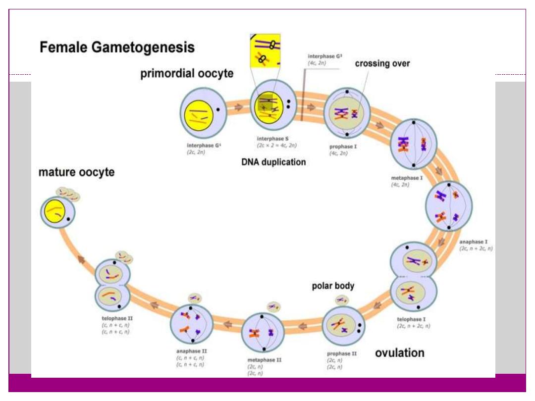
Female Gametogenesis
In females, the total number of eggs ever to be produced are
present in the newborn female.
All eggs are arrested at an early stage of the first meiotic
division as a primary oocyte (primordial follicle). Following
purberty, during each menstrual cycle, pituitary
gonadotrophin stimulates completion of meiosis 1the day
before ovulation.
In meiosis 1, a diploid cell becomes 2 haploid (23
chromosomes) daughter cells, each chromosome has two
chromatids. One cell becomes the secondary oocyte the
other cell forms the first polar body.
The secondary oocyte then commences meiosis 2 which
arrests at metaphase and will not continue without
fertilization.
At fertilization meiosis 2 completes, forming a second
polar body. Note that the first polar body may also undergo
this process forming a third polar body.
.

:
Polar Body
Human oocyte at metaphase II showing polar body at 12
o'clock position.
The breakdown of the germinal vesicle indicates a
resumption of meiosis and the extrusion of the first polar
body (1 PB) indicates completion of the first meiotic
division in human oocytes. The polar body is a small
cytoplasmic exclusion body formed to enclose the excess
DNA formed during the oocyte (egg) meiosis and following
sperm fertilization.
There are 2-3 polar bodies derived from the oocyte present
in the zygote, the number is dependent upon whether polar
body 1 (the first polar body formed during meiosis 1) divides
during meiosis 2. This exclusion body contains the excess
DNA from the reductive division (the second and third polar
bodies are formed from meiosis 2 at fertilization). These
polar bodies do not contribute to the future genetic
complement of the zygote, embryo or fetus.

.
Recent research in some species suggest that the
space formed by the peripheral polar body (between
the oocyte and the zona pellucia) can influence the
site of spermatozoa fertilization.
Assisted reproductive techniques involving
intracytoplasmic sperm injection (ICSI) have looked
at the "quality" of the polar body and found that the
morphology is related to mature oocyte viability and
has the potential to predict oocyte fertilization rates
and pregnancy achievement.

:
Female Abnormalities
Trisomy 21 female karyotype
Meiotic non-disjunction resulting in aneuploidy,
most are embryonic lethal and not seen. The
potential for genetic abnormalities increase with
maternal age.
Autosomal chromosome aneuploidy
trisomy 21 - Down syndrome
trisomy 18 - Edwards syndrome
trisomy 13 - Patau syndrome
Sex chromosome aneuploidy
monosomy X - Turner's Syndrome
trisomy X - Triple-X syndrome
47 XXY - Klinefelter's Syndrome
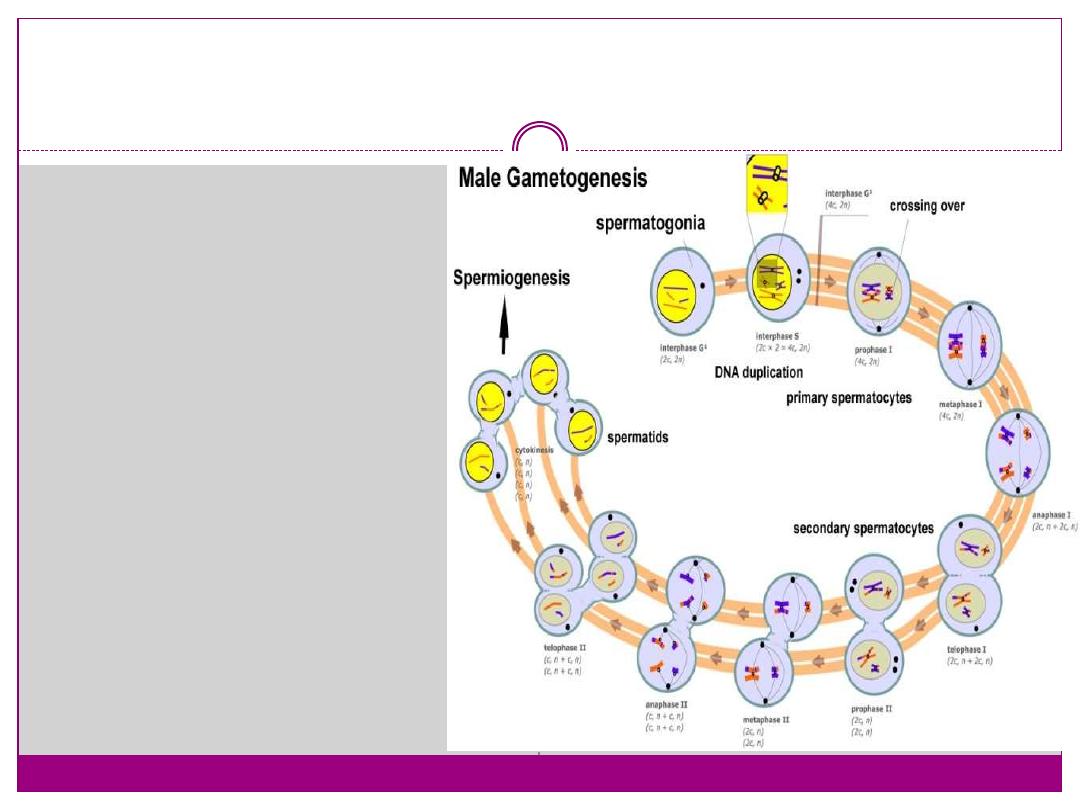
Male Gametogenesis:
In males, sperm
continues to be
generated
throughout life
from a stem cell
population in the
testis.
Spermatozoa
maturation involves
two processes
meiosis
and
spermiogenesis
Human Spermatozoa Development:
Spermatogenesis process
of spermatagonia mature
into spermatazoa
(sperm).
Continuously throughout
life occurs in the
seminiferous tubules in
the male gonad- testis
(plural testes).
At puberty
spermatagonia activate
and proliferate (mitosis).

.
about 48 days from entering meiosis until
morphologically mature spermatozoa
about 64 days to complete spermatogenesis,
depending reproduction time of spermatogonia
follicle stimulating hormone (FSH) - stimulates the
spermatogenic epithelium
luteinizing-hormone (LH) - stimulates testosterone
production by Leydig cells.
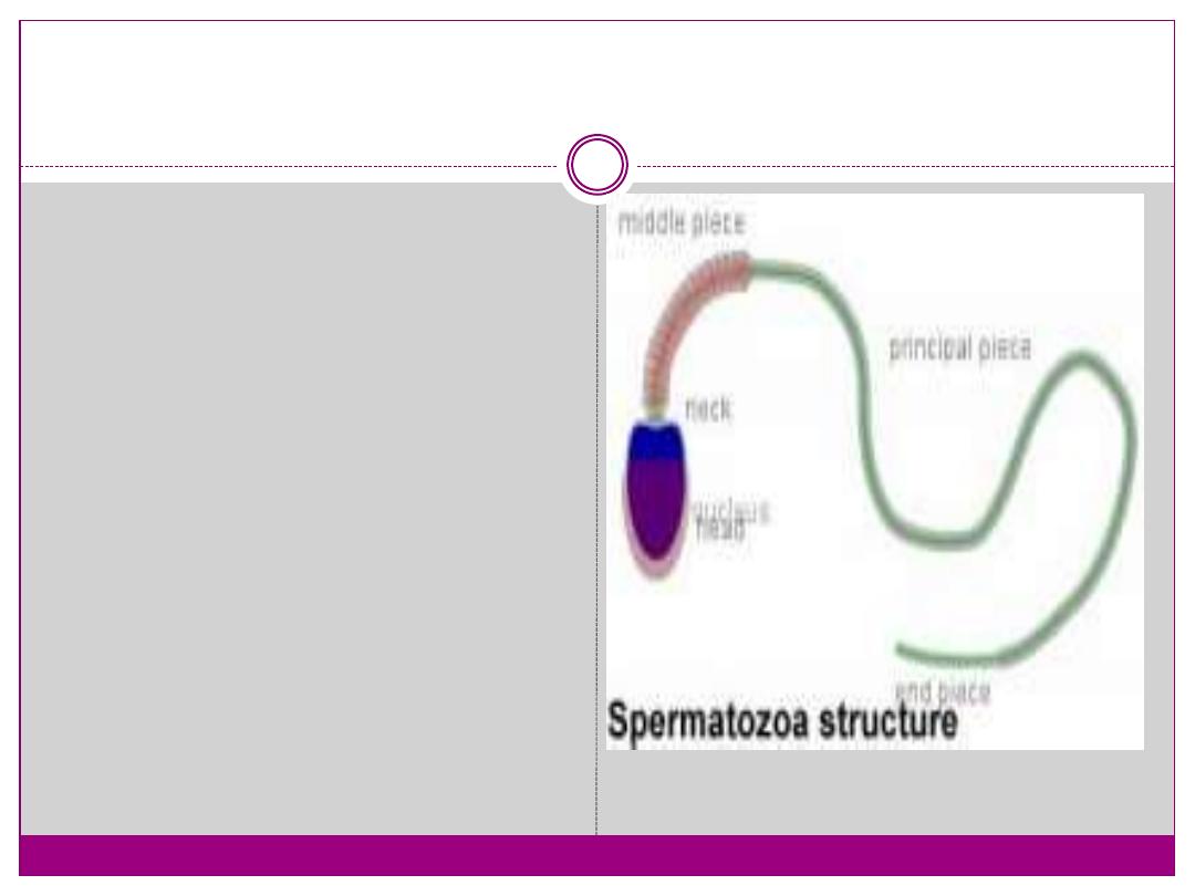
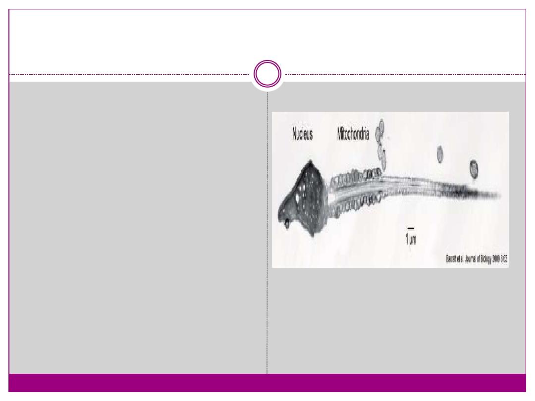
Mature human spermatozoa:
Mature human
spermatozoa60 µm
long, actively motile
divided into 3 main
regions (head, neck and
tail)
head - (flattened, 5 µm
long by 3 µm wide) the
nucleus and acrosome.
Posterior part of nuclear
membrane forms the
basal plate.
.
neck - (1 µm) attached to basal plate, transverse
oriented centriole, contains nine segmented columns
of fibrous material, continue as outer dense fibres in
tail.
tail - 3 parts a middle piece, principal piece and end
piece
middle piece - (5 µm long)
surrounded by mitochondria
principal piece - (45 µm long) fibrous sheath interconnected by
regularly spaced circumferential hoops
surrounded by small amount
of cytoplasm and plasma membrane

:
Male Abnormalities
Oligospermia - (Low Sperm Count) less than 20
million sperm after 72 hour abstinence from sex.
Azoospermia - (Absent Sperm) blockage of duct
network.
Immotile Cilia Syndrome - lack of sperm
motility.

.
Thank you