


Objectives
At the end of this lecture, the 1
st
medical student
should be able to
•
Identify the different stages of gametogenesis in
males and females
•
Outline the stages of spermiogenesis

In preparation for fertilization,
Germ cells undergo
•
Gametogenesis ( include meiosis)
•
Cytodifferentiation to complete their
differentiation

Gametogenesis
•
Is the process of formation of gametes from
germ cells in the testes and ovaries
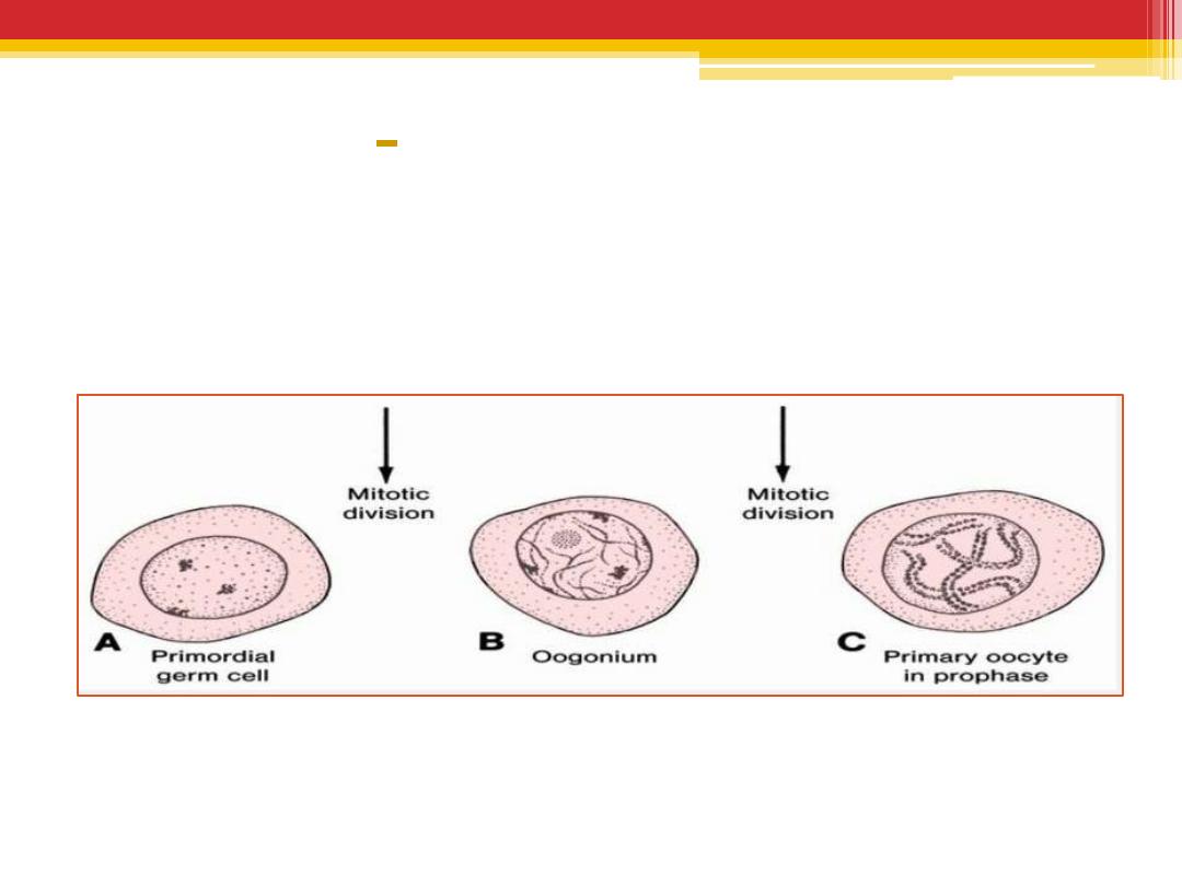
Oogenesis
Is the process where by oogonia differentiate
into mature oocytes
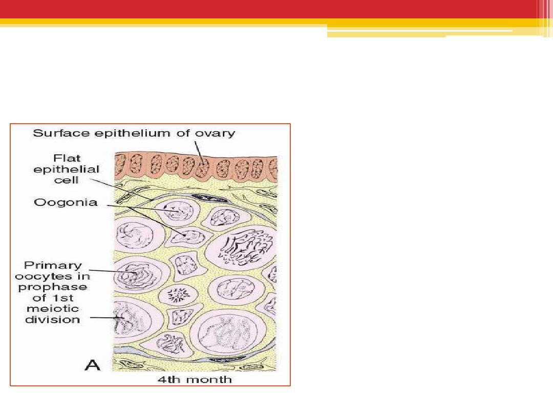
Oogonia
•
By the end of the
3
rd
month, the
majority of oogonia
continue to divide
by mitosis, but some
of them give rise to
primary oocytes that
enter prophase of
the first meiotic
division.
By
the 5
th
month of
prenatal development
,
germ cells in the ovary
reaches its maximum( 7
million). cell death begins
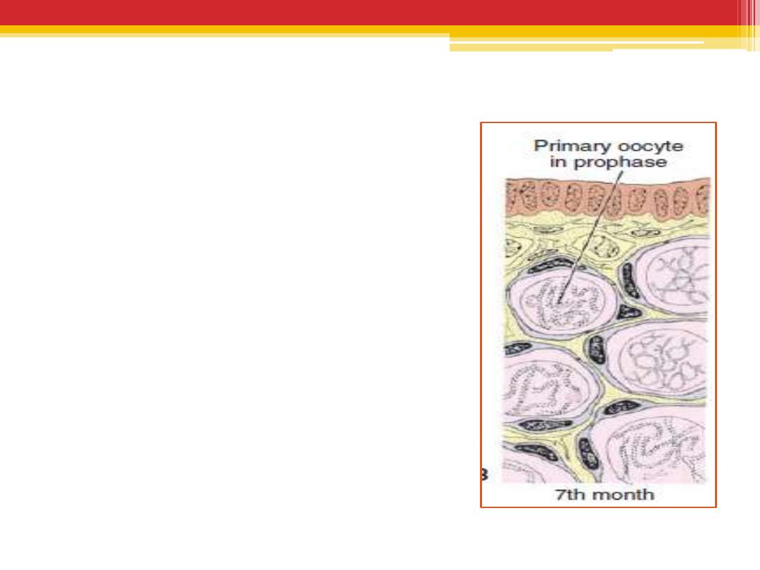
By the 7
th
month
,
•
the majority of oogonia have
degenerated,
•
All surviving primary
oocytes have entered
prophase of meiosis I, and
most of them are individually
surrounded by a layer of flat
epithelial cells
(primordial follicle ).
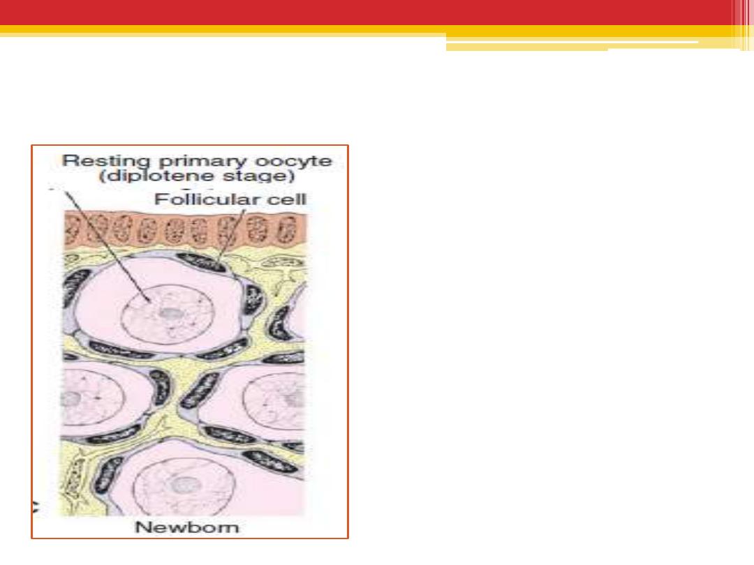
Maturation of the oocytes
At birth and during childhood
Near the time of birth,
all
primary oocytes
have started
prophase of meiosis I, but instead
of proceeding into metaphase,
they enter
the diplotene stage
,
a resting stage during prophase.
The total number of primary
oocytes at birth is estimated to
vary from 700,000 to two million
remain.
During childhood, most oocytes
become atretic
only approximately
400,000
are present by the beginning of
puberty, and fewer than 500 will
be ovulated.

At puberty
•
Each month, 15 to 20 follicles begin to mature
and passing through three stages:
(1) primary,
(2) secondary or antral, and
(3) Tertiary or mature vesicular (Graafian)
follicle.
•
Under normal conditions, only one of these
follicles reaches full maturity, and the others
degenerate and become atretic.
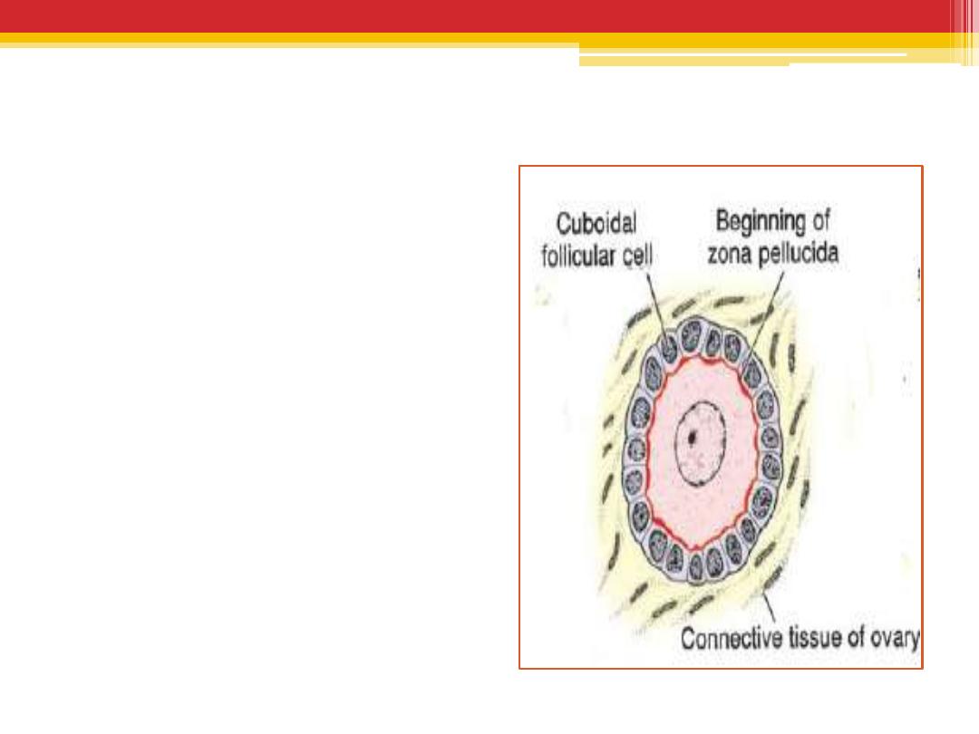
Unilaminar primary follicle
•
Primary oocyte
surrounded by a layer
of cuboidal epithelium
•
Beginning of zona
pellucida
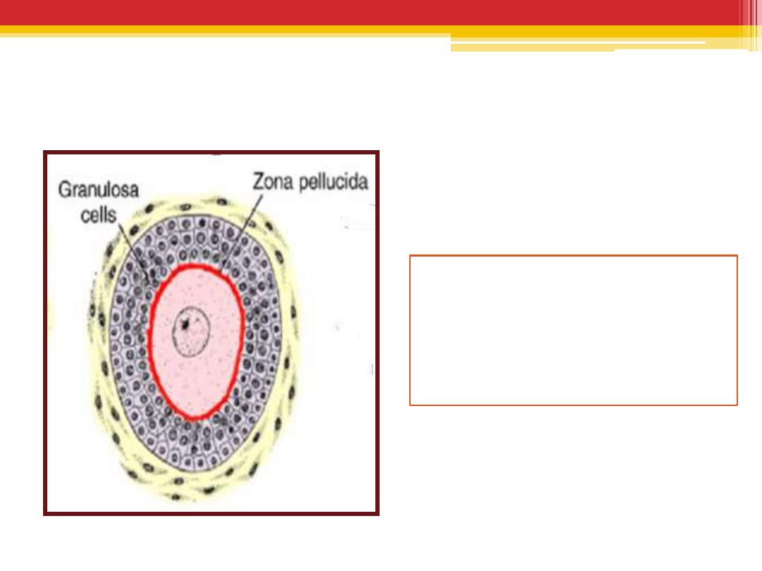
Multilaminar primary follicle
•
Follicular cells proliferate and
produce a stratified
epithelium of granulosa cells
•
Zona pellucida
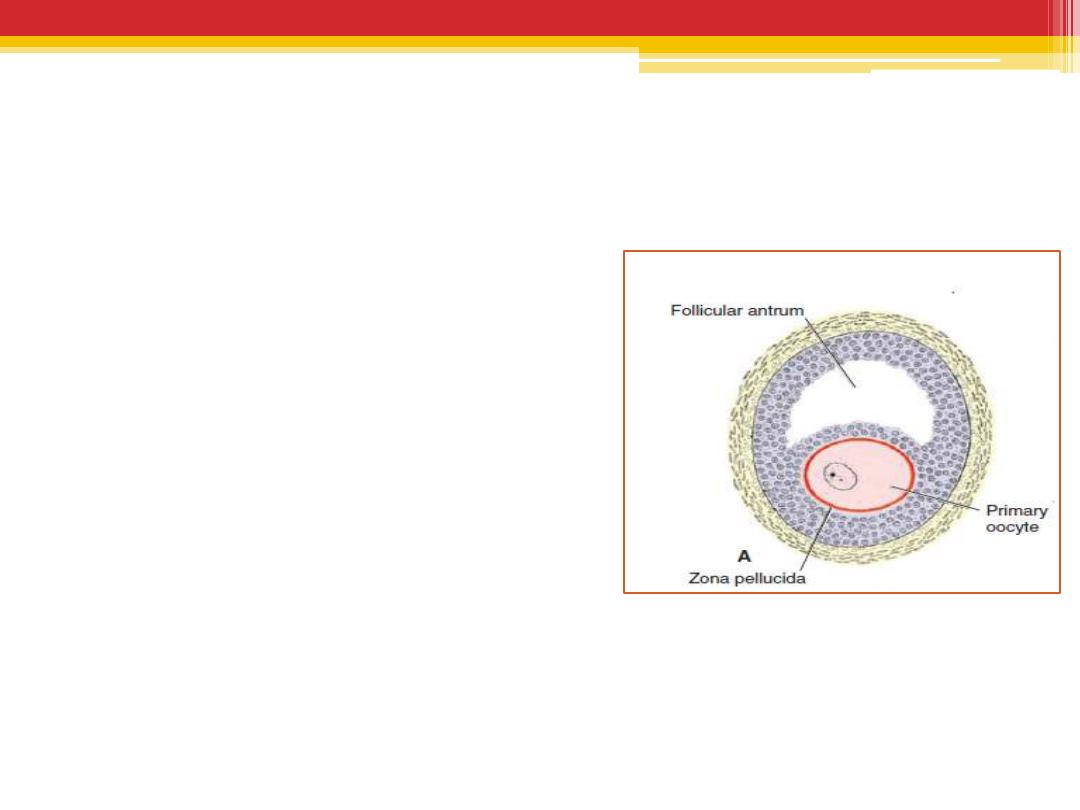
Secondary ( vesicular , antral )
•
fluid –filled spaces appear
between the granulosa cells
.coalescence of these spaces
form the antrum which is
crescent shaped, but with
time, it enlarges .
•
The follicle is surrounded by
1.
the theca interna
2.
the theca externa
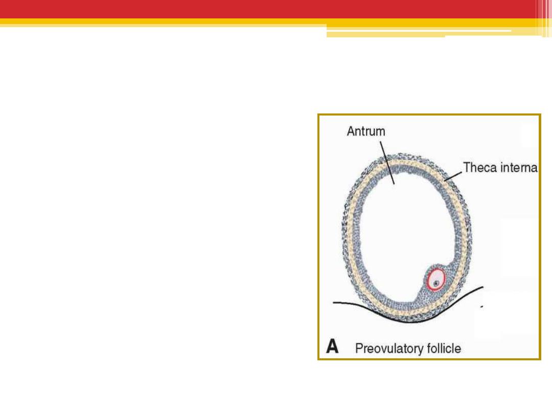
Tertiary follicle
•
Granulosa cells
surrounding the oocyte
form the cumulus
oophorus.
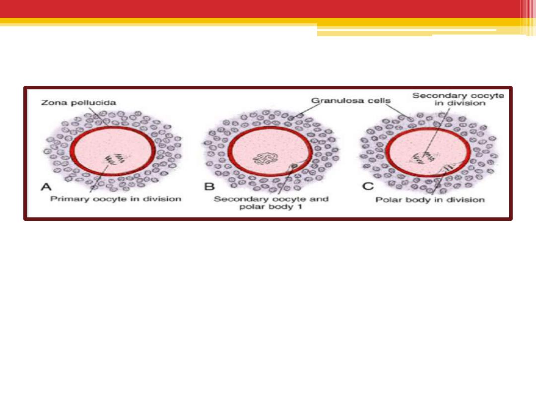
Maturation of the oocyte.
•
Meiosis I is completed, resulting in formation of two
daughter cells of unequal size, each with 23 double-
structured chromosomes . One cell, the secondary
oocyte, receives most of the cytoplasm; the other, the
first polar body, receives practically none.
•
The cell then enters meiosis II but arrests in metaphase
approximately 3 hours before ovulation.
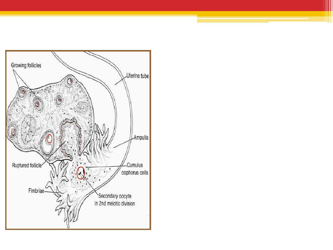
Ovulation
•
The oocyte, in metaphase of
meiosis II, is discharged from
the ovary together with a large
number of cumulus oophorus
cells.
•
Some of the cumulus oophorus
cells then rearrange
themselves around the zona
pellucida to form the corona
radiata
•
Meiosis II is completed only if
the oocyte is fertilized;
otherwise, the cell degenerates
approximately 24 hours after
ovulation.
•
The first polar body may
undergo a second division
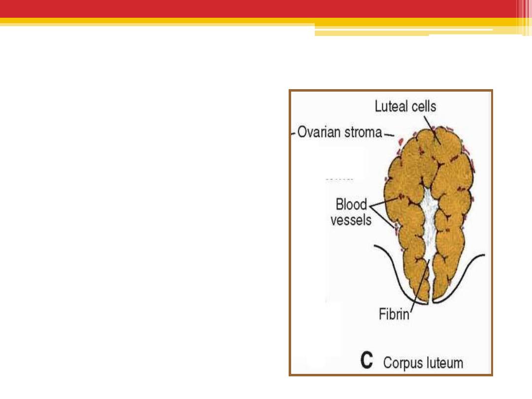
Corpus Luteum
•
After ovulation,
granulosa
cells
together with
cells
from the theca interna
,
change into lutean cells,
•
secrete the hormone
progesterone .

Fate of the corpus luteum
•
If fertilization does not
occur
, the corpus luteum
reaches maximum
development approximately 9
days after ovulation, then
shrinks and forms a mass of
fibrotic scar tissue, the
corpus albicans
.
•
If the oocyte is fertilized
,
the corpus luteum continues to
grow and forms
the corpus
luteum of pregnancy
(corpus luteum graviditatis).
•
Yellowish luteal cells continue
to secrete progesterone until
the end of the fourth month;

Spermatogenesis
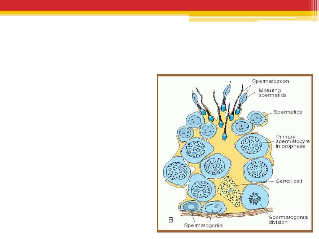
Is a complex series of changes by which spermatogonia
are transferred into spermatozoa .
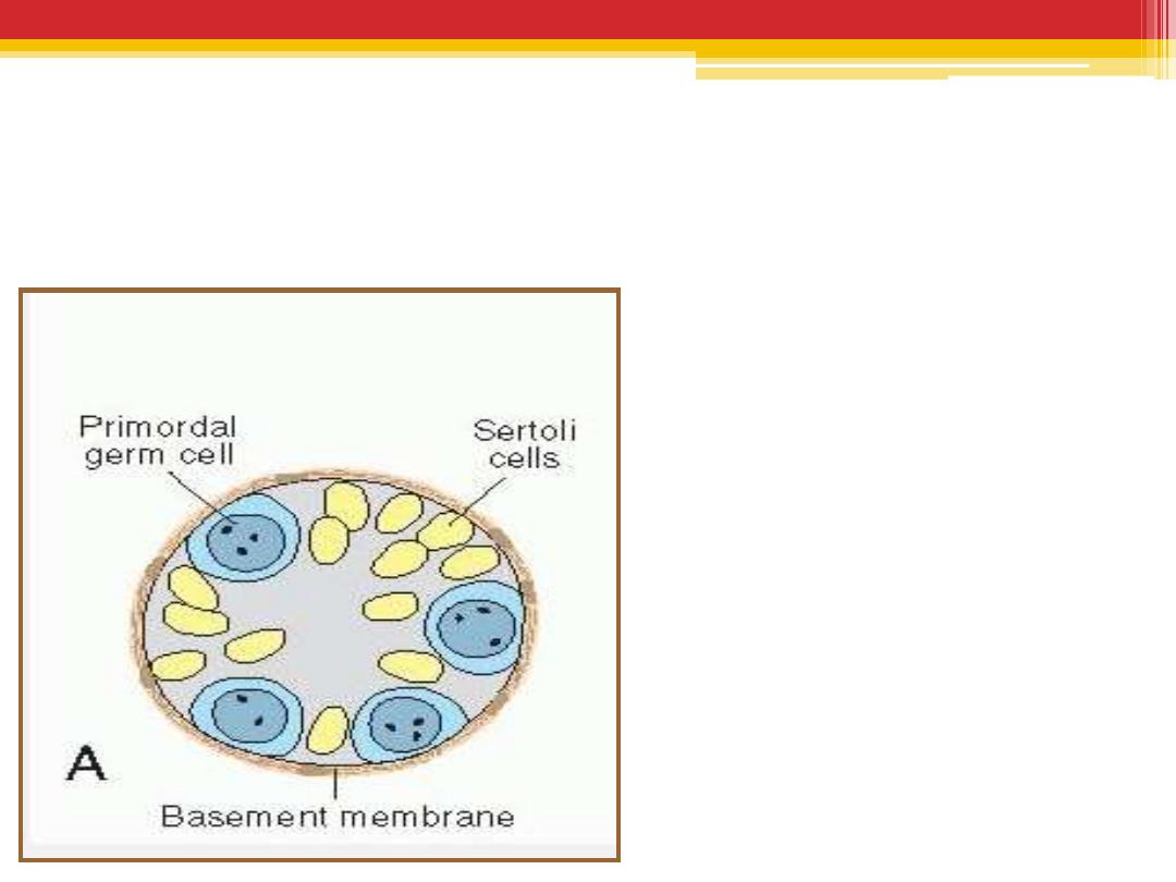
in the male infant
•
Germ cells
can be
recognized in
the sex
cords of the testis
as large,
pale cells surrounded by
supporting cells .
•
Supporting cells
become
sustentacular cells, or
Sertoli cells
.
Shortly before puberty,
•
the sex cords acquire a lumen
and become the seminiferous
tubules.
•
Maturation of Sperm begins at
Puberty
•
At about the same time,
primordial germ cells give rise
to spermatogonial stem cells.
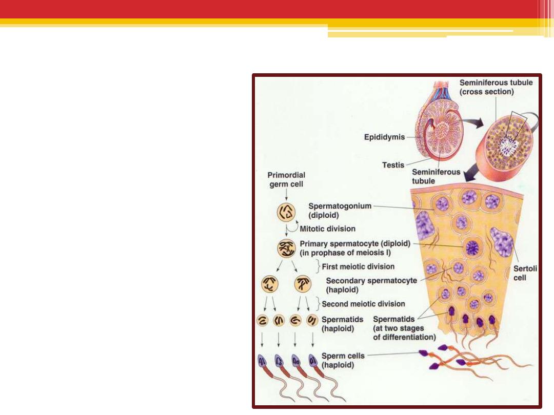
Spermatogenesis
can be divided into 3 phases :
A.
spermatocytosis
B.
meiosis
C.
spermiogenesis
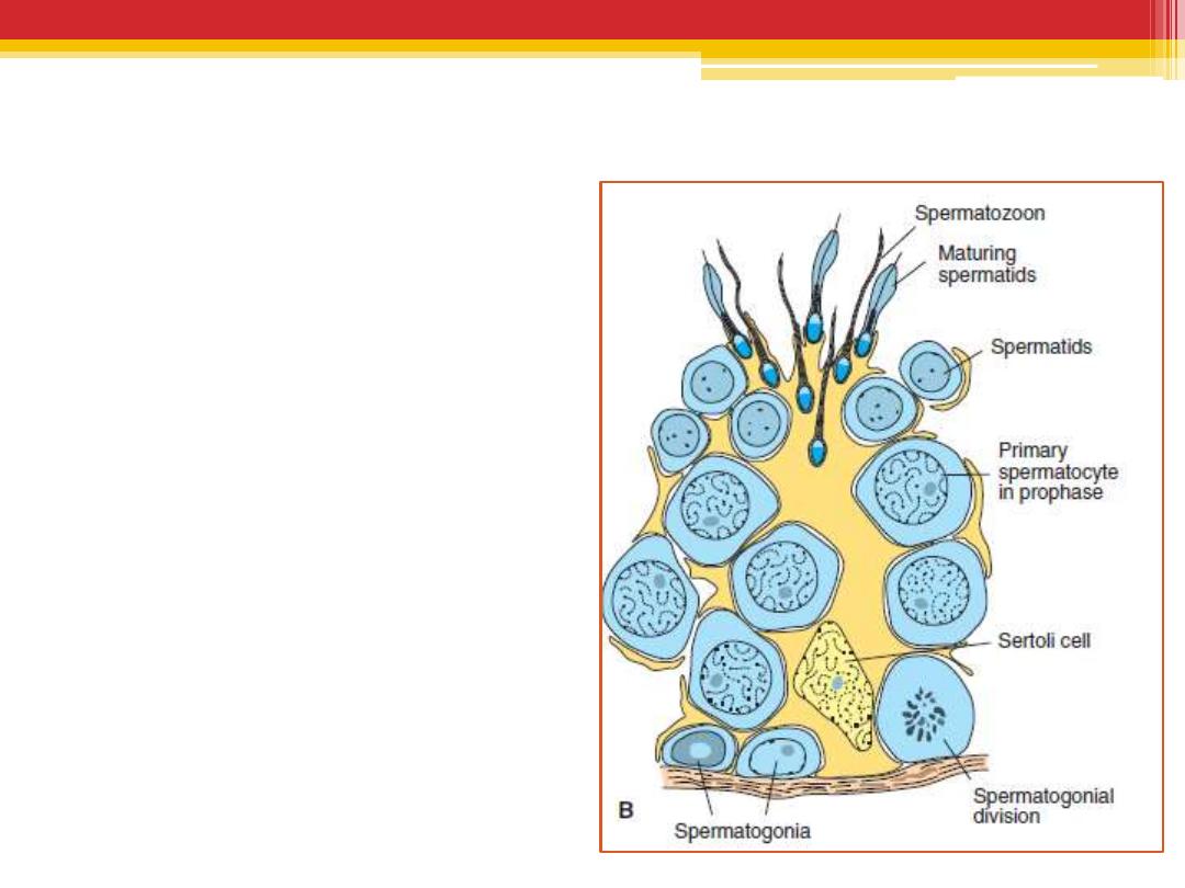
spermatocytosis
•
Spermatogonia
proliferate by mitotic
division to replace
themselves and to
produce primary
spermatocytes
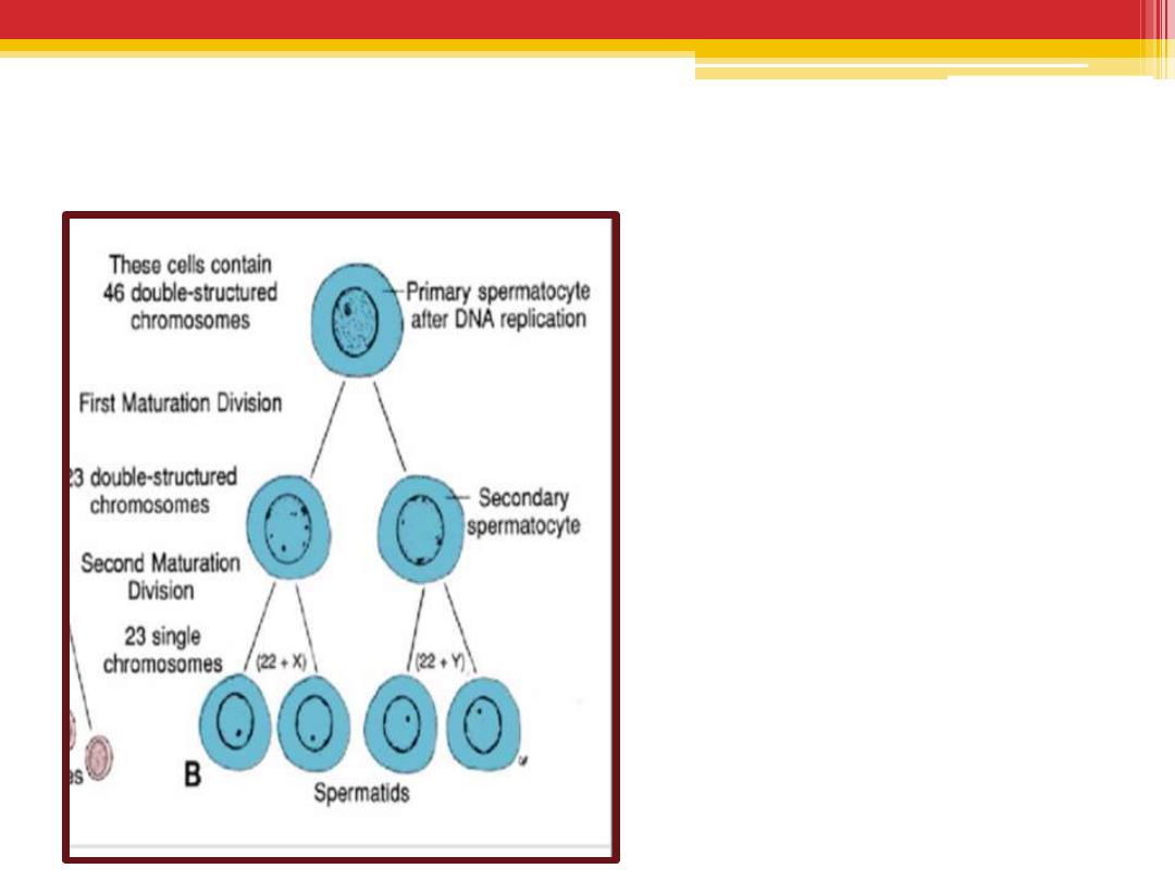
Meiosis
•
2 successive
divisions
•
Meiosis I produce
secondary
spermatocytes
•
Meiosis II produce
spermatids
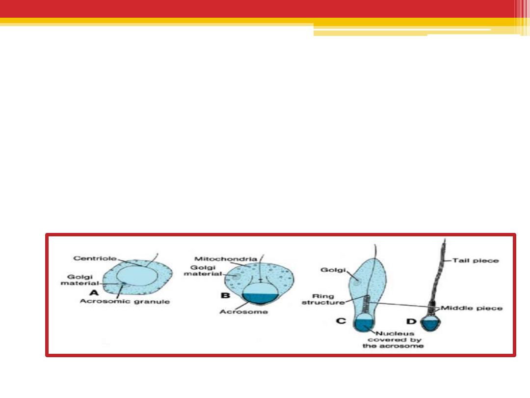
Spermiogenesis
•
The series of changes resulting in the transformation of
spermatids into spermatozoa include
(a) formation of the acrosome, which covers half of the
nuclear surface and contains enzymes to assist in penetration
of the egg during fertilization ;
(b) condensation of the nucleus;
(c) formation of neck, middle piece, and tail;
(d) shedding of most of the cytoplasm.
Clinical Correlates
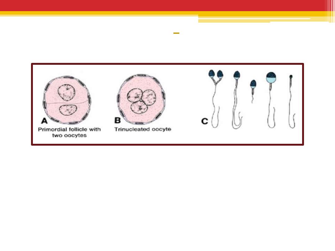
Abnormal Gametes
•
A. Primordial follicle with two oocytes.
•
B. Trinucleated oocyte.
•
C. Various types of abnormal spermatozoa.


Summary
•
Oogenesis begins before birth while spermatogenesis begins at puberty
•
At puberty , in female every month,15 to 20 follicles begin to grow, and as
they mature, they pass through three stages: (1) primary or preantral, (2)
vesicular or antral, and (3) mature vesicular or Graafi an follicle.
•
The primary oocyte remains in prophase of the first meiotic division until the
secondary follicle is mature.
•
the secondary oocyte is arrested in metaphase of meiosis II approximately
3 hours before ovulation and will not complete this cell division until
fertilization
•
In the male, primordial cells remain dormant until puberty, and only then do
they differentiate into spermatogonia. These stem cells give rise to primary
spermatocytes, which through two successive meiotic divisions produce
•
four spermatids .
•
Spermiogenesis a series of changes including (1) formation of the
acrosome; (2) condensation of the nucleus; (3) formation of neck, middle
piece, and tail; and (4) shedding of most of the cytoplasm.
•
The time required for a spermatogonium to become a mature
spermatozoon is approximately 74 days.