Lymphocytes Cell Surface Markers
ImmunologyIntroduction
B cellT cell
Monocyte
NK cell
T h
T c
Dendritic
cellNKT cell
So, how these cells can be differentiated from each other? Cell surface markersCell surface markers
• Usually proteins expressed on the surface of the cell and help in differentiation of different groups of the immune cells
• Usually given the designation of (CD) which means: cluster of designation or differentiation together with appropriate number e.g. CD19, CD3 , CD4
Introduction
B cellT cell
Monocyte
NK cell
T h
T c
Dendritic
cellNKT cell
- These markers may not be specific (shared). i.e. differentiation of one group of cells may require more than one markerIgD
IgM
CD19
CD20
CD3
CD3
CD8
CD4
CD3
CD14
CD56CD56
CD3DRC
Techniques
Immuno-histochemical stainingFlowcytometry
ELISA
Western blotting
Flowcytometry
CD3
CD56
9.4%
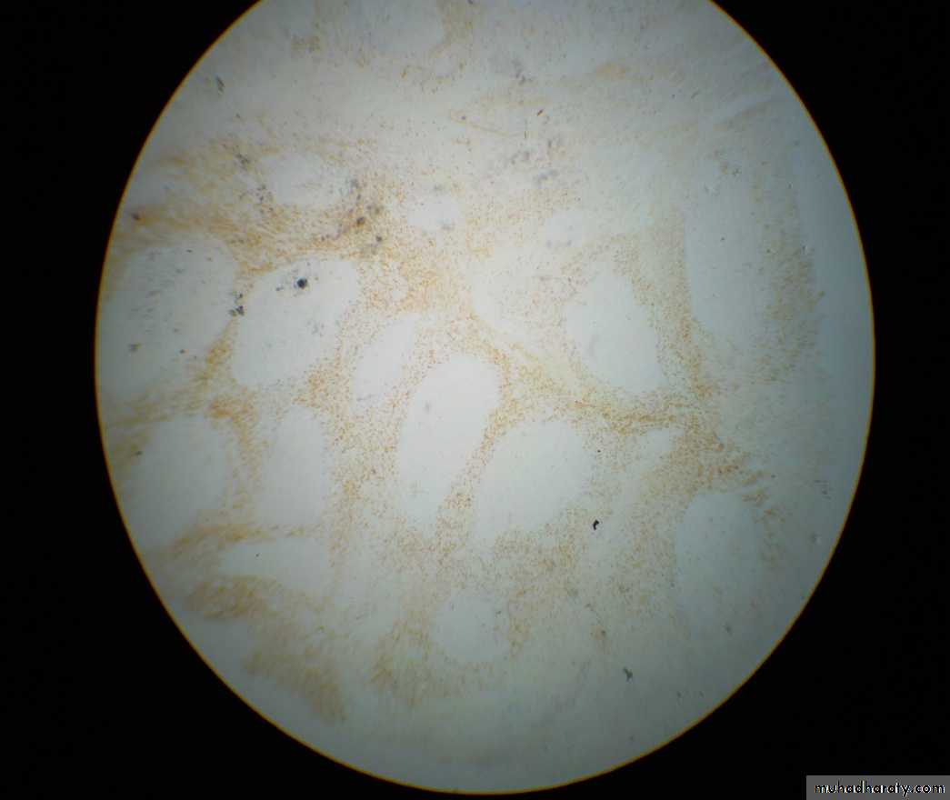
Immuno-histochemical staining
Used for the staining of cell markersApplied to identify the distribution of antigens (Ags) in the tissues
2 types:
Immuno-fluorescent
Immuno-enzymatic
Involve the addition of specific antibodies (monoclonal Abs) which bind to their target Ags in the tissues, but in order for the reaction to be visible , it should be coloured.
Green spots
Red or brown spots
Immuno-histochemical staining
PP
P
P
Direct
Indirect
Sandwich
Lymphocytes cell markers
ThymusTonsil
Lymph nodes
Spleen
Blood
Bone marrow
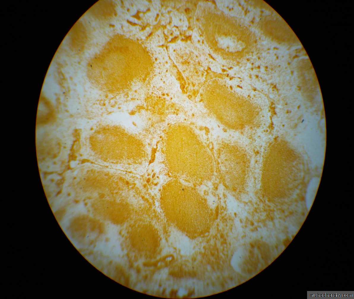
Tonsil: Histology
•Medulla
Cortex
•
•
•
•
•
•
Germinal centre
Mantle zone1ry follicle
2ry follicleDendritic cells
IFA- Majority of lymphocytes in the follicles are of B type
- Majority of lymphocytes in the IFA are of T type
B cell markers : IgM
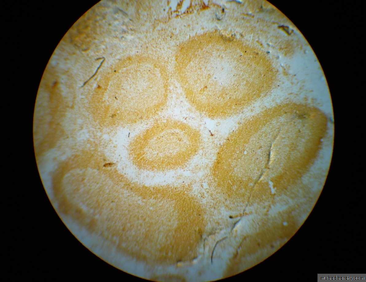
1ry follicles : Many cells are positive
2ry follicles:M.Z: Many cells are positive
G.C: Meshwork staining
- IFA : few cells are positive
B cell markers: IgD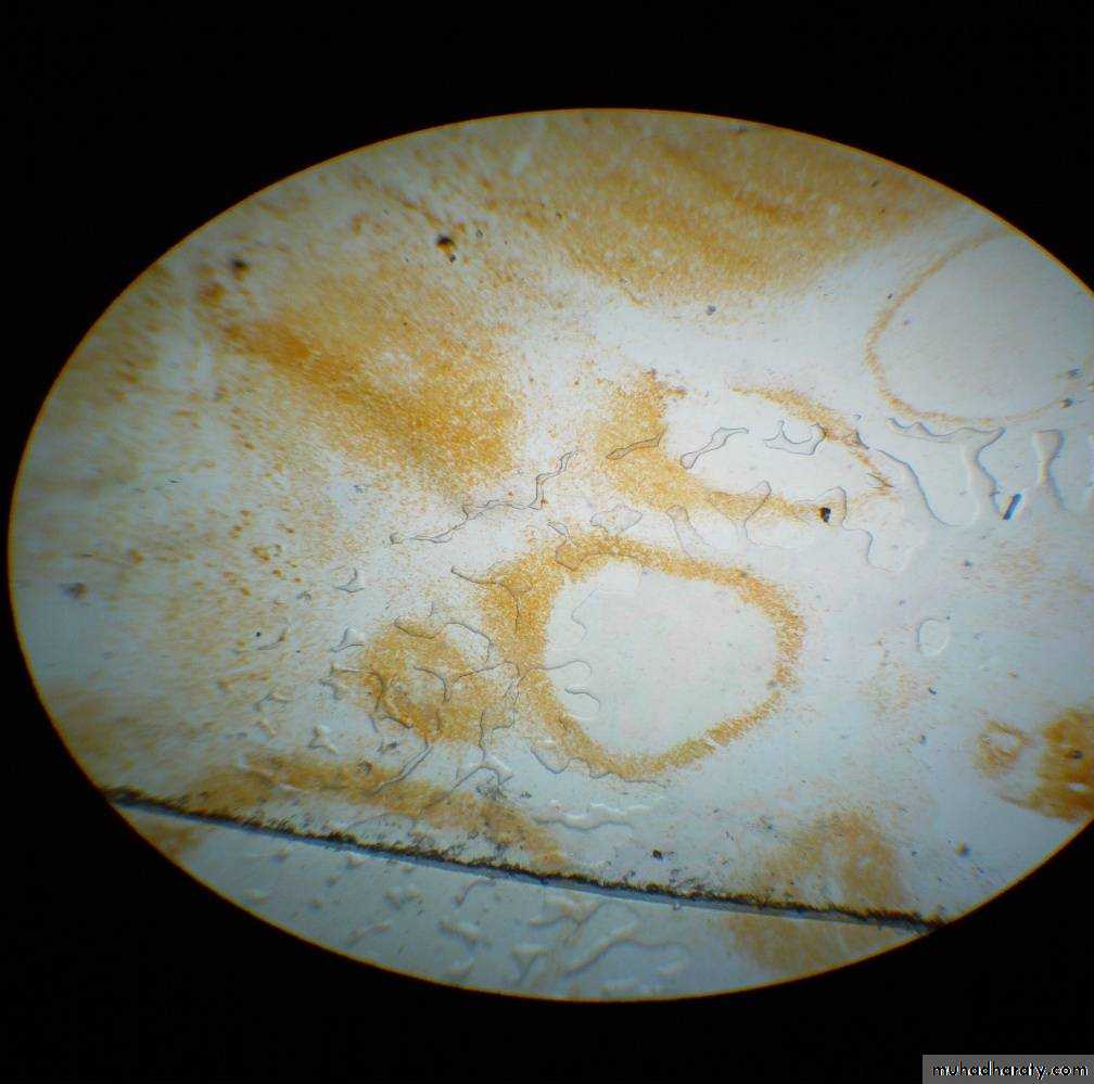
1ry follicles : Many cells are positive
2ry follicles:M.Z: Many cells are positive
G.C: Few cells are positive
- IFA : few cells are positive
Ring-like appearance
T cell markers: CD3 or CD4
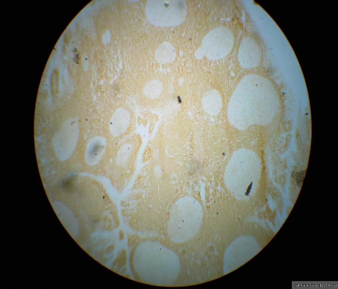
1ry follicles : Few cells are positive
2ry follicles:M.Z: Few cells are positive
G.C: Few cells are positive
- IFA : Majority of cells are positive (2/3 of the cell population)
CD3 can not be differentiated from CD4 from this slideT cell markers: CD8
1ry follicles : Few cells are positive2ry follicles:
M.Z: Few cells are positiveG.C: Few cells are positive
- IFA : Minority of cells are positive (1/3 of the cell population)
DRC marker