Face & parotid
ByDr. Adel sahib Al-Mayaly
Otolaryngologist
Facial skeleton
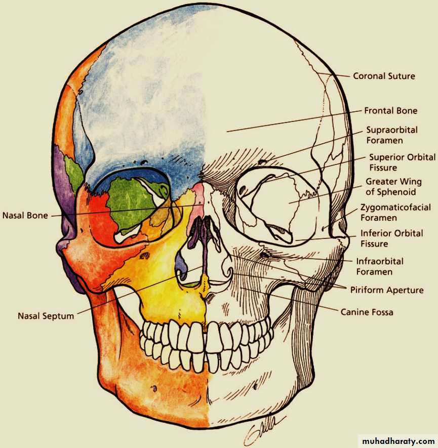
Facial Skeleton
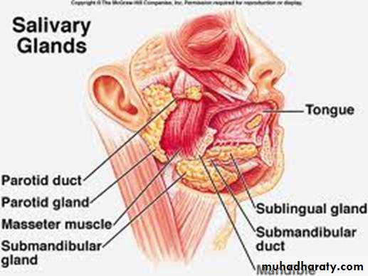
Parotid regionIt is a deep hollow that lies on the lateral surface of the face which contains the parotid gland
The parotid region is bounded by
•Superiorly – Zygomatic arch.•Inferiorly – Inferior border of the mandible.
•Anteriorly – Masseter muscle.
•Posteriorly – External ear, mastoid process and sternocleidomastoid muscle.
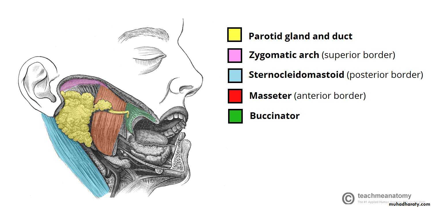
Position of the parotid gland and borders of the parotid region.
Parotid Gland- Largest salivary gland (15-30g), 6 x 3 cm
- Has broad superficial lobe and smaller deeper lobe, with facial nerve usually between both lobes- Stensen’s duct (main duct) empties into oral cavity opposite crown of second maxillary molar
- 20% have accessory parotid gland and duct, usually overlying the masseter
- Parotid gland has own fascia (capsule), which is continuous with superficial layer of deep cervical fascia
- Contains 3-24 lymph nodes (not all with complete structural organization), usually lateral to facial nerve in superficial lobe
Parotid gland
Wedge-shaped when viewed externally , with the base above & the apex behind the angle of the mandible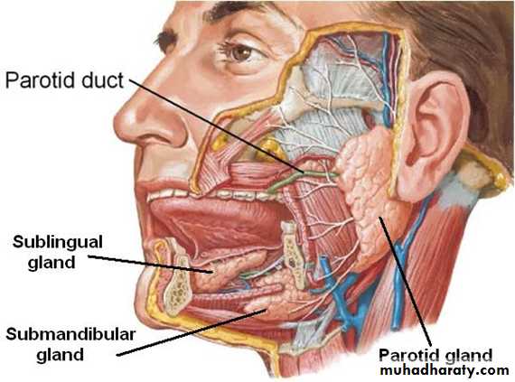

Parotid gland
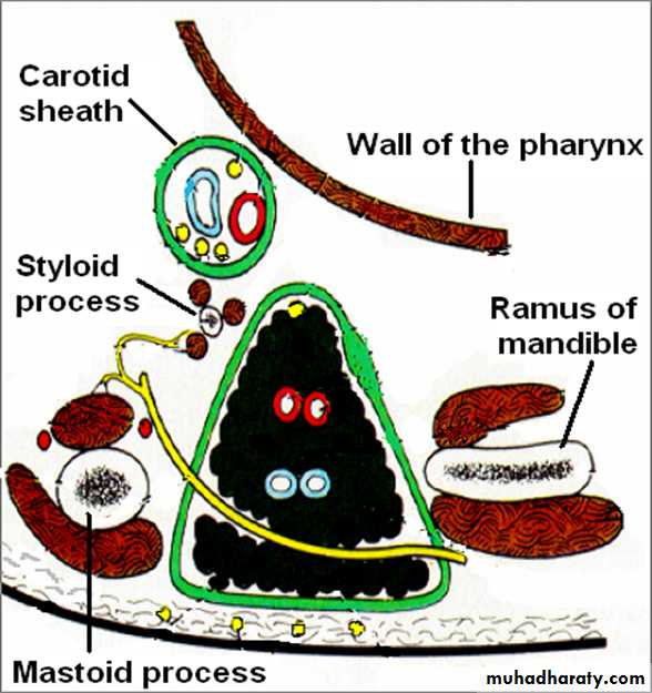
Wedge-shaped in horizontal section with the base in the lateral position and apex against the pharyngeal wall.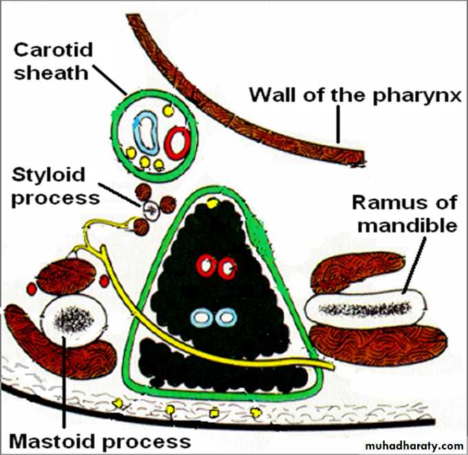
Lobes
Each parotid gland is divided by the facial
nerve into:-
1- Superficial lobe:-
is defined as the part of the gland lateral to the
facial nerve.
2- Deep lobe:-is medial to the facial nerve.
Borders
Anterior border • Separates superficial surface from anteromedial surface.Posterior Border • Separates superficial surface from posteromedial surface • Overlaps sternomastoid
Medial Border • Separates anteromedial surface from posteromedial surface • Related to lateral wall of pharynx
Surfaces
1- Superficial Surface • Covered by • Skin • Superficial fascia containing facial branches of great auricular N.2- Anteromedial Surface • Grooved by posterior border of ramus of mandible.
3- Posteromedial Surface:-
Related to mastoid process with sternomastoid and posterior belly of digastric
Capsules
The parotid gland is enclosed in two capsules:
1- An inner connective tissue capsule (true capsule)
2- An outer dense fibrous capsule derived from the investing layer of the deep cervical fascia ( false).
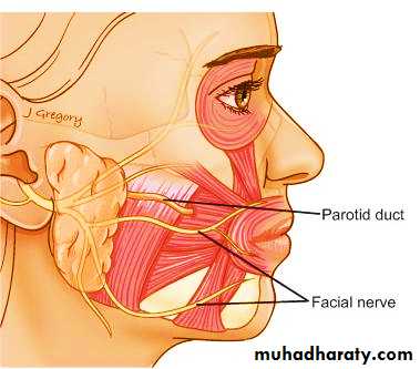
Parotid Duct
About 2 inches longEmerges from the anterior border of the glannd.
Passes forward over the lateral surface of the masseter muscle
about a fingerbreadth below the zygomatic arch
accompanied by the:
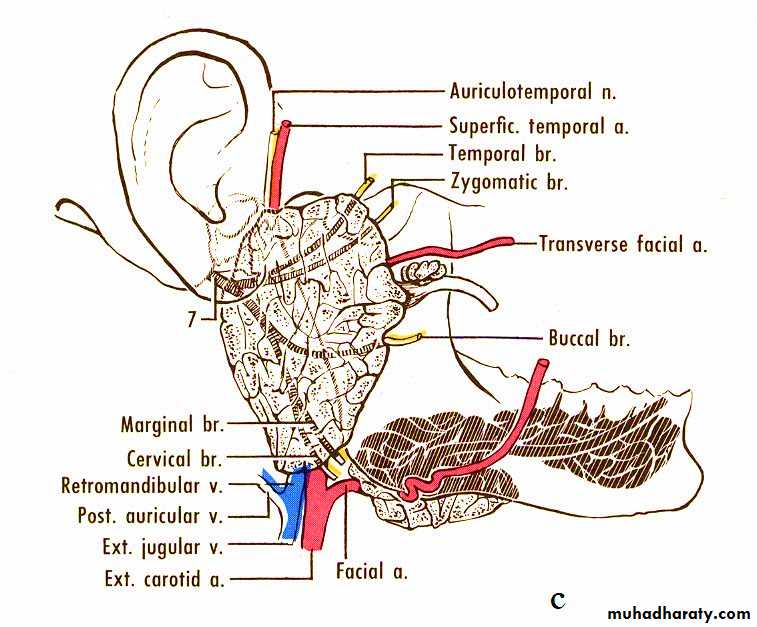
transverse facial vessels & buccal branches of facial nerve
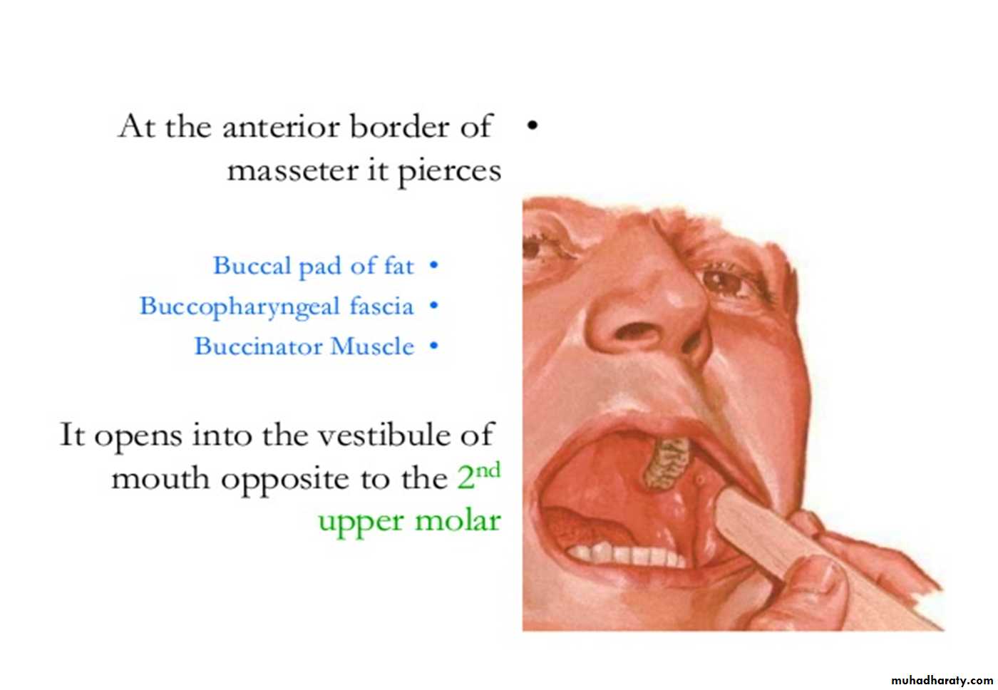
Parotid duct
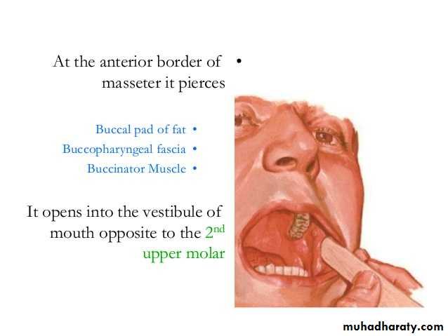
Pierces the:Buccal pad of fat
Buccopharyngeal fascia
Buccinator muscle &
Buccal mucosa
Opens into the vestibule of mouth on a small papilla, opposite the second upper molar tooth
Surface anatomy
The duct is represented by the middle 1/3 of a line extending from the tragus of the auricle to a point midway between the ala of nose & upper lip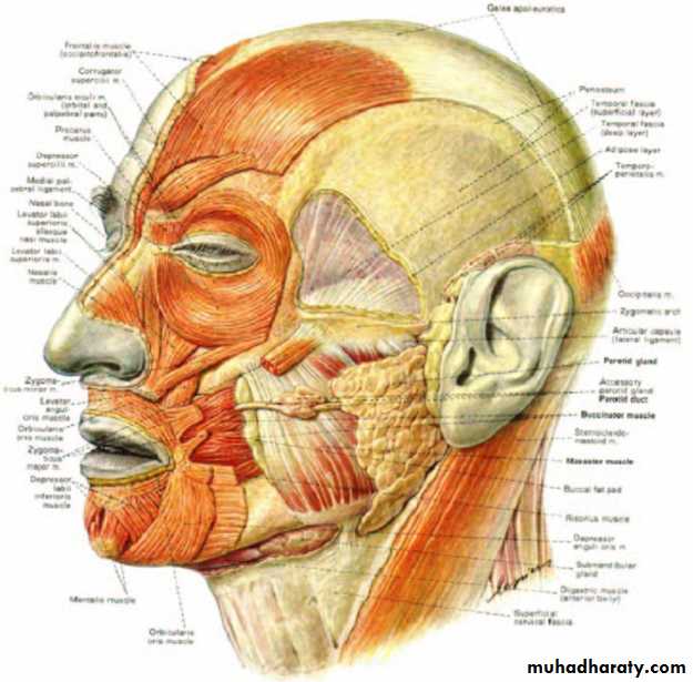

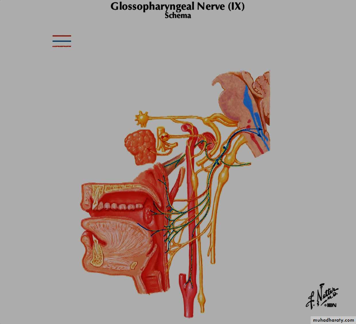
Structures Coursing Within the Parotid Gland
Auriculotemporal nerveExternal carotid artery
Retromandibular vein
Facial nerve
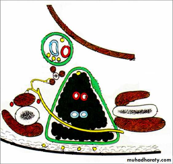

Deep

Superficial

A few lymph nodes are scattered in the substance of the gland
Facial Nerve & parotid gland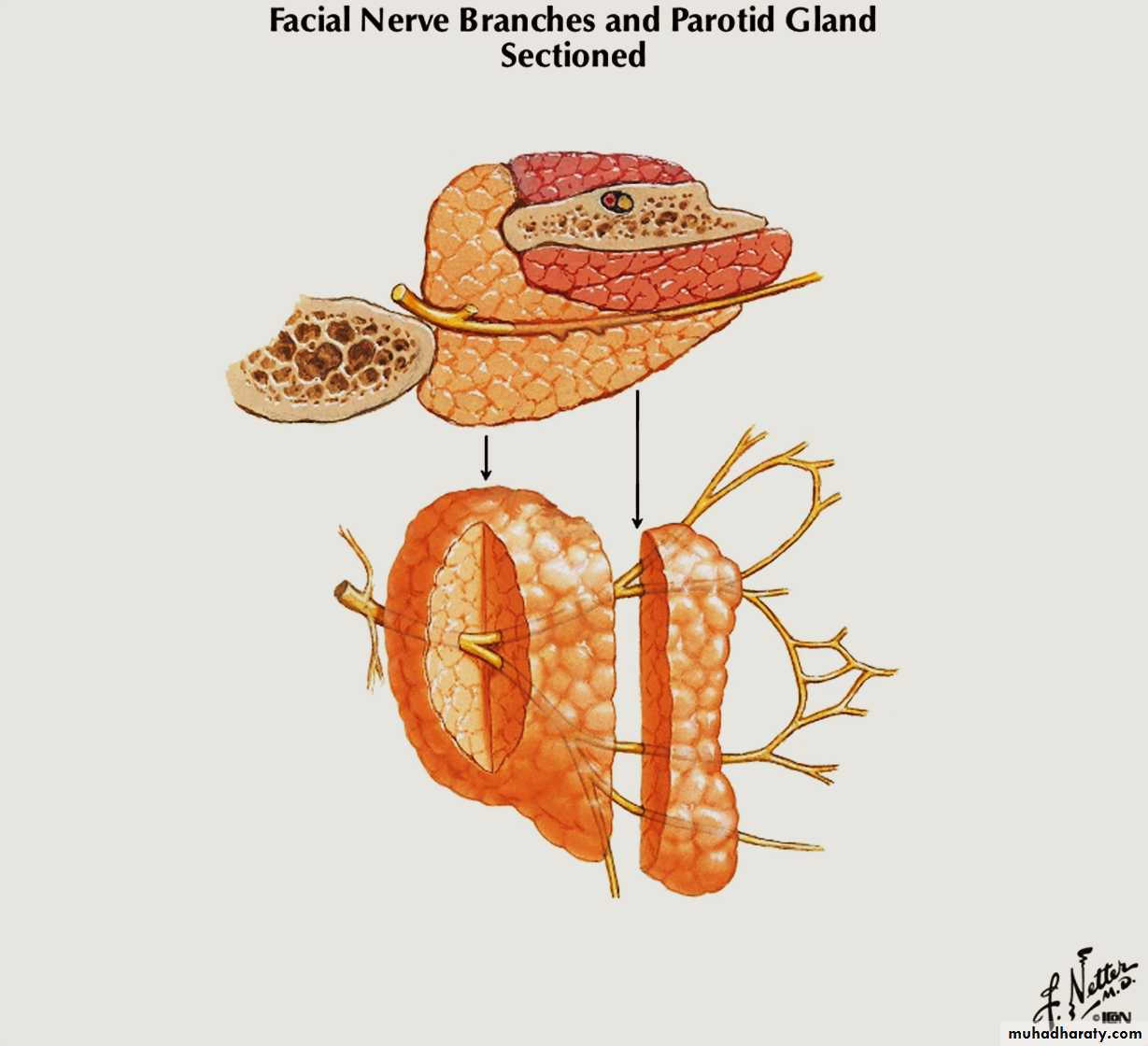
Facial Nerve & parotid gland
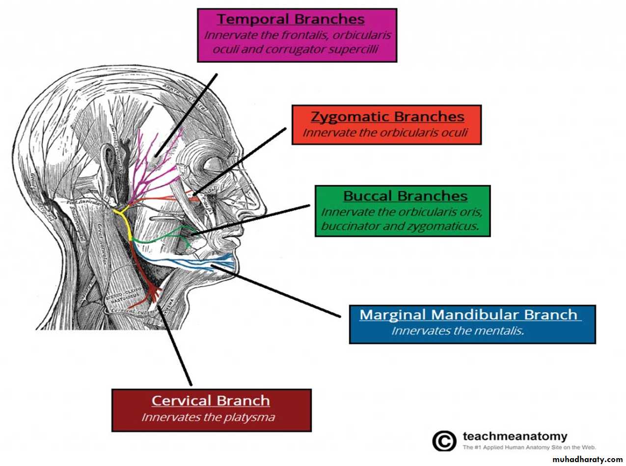
Within the parotid gland, :-•Temporal branch
•Zygomatic branch
•Buccal branch
•Marginal mandibular branch
•Cervical branch
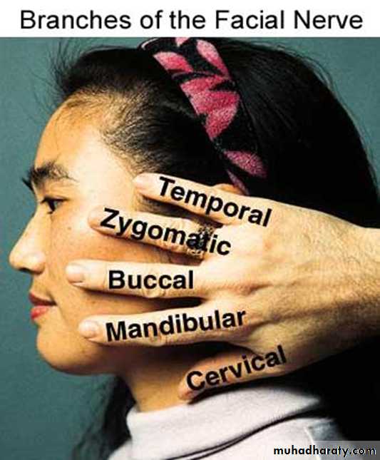
Terminal branches of facial nerve
Neurovascular supply
Arteries:- External carotid arteryVeins:- Retromandibular vein.
Lymphatics:- Superficial & deep parotid LN
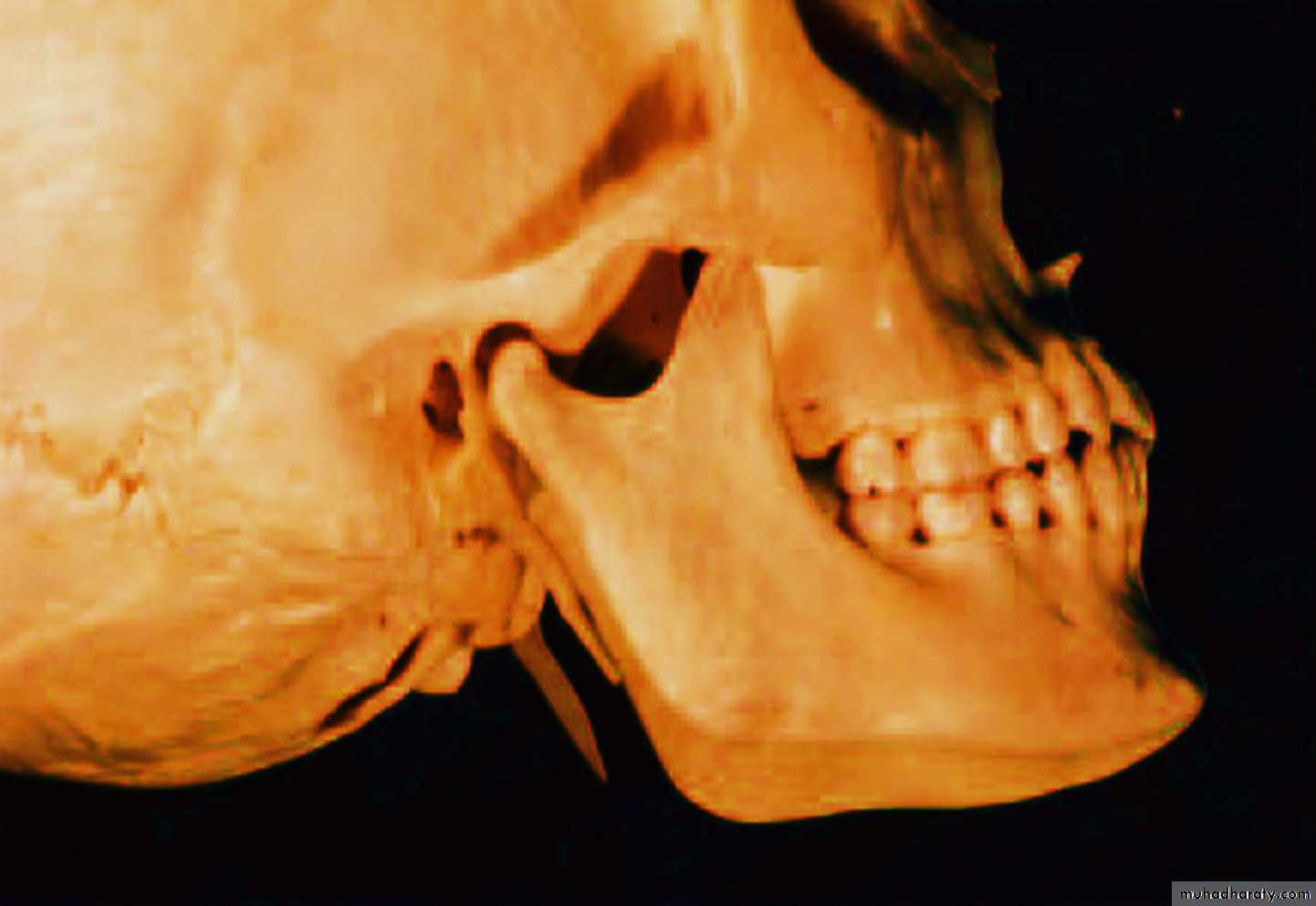
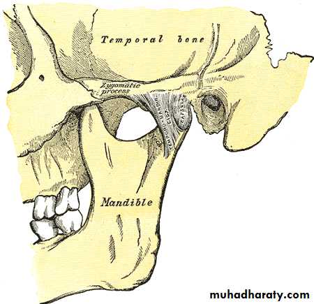
Temporomandibular jointJaw joint or TMJ
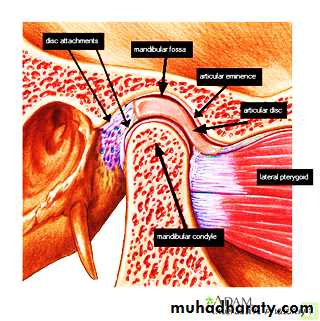
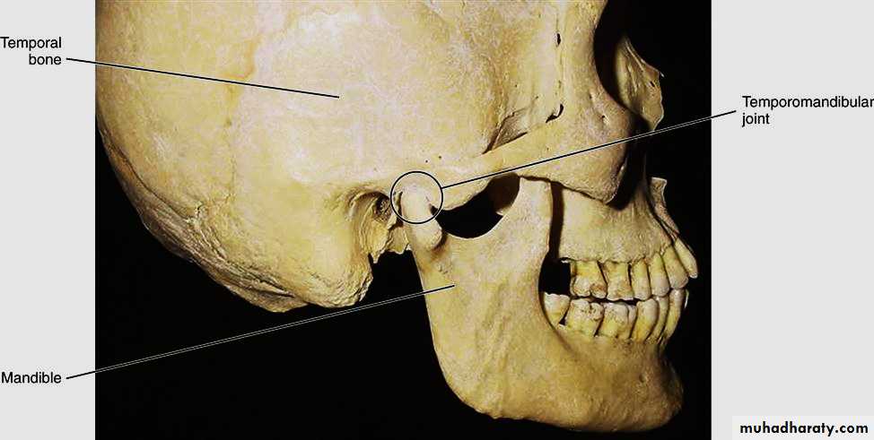
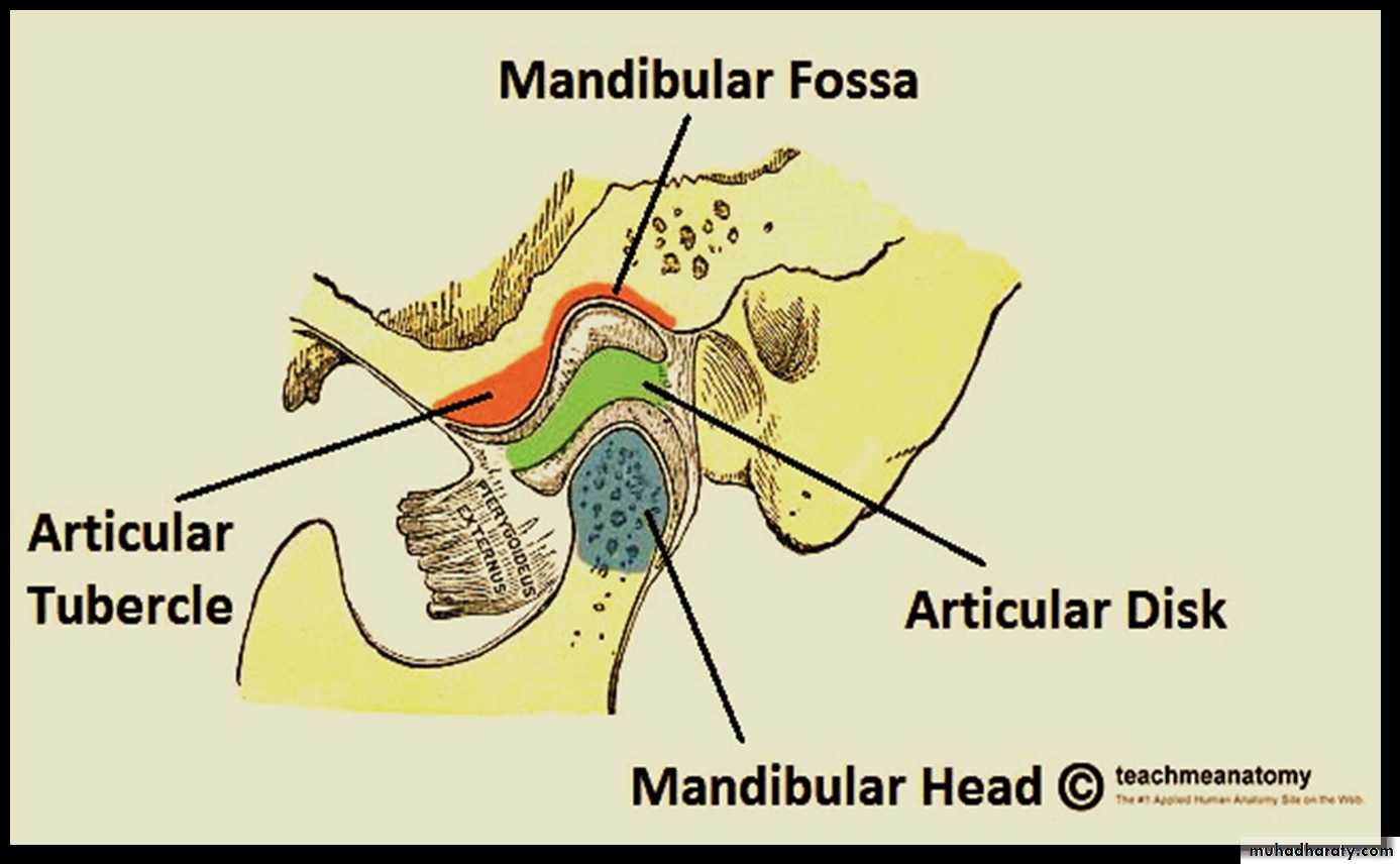
The osteology of the Temporomandibular joint
The temporomandibular jointIt is a bilateral synovial articulation between the upper temporal bone and the lower mandible; it is from these bones that its name is derived.
TMJ
The joint is atypical synovial joint of the hinge variety.
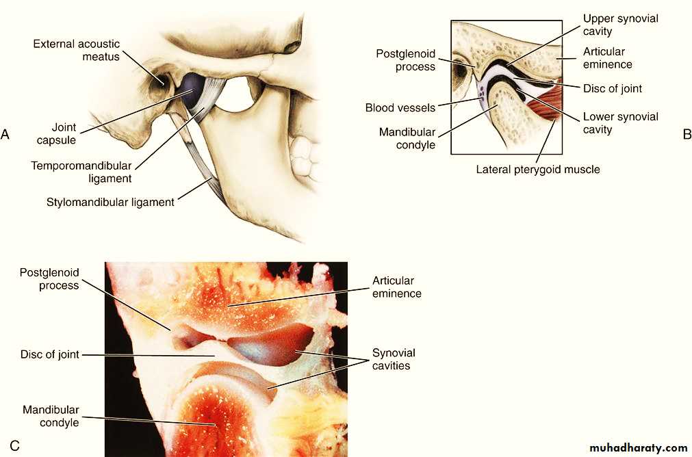
The joint cavity is divided by the articular disc (fibrocartilage ) into upper & lower cavities.
Capsule of joint
It surrounds the jointIt is attached above to the articular tubercle & margins of mandibular fossa.
Below; neck of mandible.
There are 3 extra capsular ligaments.
They act to stablise the joint.
1- lateral TM ligament:-
it strengthens the lateral aspect of the capsule.
Its fibres run from the tubercle down & backward toward neck of mandible.
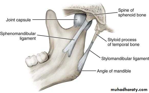
2- Sphenomandibular ligament:-
lies on the medial side of the joint.It is a thin band that is attached above to the sphenoid bone & below to the mandibular foramen.
Movements of joint
Movements at this joint are produced by the muscles of mastication, and the hyoid muscles.Protrusion and Retraction
the anterior and posterior movement.
Lateral pterygoid is responsible for this movement
the geniohyoid and digastric muscles perform retraction.
Elevation and Depression
Venous drainage
Veins follow the arteries
Osteology of parotid bed