د. زينب خالد خليل
د. زينب خالد خليل
Lec. 1
Lec. 1

Parasitology
Is a science, study the relationship between two
organisms one called parasite & the other is called
the host
.

CLASSIFICATION OF PARASITES
:
Parasitic kingdom include three phyla
1
-
Protozoa
.
2
-
Helminths
.
3
-
Arthropods
.

I- Protozoa
:
Is a phylum of the animal kingdom consisting of
unicellular parasites, divided into 4 classes according
to the organ of locomotion
:
1
-
Class sarcodina: Parasites that move by means of
pseudopodia example Entamoeba histolytica
.
2
-
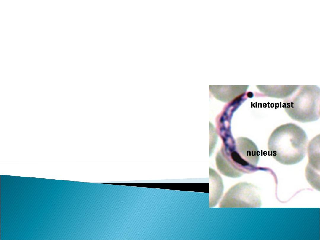
Class mastigophora : Parasites that move by
means of flagella example Giardia lamblia

3- Class ciliates : parasites that move by means
of cilia example Balantidium coli .
4- Class Sporozoa : parasites have both sexual
and asexual reproductive organs, all these
parasites are intracellular and they have no
organ of locomotion example Plasmodium
parasites causing malaria.

II- Helminths :
They are metazoa ( Multicellular parasite ) wormlike
parasite, divided into 3 classes :
1.Class Nematoda ( Roundworms ) :
a- Intestinal nematodes, e.g, Ascaris
lumbricoides .
b- Tissue nematodes, e.g, Wuchereria bancrofti .

2- Class Cestoda ( Tapeworms) :
They are flattened and segmented worms,
e.g: Taenia saginata .
3- Class Trematoda (Flukes):
They are flattened leaf- shaped worms. e.g:
Schistosoma heamatobium.

III- Arthropods
:
These parasites having exoskeleton
and jointed legs, divided into 2 classes
:
1
-
Class Insecta
:e.g. Mosquitoes, lice
and fleas
.
2
-
Class Arachnida
:e.g. Ticks and mites
.

GENERAL TERMINOLOGY
:
*
pathogenic parasite (parasitism):
A parasite
infect the host and cause tissue changes or a
disease (harmful parasite)
.
•
*
Commensal parasite (Commensalism):
The association of two different species of
organisms in which one of them is benefited and
the other neither benefited nor injured
.

* Ectoparasite:
A parasite present on or in the
exterior surface of a host.
* Endoparasite:
A parasite present within the body
of its host .

Facultative parasite:
A parasite capable of living
an independent or a parasitic existence .
Obligatory parasite:
A parasite is capable of living
as parasitic on a host, but it can not exist as
independent living.

Types of hosts
*Definitive host:
The animal or human in which
a parasite passes its adult stage and/ or the
sexual reproductive phase can take place.
*
Reservoir host
:
An animal e.g. (dogs,cats or rodents) which
carry a species of parasite from which man
become infected. The host do not get the
disease or its carried as a subclinical infection
.

* Carrier
. A host carring a parasite but not
showing any clinical sings or symptoms.
* Accidental ( or incidental ) host :
Infection of
a host other than the normal host species.

Vector:
Any arthropod or other living carrier which
transport a pathogenic micro-organism from an
infected to a non infected host. A vector may
transmit disease :
(1)passively called (mechanical vector)
e.g.housefly
(2) The vector is essential in the life cycle of the
pathogenic parasites called (biologic vector)
e.g.mosqutoes.

Ectoplasm:
The gelatinous material beneath the
cell membrane
.
*
Endoplasm:
The fluid & inner material of a
protozoal parasite
.
*Flagellum(flagella) :
An extension of ectoplasim which provides
locomotion similar to a tail .
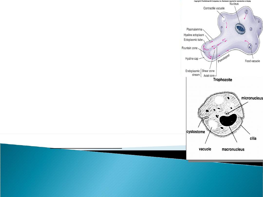
Pseudopod:
A protoplasmic
extension on the trophozoites of
amoeba allowing them to move and
engulf food .
Cilia:
Hairlike processes attached to
a free surface of a cell; function for
motility of fluids at the surface of the
cell, e.g. Balantidium coli .

د
.
زينب خالد خليل
Lec.(2)
Amoebas
Amoebas

Amoebas
:
The genus Entamoeba include many
amoebas that infect humans, but not all of
them are associated with disease,like E.
histolytica which is a pathogenic amoeba
causing intestinal and extraintestinal
Infections
.

None pathogenic amoeba
:
These parasites are commensal none pathogenic
but they are important because they may be
confused with E
.
histolytica in diagnostic investigations. These
amoebas include many free-living and parasitic
amoebas
.
The most amoebas affecting human being are
:

1
-
E.coli
.
2
-
E.gingivalis
.
3
-
Dientamoeba fraglis.
4- Endolimax nana
.
5
-
Iodoamoeba butschlii
.
6
-
Other amoebas infecting human are
morphologically very simillar to E.histolytica,
e.g, E.hartmanni and E.dispar
.

7- Free living amoebas are Negleria &
Acanthamoeba are accidental parasites of
human being .The majority of these amoeba are
non-pathogenic commensal parasites or only
cause mild infection.

ENTAMOEBA HISTOLYTICA

Morphology ( Trophozoite ):
1- Its size (12-30 µm).
2- Large finger – like pseudopdia
3- The endoplasm is granular and may
contain RBCs.
4
-
It has one nucleous, contain small central
karyosome and fine chromatin granules
arranged regularly beneath nuclear
membrane
.
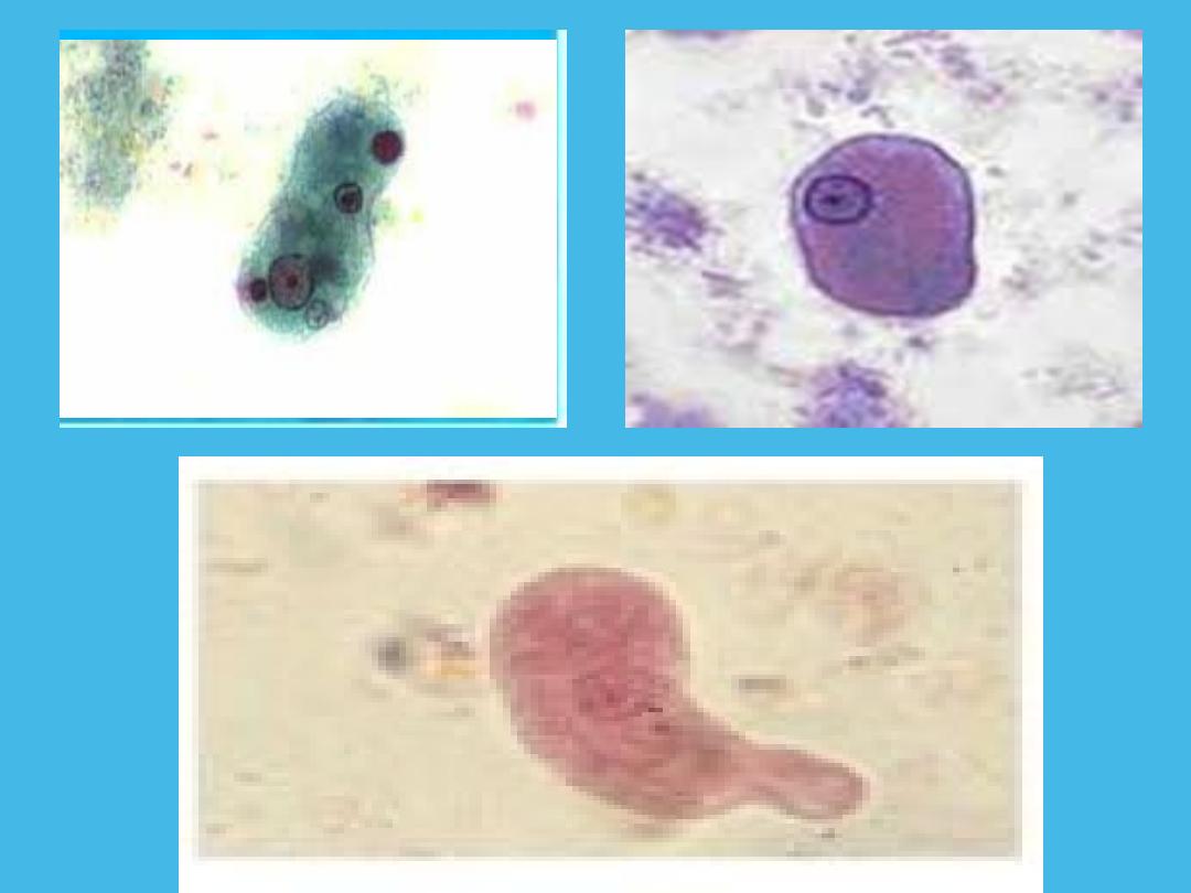
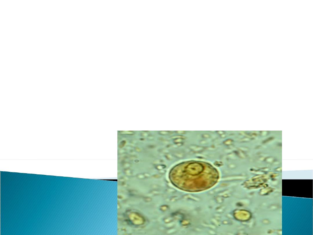
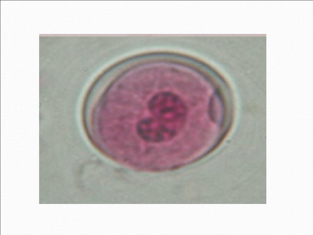
Morphology ( mature cyst) :
1- Small (10 – 20 µm) , spherical in shape,
containing 1 - 4 nuclei is usually found in feces .
Each nucleus contain similar nuclear morphology
like the trophozoite.

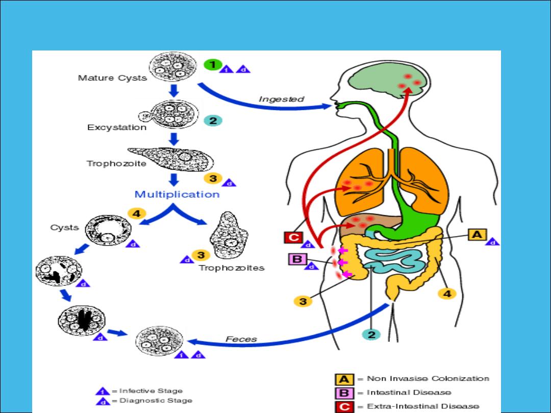
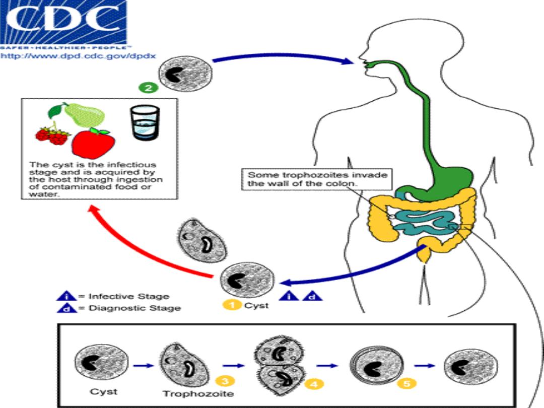
Life cycle of E. histolytica
:
Infection by E. histolytica occurs by
ingestion of mature cysts in fecally
contaminated food, water, or hands
.
Excystation occurs in the small intestine
and trophozoites are released which migrate
to the large intestine
.
The trophozoites multiply by binary fission and
produce cysts , which are passed in the feces
.

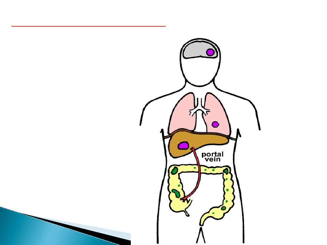
In some patients the trophozoites
invade the intestinal mucosa and
cause intestinal disease or developed
perforated ulcer and the trophozoites
migrate through the blood stream to
invade the extraintestinal organs such
as the liver, brain, and lungs and it will
cause amoebic infection in these organs
.
Life cycle of E. histolytica
:

Epidemiology :
.
* The incidence of Amebiasis is common &
high in tropical & subtropical areas especially in
areas of lower socioeconomic status due to:
(1)poor sanitation(2) overcrowding &
(3)malnutrition
It is estimated that up to 10% of the world`s
population may infected with E.histolytica.

Transmision of amoebiasis occure through:
1. Mature cyst is the main sourse of the infection
which passing with the feces of chronic patients
or asymptomatic carrier .
2. Human being acquire the infection via
contamination of food, drinks, vegetables or
hands with infective cysts especially in
restorants .
3. Flies (House fly) play an important roles in
trasmission of these cysts to the food of human .

Pathogenesis of E.histolytica
:
The Pathogenic activity of E. histolytica
depend upon :

1
-
The resistant of the host
.
2
-
The number of the amebas
.
3
-
Presence of pathogenic bacteria
.
4
.
Presence of physical & chemical injury
of the mucosa
.

The lesions produced by E. histolytica are
primarily in large intestine and
seconderily extraintestinal especially the
liver, brain or any organ of the body may
be affected .

Pathogenisis of Intestinal lesion :
1.The lesion vary from small ulcer to a large typical
flask shape ulcer.
2.The ulcer has a wide base and narrow opening
with irregular elevated edges .
3.The ulcer charecterized with large area of tissue
necrosis, cell infiltration & rapid lysis of
inflamatory cells.
4.The amoebas usually found on the floor of the base
of ulcer.
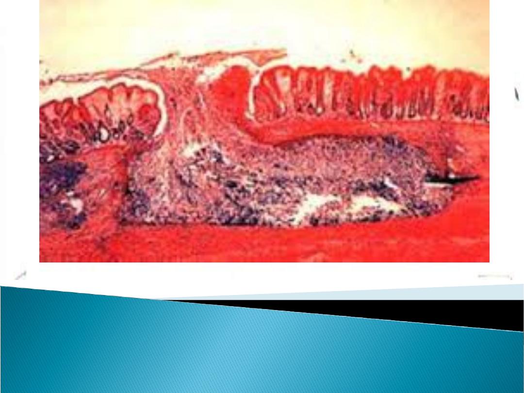
E. histolytica in the large intestine
( Flask shape ulser )

Clinical features of intestinal lesion :
1- The incubation period range from 2 – 4
weeks .
2- The majority of infections with E.histolytica
show no symptoms or show symptoms which
varies from mild to intense and long lasting .

The typical symptoms include :
1- Diarrohea, The diarrohea frequently
alternates with constipation or soft stools may
contain mucous but no visible blood .
2-Abdominal cramps.
3-Nausia.
4-Anoroxia.

5- Dysentery :
Which is usually starts slowly with abdominal
cramps and associated with loose stool and
diarrhea with blood , mucus and necrotic
tissues.
6- Few patients especially children may show
fever, vomiting, abdominal tenderness .

The complications of intestinal amoebiasis:
1- Appendicitis .
2- Intestinal perforation .
3- Hemorrhage .
4- Liver abscess.
5- Ameboma (Granulomas).

Extraintestinal Amoebiasis :
1.The metastasis of amoeba usually via blood
streem or by direct extension after intestinal
perforation to the peritonium. The amoeba may
cause local abcsess or peritonitis or
migrate to the liver which is the most
commonly affected than other organs e.g,
lungs, perianal skin or brain.
•
Extraintestinal Amebiasis

2.Amoebic liver abscesses:
Are the most common
extraintestinal amebiasis and
characterised By:
a.Hepatomegaly, Liver tenderness, fever
and anorexia .
b. Liver function tests are usually normal or
slightly abnormal .
c. Liver abscesses will occasionally rupture
into the peritoneum causing peritonits

3.Pulmonery
amoebiasis :
a.The clinical symptoms are:
cough, chest pain, dysnea and fever .
b.The sputum may be purulent or contain
blood and trophozoites of E. histolytica.

4. Cutaneous amoebiasis :
It is caused by contact of the skin with
amoebic abscess which lead to fistula
formation in the skin.

Diagnosis of Amoebiasis :
1- Stool of patient should be examined
microscopically :
a- The typical amoebic stool is contain blood,
mucous , WBC & Bacteria .
b-Direct method with saline for motile
trophozoite .
C-Stool specimens should be stained
usually with ioden and microscopically examined
for cysts of E.histolytica .

2- Culture of stool.
3- Sigmoidoscopy may reveal the
charecteristic flask-shaped ulcers
especially in sever cases .

4- Biopsy & fluid from large intestine
aspirates also be examined
microscopically for trophozoites .
5- Serology, is very important for the
diagnosis of extraintestinal amoebiasis
e.g: Indirect haemagglutination (IHA) &
Polymirase Chain Reaction (PCR test) .

6- Ultrasound, CTscan, MRI can be used to
detect hepatic abscesses .

Treatment :
1. A symptomatic (source of infection)
patients can be treated with
Diiodohydroxyquine with tetracycline.

Treatment Cont… :
2. Symptomatic patients with diarrhoea or
dysentary or extraintesinal amebiasis
should be treated as follows :
a- Supportive methods
Patients should remain in bed and
receive a high protein and high vitamin
with adequate fluids .

b. Chemotherapy for sever amoebiasis:
1.Metronidazol (Flagel) is the drug of choice :
750 mg three times a day, orally for 5 – 10 days.
2.Tetracycline & diiodohydroxyquine are
recommended to be given to the patient since
metronidzol may not always cure the intestinal
infection .

Prevention & Control
:
1
-
All human infections should be treated
2
-
A symptomatic carriers should be treated
especially those working in restorants
.

3- Effective enviromental sanitation is necessary
to prevent water ,food , and vegitable
contamination, e.g. Sewage disposal should be
treated with chemical before used as fertiliser
in gardens.
4- Chlorination & filtered water supply are
important to kill the cyst of
E.histolytica.

5- Insects should be controlled by insecticides.
6- Uncooked vegetables should be washed with
running water.
.

Lec.(3)
د. زينب خالد خليل
Balantidium coli
Balantidium coli

*
Primarily a zoonotic intestinal parasite
:
animals that represent a source of
infection include Horses, cows, pigs
*
The most risky people are farm workers
*
Symptoms similar to amoebiasis except,
No
extraintestinal infection
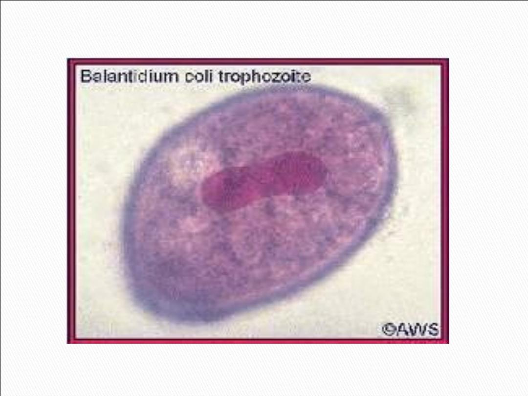
Balantidium
coli

Balantidium is the largest protozoan and only ciliate
known to parasitize humans
Morphology
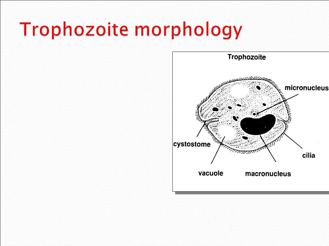
50-150 mic
Cilliated parasite
Oval shape
Greenish yellow color
Kidney or bean shape
Macronucleus
Small micronucleus
Retractile food vacule
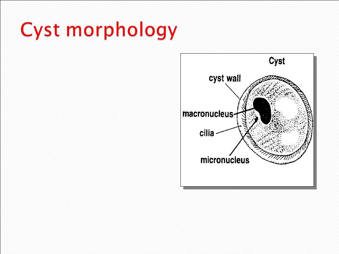
45-55 mic
Spherical shape
Cyst wall is thick
consist of 1-2 layers
No phagosome
Macronucleus
Conractile vacules
No cilia

Life cycle & Pathogenicity
:
Infection is happened by consumption of material
contaminated with feces of some farm animals
cotaining cyst (the infective stage)
.
Exystation happened in the small intestine
releasing trophozoites that migrate to the large
intestine. Trophozoites reside in the lumen of large
intestine Invade mucosa and submucosa
.
Feed on mucosal cells, RBC, leukocyte
where they divide by transverse binary fission
.
Encystation is triggered by dehydration of intestinal
content and cysts passed with stool
.
Life cycle

*Parasite live in L.I specially cecal region
*Cyst formed in large intestine or in outer
envirnment
1)
Intermitent periods of diarrhea and constipation
2)
Bloody diarrhea
3)
Abdominal pain
4)
Anorexia
5)
Ulceration of large intestine
6)
Tender colon
7)
Cachexia
8)
Gangrenous lesions could occur

1
.
History: if there any animal contact
.
2
.
Symptoms
Clinical signs could confused by E.
histolytica infection
3
.
Laboratory tests: finding the typical
trophozoites and cysts in the stoo
l
1
.
History: if there any animal contact
.
2
.
Symptoms
Clinical signs could confused by E.
histolytica infection
3
.
Laboratory tests: finding the typical
trophozoites and cysts in the stoo
l
Diagnosis

Laboratory methods to detect (cyst or trophozoite)
in stool by
•
Direct wet mount preparation methode
•
Stained smear by iodin
•
Looking for characteristic kidney shape nucleos
and retractile food vacule

Prevention & control
Avoid ingestion of food and drinks contaminated
.by animal feces

Treatment
1
.
Tetracycline
2
.
Iodoquinol
3
.
Metronidazole