
Acute hepatitis& Chronic hepatitis TUCOMInternal Medicine 4th classDr. Hasan.I.Sultan
Acute hepatitisLearning objectives;
• 1- Define hepatitis.
• 2- Define the acute hepatitis.
• 3-List the causes of acute hepatitis.
• 4-Discribe the various types of viral hepatitis. including viral hepatitis A, B, C, D and E.
• 5-Clarify the clinical features of acute viral hepatitis.
• 6-List the complications of acute viral hepatitis.
• 7-Discuss the investigations of acute viral hepatitis.
• 8-Identify the persons of high risk of viral hepatitis.
• 9-Explain the preventive measures of viral hepatitis.
• 10-Summarize the treatment options of acute viral hepatitis.
Acute hepatitis
Hepatitis; is clinicopathologic conditions due to liver damage by viral, toxic, metabolic, pharmacologic, or immune-mediated attack.Acute hepatitis; inflammation of liver lasting less than 6 months.
Fate: Either complete resolution or rapid progression to fulminant hepatitis and acute liver failure or slowly progress to chronic hepatitis.
Clinical features:
• Subclinical; Just abnormal LFT.
• Mild to moderate; Jaundice, enlarged tender liver + abnormal LFT.
• Severe; hepatocellular dysfunction with impairment of coagulation, marked jaundice, and disturbance of neurologic function.
Lab; Elevated aminotransferases levels 20- to 100-fold.
Histophathology; Characterized by infiltration of portal tract by lymphocytes, swelling and destruction of hepatocytes which may leads to intrahepatic cholestasis.


Important liver functions

Liver Histology: A. Infiltration of a portal tract, by lymphocytes and a few eosinophils, and lymphocytic infiltration of the epithelium of a bile duct. B. Infiltration by lymphocytes in a portal tract, and the lobular parenchyma, with swelling of hepatocytes, and mild lobular disarray. C. A focus of Kupffer cell clustering, containing PAS-positive membrane debris and intrahepatic cholestasis (black arrow). D. Staining for collagen showing mild pericellular fibrosis.
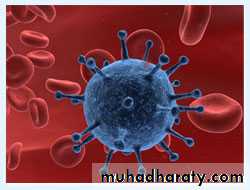
• Viral Hepatitis; Hepatitis A virus, Hepatitis B virus Hepatitis C virus, Hepatitis D virus ("delta agent") and Hepatitis E virus. Other: Epstein-Barr virus Cytomegalovirus.
• Alcohol
• Toxins; Amanita phalloides mushroom poisoning and Herbal preparations.
• Drugs; Acetaminophen, Isoniazid, Halothane Phenytoin, Ketoconazole.
• Other; Autoimmune hepatitis, Wilson's disease.
Causes of Acute Hepatitis

Acute Viral Hepatitis
This is a common cause of jaundiceHepatitis A
Hepatitis BHepatitis C
Hepatitis D
Hepatitis E
Group
Enterovirus
Hepadna
Flavivirus
Incomplete virus
Calicivirus
Nucleic acid
RNA
DNA
RNA
RNA
RNA
Size (diameter)
27 nm
42 nm
30-38 nm
35 nm
27 nm
Incubation (weeks)
2-4
4-20
2-26
6-9
3-8
Spread
Faeces
Yes
No
No
No
Yes
Blood
Uncommon
Yes
Yes
Yes
No
Saliva
Yes
Yes
Yes
?
?
Sexual
Uncommon
Yes
Uncommon
Yes
?
Vertical
No
Yes
Uncommon
Yes
No
Chronic infection
No
Yes
Yes
Yes
No
Prevention
Active
Vaccine
Vaccine
No
Prevented by
No
Passive
• Immune serum globulin
• Hyperimmune serum globulin
No• hepatitis B vaccination
No
Anti-HAV of IgM type, indicating a primary immune response, is already present in the blood at the onset of the clinical illness and is diagnostic of an acute HAV infection.
Hepatitis A

acute infection the hepatitis B surface antigen (HBs Ag) is a reliable marker of HBV infection
Anti-HBs Ab usually appears after about 3-6 months and persists for many years or perhaps permanently
The hepatitis B core antigen (HBc Ag) is not found in the blood, but antibody to it (anti-HBc) appears early in the illness and rapidly reaches a high titre which then subsides gradually but persists. Anti-HBc is initially of IgM type while IgG antibody appearing later.
The hepatitis B e antigen (Hbe Ag) appears only transiently at the outset of the illness and is followed by the production of antibody (anti-HBe). The Hbe Ag reflects active replication of the virus in the liver.
The persistence of HBs Ag for longer than 6 months indicates chronic infection.
Hepatitis B

Chronic HBV infection is marked by the presence of HBs Ag and anti-HBc (IgG) in the blood
HBV-DNA can be measured by polymerase chain reaction (PCR) in the blood. Viral loads are usually in excess of 105 copies/ml in the presence of active viral replication.Hepatitis B

HDV contains a single antigen to which infected individuals make an antibody (anti-HDV) initially IgM and later IgG.

Eighty per cent of individuals exposed to the virus will become chronically infected
hepatitis C RNA can be identified in the blood as early as 2-4 weeks after infection.Active infection is confirmed by the presence of serum hepatitis C RNA in anyone who is antibody positive.
Hepatitis C
In acute infection: IgM antibodies to HEV are positive.

HEV
1-Prodromal phase; Which last for several days and characterized by malaise, fatigue, anorexia, nausea, vomiting, myalgia, headache, and mild fever.
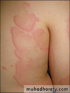
Arthritis and urticaria (serum sickness) due to immune complex deposition, may be present in 5 to 10% of cases of acute hepatitis B and C.
Taste and smell alteration.
Clinical features
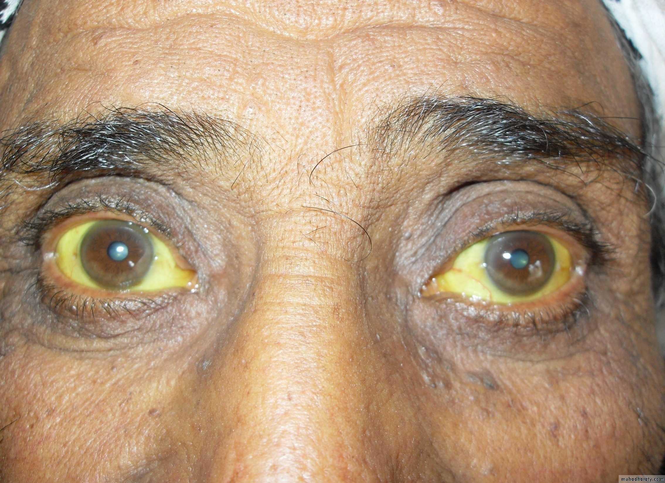
2-Icteric phase;
Jaundice appear after few days to 2 weeks.Dark urine and pale stools.
Improvement in the patient's well-being.
Splenomegaly is found in about one-fifth of patients.
Anicteric phase; Symptoms without jaundice.


Laboratory;
• Aminotransferases (ALT & AST) serum levels rise to greater than 20- 100 fold normal.• An elevated serum bilirubin (>2.5 to 3.0 mg/dL) or higher than 20 mg/dL .
• Serum alkaline phosphatase when it rise to more three times of normal levels indicate cholestatic hepatitis.
• Prolongation of the prothrombin time in sever hepatitis.
• Serological tests confirm the aetiology of the infection.
3-Recovery phase; Gradual resolution of symptoms and laboratory values.
Complications
• Cholestatic hepatitis.
• Acute liver failure.
• Chronic hepatitis.
• Aplastic anemia.
• Relapsing hepatitis.
Management
1-Supportive treatment;Rest
Maintenance of hydration and adequate dietary intake.
Alcohol should be avoided.
Hospitalization if severe nausea and vomiting or deterioration of liver function.
Drugs such as sedatives and narcotics should be avoided.
Elective surgery should be avoided.
Acute hepatitis C; α-interferon may decrease the risk of chronicity.
Vitamin K indicated if prolonged cholestasis.
2-Liver transplantation;
Very rarely indicated, when the patient developed acute liver failure.
Chronic Hepatitis
Learning objectives;• Define chronic hepatitis.
• List the causes of chronic hepatitis.
• Summarize the classification of chronic hepatitis.
• Describe the various types of chronic viral hepatitis including hepatitis B, C and D.
• Summarize the other forms of chronic hepatitis including nonalcoholic steatohepatitis, autoimmune hepatitis and metabolic (genetic) hepatitis.
• Interpret laboratory test results in chronic hepatitis.
• Discuss treatment options of chronic hepatitis.
Chronic Hepatitis
Defined as a hepatic inflammatory process lasting more than 6 months.Causes of Chronic Hepatitis;
• Viral Hepatitis; B, C and Hepatitis B with superimposed hepatitis D.
• Alcoholic hepatitis.
• Nonalcoholic steatohepatitis.
• Drugs and Toxins; Methyldopa, Nitrofurantoin, Amiodarone, Captopril, Propylthiouracil.
• Autoimmune
• Genetic and Metabolic Disorders; Wilson's disease, and α1-Antitrypsin deficiency.
Classification
1-Initial classification; which depend on histopathology only;
• Chronic persistent hepatitis; inflammatory activity confined to portal areas.
• Chronic lobular hepatitis; inflammatory activity and necrosis scattered throughout the lobule -- good prognosis.
• Chronic active hepatitis; inflammation that spilled into the adjacent lobule associated with necrosis and fibrosis, which progress to cirrhosis and liver failure.
2-Recent classification; which depend on;
• Etiologic agent• Grade of injury (numbers and location of inflammatory cells)
• Stage of disease (degree and location of fibrosis)

Chronic persistent hepatitis; inflammatory activity confined to portal areas.

Chronic lobular hepatitis; inflammatory activity and necrosis scattered throughout the lobule
Chronic active hepatitis; inflammation that spilled into the adjacent lobule associated with necrosis and fibrosis.

Chronic hepatitis B
Route of transmission and risk of chronic infectionHorizontal transmission (10%); Injection drug use, infected unscreened blood products, tattoos/acupuncture needles, sexual (homosexual and heterosexual).
Vertical transmission (90%); From HBs Ag positive mother.

Clinical features: Asymptomatic, fatigue, jaundice, malaise and anorexia, may progress to liver failure.
Extrahepatic features; arthralgias and arthritis, glomerulonephritis, and vasculitis , due to immune complexes deposition.
Invx; Aminotransferases elevated. HBs Ag +ve, HBe Ag +ve, and anti-HBc IgG Ab +ve.
Treatment
Indication for treatment; High viral load, elevated serum transaminases and/or histological evidence of inflammation.• Alfa-interferon or longer-acting pegylated interferons; that augmenting immune response.
• Lamivudine; Nucleoside analogue which inhibits DNA polymerase and suppresses HBV-DNA levels.
• Adefovir; Nucleotide analogue which inhibits HBV-DNA polymerase.
• Liver transplantation.
Chronic Hepatitis D
Chronic hepatitis B plus D has similar clinical and laboratory features to those seen in chronic hepatitis B alone– but more severe chronic hepatitis or cirrhosis.
Preventing hepatitis B effectively prevents hepatitis D.
Anti-HDV IgG titer is high.
Chronic Hepatitis C
75% will become chronically infected .Clinlcal features;
Similar to chronic hepatitis B, fatigue common, but jaundice is rare.
Extrahepatic features; Essential mixed cryoglobulinemia , Sjögren's syndrome, lichen planus, and porphyria cutanea tarda.
Progress to cirrhosis and hepatocellular carcinoma.
Invx;
Presence of serum hepatitis C RNA in anyone who is antibody-positive.Liver histology degree of liver fibrosis
Treatment;
Pegylated α-interferon together with oral ribavirin, a synthetic nucleotide analogue.
Liver transplantation
Non-alcoholic steatohepatitis
Most common in persons who are overweight, diabetes, and hyperlipidemia.Clinical features;
Asymptomatic; As abnormal LFTs.
Complication ; Cirrhosis ,variceal haemorrhage or hepatocellular carcinoma.
U/S; liver will appear bright.
Liver biopsy ; fat deposition is usually macrovesicular ,neutrophil infiltration and fibrosis.

Treatment;
Weight reductionVitamin E (as an antioxidant)
Lipid-lowering agents, particularly gemfibrozil.
Drugs that improve insulin resistance like metformin.
Autoimmune hepatitis
Is a liver disease of unknown aetiologyAssociation with other autoimmune diseases
Hypergammaglobulinaemia and autoantibodies
Young women
Insidious, with fatigue, anorexia, jaundice, fever, arthralgia, and vitiligo.
Spider naevi ,hepatosplenomegaly, 'cushingoid' face with acne, hirsutism and pink cutaneous striae.
Investigations;
Antinuclear antibodies, anti-smooth muscle antibody and antimicrosomal antibodies (anti-LKM) are positive. Increase IgG level.Treatment;
with corticosteroids, Azathioprine for maintenance.
GENETIC AND METABOLIC HEPATITIS
Wilson's disease and α1-antitrypsin deficiency
Before the age of 35 years
Family history of liver disease
Wilson's disease
Wilson's disease is a rare, autosomal recessive disorder of copper metabolism that is caused by a variety of mutations in the gene ATP7B on chromosome 13. Total body copper is increased, with excess copper deposited in, and causing damage to, several organs.Hepatic disease occurs predominantly in childhood and early adolescence. Neurological damage causes basal ganglion syndromes and dementia which tends to present in later adolescence. Others include renal tubular damage and osteoporosis. Kayser-Fleischer rings the most important single clinical clue to the diagnosis and can be seen in 60% of adults.


low serum caeruloplasmin is the best single laboratory clue to the diagnosis. high free serum copper concentration. high urine copper excretion of greater than 0.6 μmol/24 hrs and a very high hepatic copper content
The copper-binding agent penicillamine is the drug of choice.
13
Alpha1-antitrypsin deficiency
Alpha1-antitrypsin (α1-AT) is a serine protease inhibitor (Pi) produced by the liver. Homozygous individuals (PiZZ) have low plasma α1-AT concentrations, these forms cannot be secreted into the blood by liver cells because it is polymerised within the endoplasmic reticulum of the hepatocyte, that leads to hepatic and pulmonary disease.Liver disease includes chronic hepatitis and cirrhosis in adults, and in the long term hepatocellular carcinoma. Lung disease including emphysema in adult life.

Diagnosis is made from the low plasma α1-AT concentration and the PiZZ genotype.
No specific treatment is available; the concurrent risk of severe and early-onset emphysema means that all patients should be advised to stop cigarette smoking.