Lecture 12 (Immune system) [4-9th March 2017]
The immune system is a host defense system made up of many biological structures and processes within an organism that protects against disease. To function properly, an immune system must detect a wide variety of agents, known as pathogens, from viruses to parasitic worms, and distinguish them from the organism's own healthy tissue.Pathogens can rapidly evolve and adapt, and thereby avoid detection and neutralization by the immune system; however, multiple defense mechanisms have also evolved to recognize and neutralize pathogens. Humans, have sophisticated defense mechanisms, including the ability to adapt over time to recognize specific pathogens more efficiently. Adaptive (or acquired) immunity creates immunological memory after an initial response to a specific pathogen, leading to an enhanced response to subsequent encounters with that same pathogen. This process of acquired immunity is the basis of vaccination.
Disorders of the immune system can result in autoimmune diseases, inflammatory diseases and cancer. Immunodeficiency occurs when the immune system is less active than normal, resulting in recurring and life-threatening infections. In humans, immunodeficiency can either be the result of a genetic disease such as severe combined immunodeficiency, acquired conditions such as HIV/AIDS, or the use of immunosuppressive medication. There are a few immune ozrgans in the body which function to protect the body against the diseases:
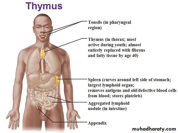
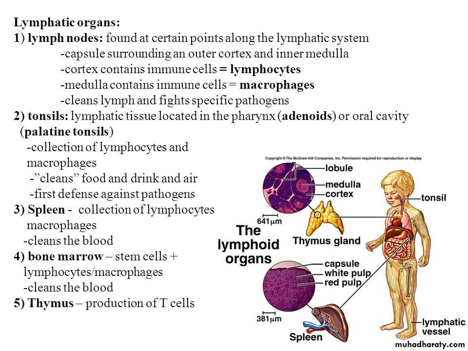
(1). Thymus: is a specialized primary lymphoid organ of the immune system and is an endocrine gland located behind the sternum above and front of the heart (Fig.1). Within the thymus, T-cells or T-lymphocytes mature. largest and most active during the neonatal and pre-adolescent periods. By the early teens, the thymus begins to atrophy and thymic stroma is mostly replaced by adipose (fat) tissue (Fig.2). Nevertheless, residual T- HYPERLINK "https://en.wikipedia.org/wiki/Lymphopoiesis" \o "Lymphopoiesis" lymphopoiesis continues throughout adult life. T cells are critical to the adaptive immune system, where the body adapts specifically to foreign invaders. The thymus is composed of two identical lobes and is located anatomically in the anterior superior HYPERLINK "https://en.wikipedia.org/wiki/Mediastinum" \o "Mediastinum" mediastinum, in front of the heart and behind the sternum.
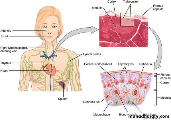
(Fig. 1): Location of thymus in the body, schematic diagram and histological structure.
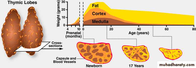
(Fig. 2): development and involution of Thymus gland in human.
Histologically, each lobe of the thymus can be divided into a central medulla and a peripheral cortex which is surrounded by an outer capsule. The cortex and medulla play different roles in the development of T-cells. Cells in the thymus can be divided into thymic HYPERLINK "https://en.wikipedia.org/wiki/Stromal_cell" \o "Stromal cell" stromal cells and cells of hematopoietic origin (derived from bone marrow resident hematopoietic stem cells). Developing T-cells are referred to as HYPERLINK "https://en.wikipedia.org/wiki/Thymocyte" \o "Thymocyte" thymocytes and are of hematopoietic origin. Stromal cells include epithelial cells of the thymic cortex and medulla, and HYPERLINK "https://en.wikipedia.org/wiki/Dendritic_cell" \o "Dendritic cell" dendritic cells (Fig.3).The thymus provides an inductive environment for development of T cells from hematopoietic progenitor cells. In addition, thymic stromal cells allow for the selection of a functional and self-tolerant T cell repertoire. Therefore, one of the most important roles of the thymus is the induction of central tolerance.
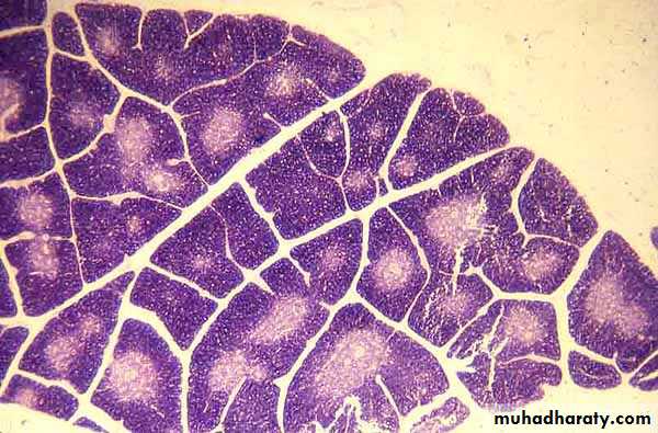
(Fig. 3): Histological configuration of Thymus gland of human.
A thymoma is a tumor originating from the epithelial cells of the thymus that may be benign or malignant and frequently associated with the neuromuscular disorder, myasthenia gravis. The latter is a long term neuromuscular disease that leads to varying degrees of skeletal muscle weakness. The most commonly affected muscles are those of the eyes, face, and swallowing. It can result in double vision, drooping eyelids, trouble talking, and trouble walking. Onset can be sudden. Those affected often have a large thymus gland or develop a HYPERLINK "https://en.wikipedia.org/wiki/Thymoma" \o "Thymoma" thymoma. Thymoma is found in 20% of patients with myasthenia gravis.[2] Once diagnosed, thymomas may be removed surgically. In the rare case of a malignant tumor, chemotherapy may be used.2). Lymph nodes: or lymph gland, is an ovoid or kidney-shaped organ of the lymphatic system, and of the adaptive immune system, that is widely present throughout the body (Fig.4). They are linked by the lymphatic vessels as a part of the circulatory system. Lymph nodes are major sites of B and T lymphocytes, and other white blood cells. Lymph nodes are important for the proper functioning of the immune system, acting as filters for foreign particles and cancer cells. Lymph nodes do not have a detoxification function, which is primarily dealt with by the liver and kidneys. In the lymphatic system the lymph node is a secondary lymphoid organ.
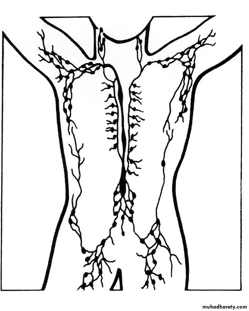
(Fig.4): distribution of lymph nodes in the body.
Lymph nodes are kidney or oval shaped and range in size from a few millimeters to about 1–2 cm long. Each lymph node is surrounded by a fibrous capsule, which extends inside the lymph node to form HYPERLINK "https://en.wikipedia.org/wiki/Trabecula" \o "Trabecula" trabeculae (Fig.5). The substance of the lymph node is divided into the outer cortex and the inner medulla. The cortex is continuous around the medulla except where the medulla comes into direct contact with the hilum. Thin reticular fibers of reticular connective tissue, and HYPERLINK "https://en.wikipedia.org/wiki/Elastin" \o "Elastin" elastin form a supporting meshwork called a reticulin inside the node. B-cells, are mainly found in the outer (superficial cortex) where they are clustered together as follicular B cells in lymphoid follicles and the T cells are mainly in the paracortex. The lymph node is divided into compartments called lymph nodules (or lobules) each consisting of a cortical region of combined follicle B cells, a paracortical region of T cells, and a basal part of the nodule in the medulla (Fig.6).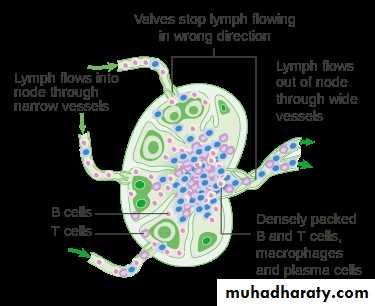
(Fig. 5) Section through the lymph node of human.
Lymph enters the convex side of the lymph node through multiple afferent lymphatic vessels, and flows through spaces called sinuses. A lymph sinus which includes the subcapsular sinus, is a channel within the node, lined by endothelial cells along with fibroblastic reticular cells and this allows for the smooth flow of lymph through them. The endothelium of the subcapsular sinus is continuous with that of the afferent lymph vessel and is also with that of the similar sinuses flanking the trabeculae and within the cortex. All of these sinuses drain the filtered lymphatic fluid into the medullary sinuses, from where the lymph flows into the efferent lymph vessels to exit the node at the hilum on the concave side. These vessels are smaller and don't allow the passage of the macrophages so that they remain contained to function within the lymph node. In the course of the lymph, lymphocytes may be activated as part of the adaptive immune response.Lymph nodes also have clinical significance. They become inflamed or enlarged in various diseases which may range from trivial throat infections, to life-threatening cancers. The condition of the lymph nodes is very important in cancer staging, which decides the treatment to be used, and determines the prognosis. When swollen, inflamed or enlarged, lymph nodes can be hard, firm or tender.
The number and composition of follicles can change especially when challenged by an antigen, when they develop a germinal center. Elsewhere in the node, there are only occasional leucocytes. As part of the reticular network there are follicular dendritic cells in the B cell follicle and fibroblastic reticular cells in the T cell cortex. The reticular network not only provides the structural support, but also the surface for adhesion of the HYPERLINK "https://en.wikipedia.org/wiki/Dendritic_cell" \o "Dendritic cell" dendritic cells, macrophages and lymphocytes. It allows exchange of material with blood through the high endothelial venules and provides the growth and regulatory factors necessary for activation and maturation of immune cells.
(Fig. 6): Section through a lymph node. 1) Capsule; 2) Subcapsular sinus; 3) Germinal centre; 4) Lymphoid nodule; 5) Trabeculae.
Spleen:
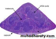
The spleen is an organ found in all vertebrates and in humans, is brownish in color and is located in the left upper quadrant of the abdomen. The spleen, is derived from HYPERLINK "https://en.wikipedia.org/wiki/Mesenchyme" \o "Mesenchyme"mesenchymal tissue and in healthy adult humans, is approximately 7-14 cm (2.8-5.5 in") in length. It usually weighs between 150-200 gm.
It has a similar structure to a large lymph node, does act as primarily as a blood filter and plays important roles in regard to red blood cells (RBC) and the immune system. It removes old RBC and holds a reserve of blood, which can be valuable in case of hemorrhagic shock, and also recycles iron. As a part of the mononuclear phagocyte system, it metabolizes hemoglobin removed from senescent RBC. The HYPERLINK "https://en.wikipedia.org/wiki/Globin" \o "Globin" globin portion of hemoglobin is degraded to its constitutive amino acids, and the HYPERLINK "https://en.wikipedia.org/wiki/Heme" \o "Heme" heme portion is metabolized to HYPERLINK "https://en.wikipedia.org/wiki/Bilirubin" \o "Bilirubin" bilirubin, which is removed in the liver.
The spleen synthesizes antibodies in its white pulp and removes antibody-coated bacteria and antibody-coated blood cells by way of blood and lymph node circulation. The red pulp of the spleen forms a reservoir that contains over half of the body's monocytes. These monocytes, upon moving to injured tissue, turn into HYPERLINK "https://en.wikipedia.org/wiki/Dendritic_cell" \o "Dendritic cell" dendritic cells and macrophages while promoting tissue healing. The spleen is a center of activity of the mononuclear phagocyte system and can be considered analogous to a large lymph node, as its absence causes a predisposition to certain infections.
Blood supply:
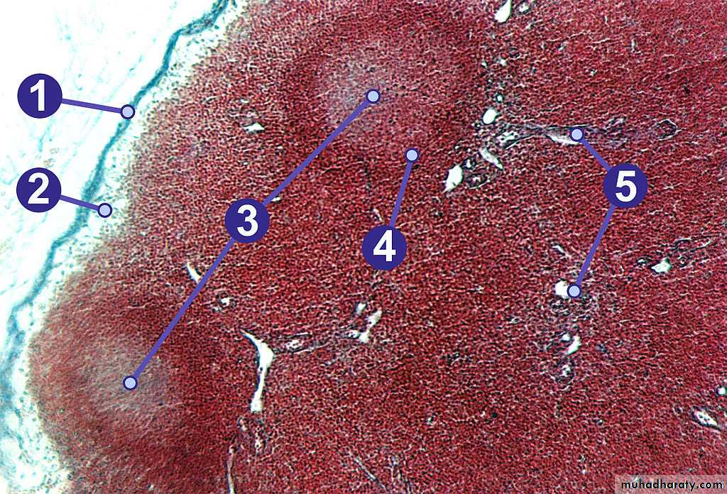
Near the middle of the spleen lies a long fissure, the splenic hilum. The HYPERLINK "https://en.wikipedia.org/wiki/Hilum_(anatomy)" \o "Hilum (anatomy)" hilum is the point of attachment for the gastrosplenic ligament and the point of insertion for the HYPERLINK "https://en.wikipedia.org/wiki/Splenic_artery" \o "Splenic artery" splenic artery and HYPERLINK "https://en.wikipedia.org/wiki/Splenic_vein" \o "Splenic vein" splenic vein. There are other openings present for lymphatic vessels and nerves. Like the thymus, the spleen possesses only efferent lymphatic vessels. The spleen is part of the lymphatic system. Both the short gastric arteries and the splenic artery supply it with blood. The germinal centers are supplied by arterioles called penicilliary radicles.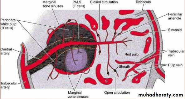
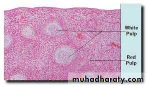
Splenic tissue shows the red pulp (red), white pulp (blue) and a thickened inflamed capusule (mostly pink - top of image) [H&E stain].
Area
Function
Composition
Red pulp
Mechanical filtration of red blood cells. In mice: Reserve of HYPERLINK "https://en.wikipedia.org/wiki/Monocyte" \o "Monocyte" monocytes[4]
Sinuses (sinusoids) are filled with blood
Splenic cords of reticular fibers
Marginal zone bordering on white pulp.
White pulp
Active immune response through humoral and cell-mediated pathways.
Composed of nodules, called HYPERLINK "https://en.wikipedia.org/wiki/White_pulp" \o "White pulp" Malpighian corpuscles. These are composed of:
lymphoid follicles (follicles) rich in B-lymphocytes
Periarteriolar lymphoid sheaths (PALS), rich in T-lymphocytes
Summary of Immune system
Tonsils are collections of lymphoid tissue facing into the aerodigestive tract. The set of lymphatic tissue known as HYPERLINK "https://en.wikipedia.org/wiki/Waldeyer%27s_tonsillar_ring" \o "Waldeyer's tonsillar ring" Waldeyer's tonsillar ring includes the adenoid tonsil, two tubal tonsils, two palatine tonsils, and the lingual tonsil.
The palatine tonsils, are masses of lymphatic material situated at either side at the back of the human throat. The palatine tonsils and the nasopharyngeal tonsil are lymphoepithelial tissues located near the HYPERLINK "https://en.wikipedia.org/wiki/Oropharynx" \o "Oropharynx" oropharynx and HYPERLINK "https://en.wikipedia.org/wiki/Nasopharynx" \o "Nasopharynx" nasopharynx (parts of the throat). Tonsils tend to reach their largest size near puberty, and they gradually undergo atrophy thereafter. However, they are largest relative to the diameter of the throat in young children.
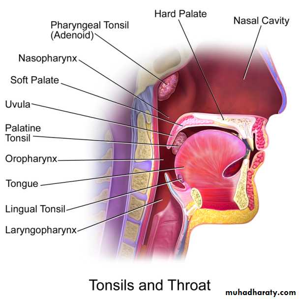
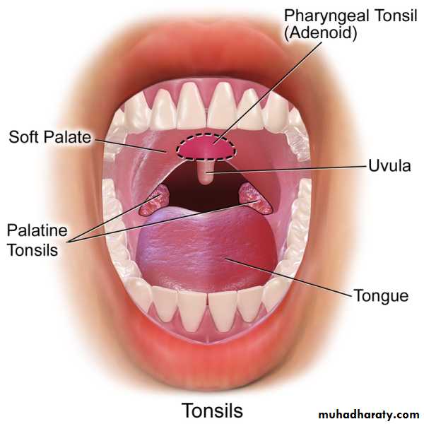
Tonsils in humans include, from anterior (front), superior (top), posterior (back), and inferior (bottom):
Type
Epithelium
capsule
Crypts
Location
Adenoids (pharyngeal tonsils)
Ciliated pseudostratified columnar (respiratory epithelium)
Incompletely encapsulated
No crypts, but small folds
Roof of pharynx
Tubal tonsils
Ciliated pseudostratified columnar (respiratory epithelium)
Roof of pharynx
Palatine tonsils
Non-keratinized stratified squamous
Incompletely encapsulated
Long, branched[2]
Sides of oropharynx between palatoglossaland palatopharyngeal arches
Lingual tonsils
Non-keratinized stratified squamous
Incompletely encapsulated
Long, branched
Behind terminal sulcus (tongue)
These HYPERLINK "https://en.wikipedia.org/wiki/Immunocompetent" \o "Immunocompetent" immunocompetent tissues are the immune system's first line of defense against ingested or inhaled foreign pathogens. Tonsils have on their surface specialized antigen capture cells called M-cells that allow for the uptake of antigens produced by pathogens. These M-cells then alert the underlying B-cells and T-cells in the tonsil that a pathogen is present and an immune response is stimulated. The B-cells are activated and proliferate in areas called germinal centres in the tonsil. These germinal centres are places where B memory cells are created and secretory antibody (IgA) is produced. Recent studies have provided evidence that the tonsils produce T lymphocytes, also known as T-cells, in a manner similar to the way the thymus does.
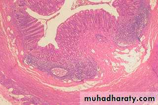
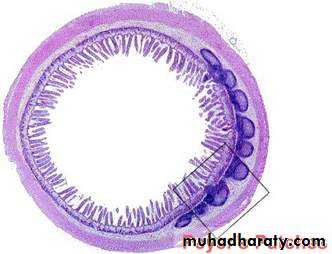
The Solitary lymphatic nodules (solitary follicles) are structures found in the small intestine and large intestine. The solitary lymphatic nodules are found scattered throughout the mucous membrane of the small intestine, but are most numerous in the lower part of the ileum. Their free surfaces are covered with rudimentary HYPERLINK "https://en.wikipedia.org/wiki/Intestinal_villus" \o "Intestinal villus" villi, except at the summits, and each gland is surrounded by the openings of the intestinal glands.
Each consists of a dense interlacing HYPERLINK "https://en.wikipedia.org/w/index.php?title=Retiform&action=edit&redlink=1" \o "Retiform (page does not exist)" retiform tissue closely packed with lymph-corpuscles, and permeated with an abundant capillary network. The interspaces of the retiform tissue are continuous with larger lymph spaces which surround the gland, through which they communicate with the lacteal system. They are situated partly in the submucous tissue, partly in the mucous membrane, where they form slight projections of its epithelial layer.
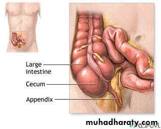
The solitary lymphatic nodules of the large intestine are most abundant in the cecum and vermiform process (appendix), but are irregularly scattered also over the rest of the intestine. They are similar to those of the small intestine.