hhhh

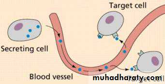
Hormones are chemical messengers that are secreted (released) from endocrine cells (glands) into the blood and coordinate the activities of different cells in another part of the body (multicellular organism).
-It is synthesized by specific tissues (glands).-They are secreted directly into the blood which carries them to their sites of action.
They specifically alter the activities of response tissues (target organs or target cells.
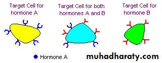
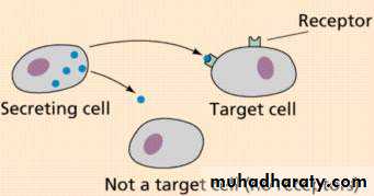
Hormones act by binding to specific receptors in the target tissue of interest.
Or a hormone is a class of regulatory biochemical produced in all multicellular organisms by glands and transported by the circulatory system to a distant target organ to coordinate its physiology and behavior .Thus hormones serve as a major form of communication between different organs and tissues.
Hormones regulate a variety of physiological and behavioral activities, including :
Digestion, metabolism ,respiration, tissue function,
lactation, stress, sensory perception, sleep, love, movement, excretion , growth and development , reproduction, and mood.
Generally, only a small amount of hormone is required to alter cell metabolism.

The Endocrine System regulates, coordinates and controls:
Growth and development.Male and female development.
How your body uses energy.
Levels of salts and sugars in your blood.
The amount (volume) of fluid in your body.
Appetite.
Many other body functions.

Gland
What it RegulatesPituitary
“Master Gland” that regulates all other Endocrine Glands, also releases growth hormone
Thyroid
Metabolism, body heat, bone growth
Parathyroids
Use of Calcium and Phosphorous
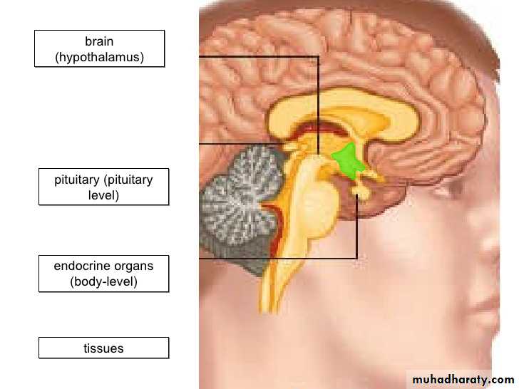
Hypothalamus
Links nervous system to endocrine system
AdrenalResponse in emergency or stressful situations, metabolism, blood pressure, salt balance
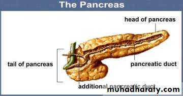
Pancreas
Blood sugar
Ovaries
Production of eggs; female characteristics
Testes
Production of sperm; male characteristics
Thymus
Parts of the immune system

The answer is Hormones!
Where are they secreted from?Hormone are secreted from the endocrine glands in the body. The glands are ductless, so hormones are secreted directly into the blood stream rather than by way of ducts.
The Endocrine System
Controls many body functionsexerts control by releasing special chemical substances into the blood called hormones
Hormones affect other endocrine glands or body systems
Derives its name from the fact that various glands release hormones directly into the blood, which in turn transports the hormones to target tissues via ducts.
The Endocrine and Exocrine Systems
Exocrine glandsRelease products indirectly into ducts .
Ex/ sweat, tears, digestive juices
Endocrine glands
Secretes directly to blood stream.No ducts.
Ex/ hormones
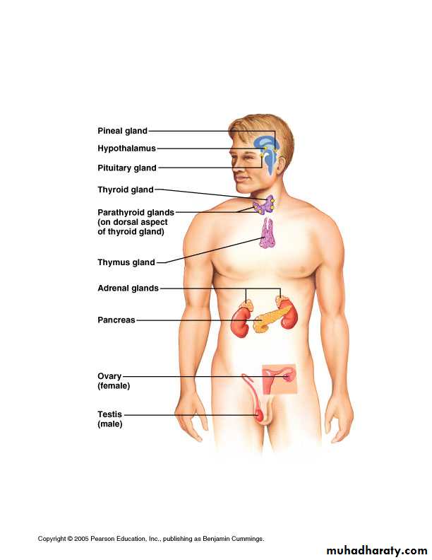
Endocrine Organs
Purely endocrine organs
Pituitary gland
Pineal gland الصنوبرية
Thyroid gland
Parathyroid glands
Adrenal: 2 glands
Cortex
Medulla
Endocrine cells in other organs
Pancreas
Thymus
Gonads
Hypothalamus
13
How do hormones work?
Hormone travel through the blood and influence the activity of other glands and organs.They produce short-and long term changes in various cells and organs by acting like neurotransmitters at metabotropic receptors.
A hormone can only influence cells that have specific target receptors for that particular hormone.
.
• frequency.
•
Hormones can classified by the location of the receptor and by the nature of the signal or second messenger used to mediate hormone action within the cell.
In animals, the brain is often a target organ for many of these hormones, and the brain in turn regulates the secretion of these hormones.
Hormones have the following effects on the body:
1-stimulation or inhibition of growth.2-wake-sleep cycle.
3-mood swings.
4-induction or suppression(inhibition) of apoptosis (programmed cell death).
5-activation or inhibition of the immune system.
6-regulation of metabolism.
7-preparation of the body for mating, fighting, fleeing, and other activity.
8-preparation of the body for a new phase of life, such as puberty, parenting, and menopause.
9-control of the reproductive cycle.
10-hunger cravings.
11-sexual arousal.
The coupling between signal and hormone occurs in two ways:
poly peptide and protein hormones bind to receptors located in the plasma membrane and generated a signal that regulate various interacellular function by changing the activity of enzyme.
steroid and thyroid hormones interact with interacellular receptors and this complex provide the signal.
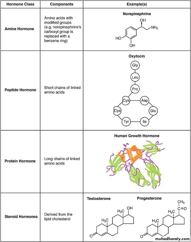
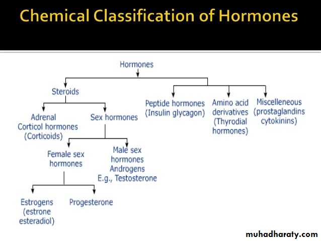
Classification of hormones according to chemical nature
1- Hormones derivative from amino acids as: Catechol amine derivative from tyrosine.Thyroid derived from tyrosine.
2- Hormones derivative from lipids as: Steroid (testosterone and cortisol). 1,25-dihydroxy cholcalciferol.
Prostaglandins hormones.
3-Hormones produce from simple peptides as: TRH (3 amino acids) Glucagon (29 amino acids) ACTH (39 amino acids) Vasopressine
4- Hormones composed from proteins as:
Parathyroid hormone(48 amino acid)
Growth hormone(191 amino acid)
Insuline
Chemical classes
Hormones fall into four main chemical classes:1-Amino acid derived – Examples include melatonin and thyroxine.
2-Peptides, polypeptides and proteins – Small peptide hormones include TRH and vasopressin. Peptides composed of scores or hundreds of amino acids are referred to as proteins. Examples of protein hormones include insulin and growth hormone. More complex protein hormones bear carbohydrate side-chains and are called glycoprotein hormones. Luteinizing hormone, follicle-stimulating hormone and thyroid-stimulating hormone are examples of glycoprotein hormones.
3-hormones derive from lipids such as arachidonic acid, lipoxins and prostaglandins.
4-Steroid Examples of steroid hormones include the sex hormones estradiol and testosterone as well as the stress hormone cortisol.
Hormones and their receptors
HormoneClass of hormone
LocationAmine (epinephrine)
Water-solubleCell surface
Amine (thyroid hormone)
Lipid soluble
IntracellularPeptide/protein
Water solubleCell surface
Steroids and Vitamin DLipid Soluble
IntracellularMechanism of action on target cells
Hormones: the body's chemical messengers
The human body secretes and circulates some 50 different hormones. These chemical substances are produced by endocrine cells, most of which are in glands. The hormones then enter the blood system to circulate throughout the body and activate target cells. The endocrine system, tightly linked to the nervous system, controls a large number of the body’s functions.Types of receptors
Receptors for the water soluble hormones are found on the surface of the target cell, on the plasma membrane.These types of receptors are coupled to various second messenger systems which mediate the action of the hormone in the target cell.
Receptors for the lipid soluble hormones reside in the nucleus (and sometimes the cytoplasm) of the target cell.
Because these hormones can diffuse through the lipid bilayer of the plasma membrane, their receptors are located on the interior of the target cell
Peptide Hormones
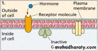
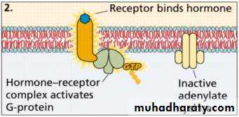
Peptide hormones do not enter the cell directly. These hormones bind to receptor proteins in the cell membrane.When the hormone binds with the receptor protein, a secondary messenger molecule initiates the cell response.
Because peptide hormones are water soluble, they often produce fast responses.
The hormones which are bind to receptors located in the plasma membrane of target cell(surface of cell).
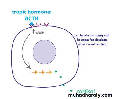
Hormone bind to the surface of cells communicate with intracellular metabolic processes through intermediary molecules called second messenger which are generated as a consequence of the ligand receptor interaction.
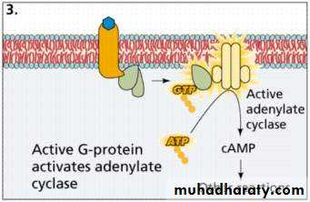
The complex was activation the adenylate cyclase enzyme (on membrane of cell also) to conversion the:
PPi
ATP cyclic-AMP
second messenger (as mediator)
The important feature of the second messenger is that the hormone needed cannot enter to the cell.
phosphodiesterase
C-AMP AMP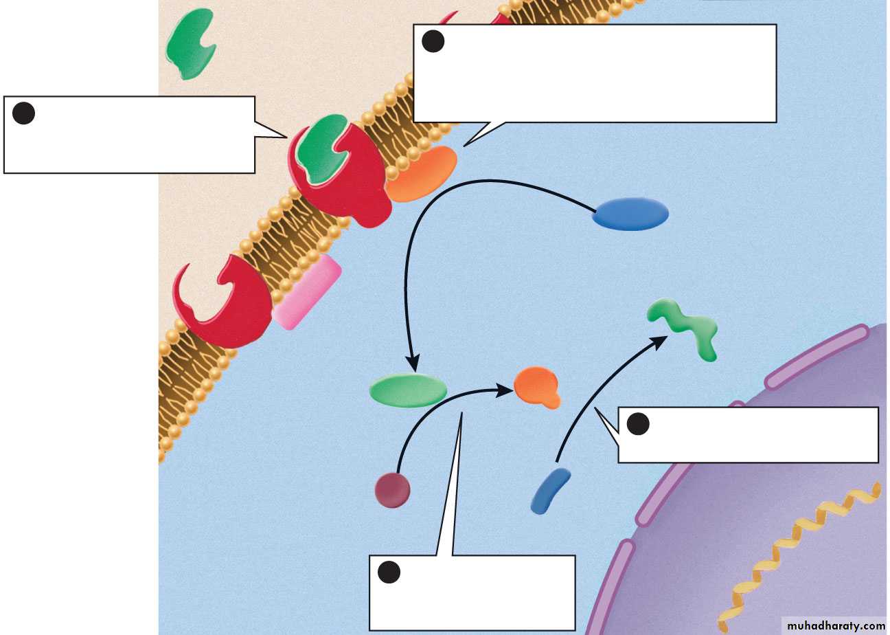
(cytoplasm)
(nucleus)peptide or amino
acid-derived
hormone
(first messenger)
(Extracellular fluid)
cyclic AMP-
synthesizing
enzyme
cyclic AMP
ATP
inactive
enzyme
(second messenger)
active
enzyme
reactant
product
plasma membrane
nuclear
envelope
receptor
The hormone binds to
a receptor on the plasma
membrane of a target cell
1
The activated enzymes
catalyze specific reactions
4
The second
messenger activates
other enzymes
3
Hormone–receptor binding
activates an enzyme that catalyzes
the synthesis of a second messenger,
such as cyclic AMP
2
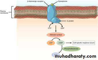
ACTH , LH , FSH , Secretine , Adrenaline , Glucagon
Adenylate cyclaseATP c-AMP
Hormones(polypeptide, proteins, glycoproteins, catecholamines)
(water soluble)Plasma membrane receptor
Second messenger
Intracellular metabolic process
Gene expression
Second messenger could be:c-AMP, c-GMP, Ca++, IP3.
The important feature of the second messenger that the hormone needed not enter to the cell.
Phosphodiesterase was inhibited by caffeine which present in coffee and theophyline which present in tea. The inhibition will keep the c-AMP in long duration, so the different activities will continue until inhibition removed.
Second messengers for cell-surface receptors
Second messenger systems include:Adenylate cyclase which catalyzes the conversion of ATP to cyclic AMP.
Guanylate cyclase which catalyzes the conversion of GMP to cyclic GMP (cyclic AMP and cyclic GMP are known collectively as cyclic nucleotides).
Calcium and calmodulin; phospholipase C which catalyzes phosphoinositide turnover producing inositol phosphates and diacyl glycerol.
Actions of second messenger:
1- Activation the transport across the intracellular membrane.2- Protein synthesis inside the endoplasmic reticulum.
3- Nuclear reactions (synthesis DNA and RNA).
4- Lipolysis and fatty acid synthesis from T.G.
5- Glycogenolysis.
6- Phosphorylation and dephosphorylation.
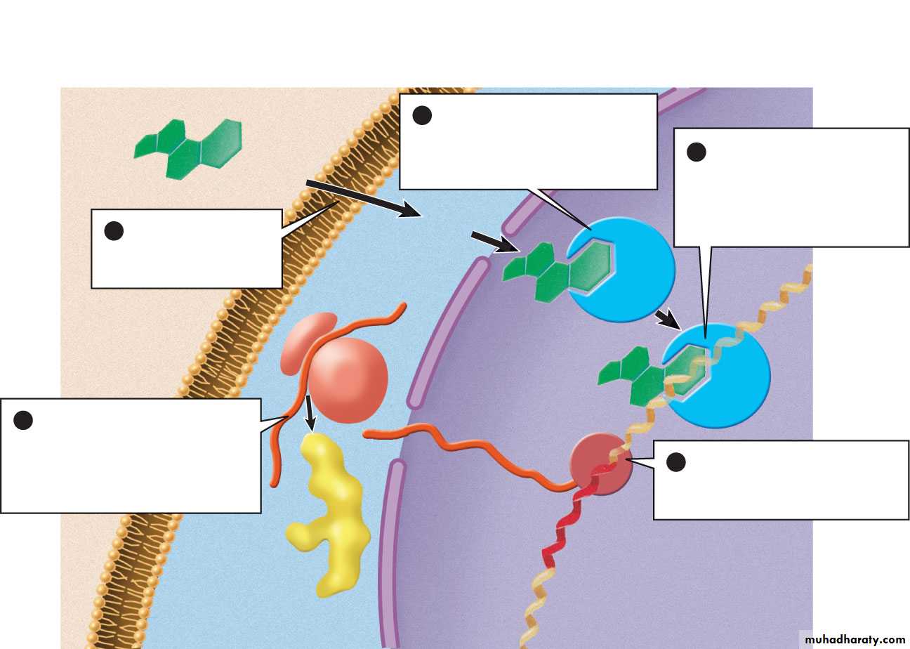
Steroid HormonesHormonal interact with intracellular receptor
Steroid hormones enter through the cell membrane and bind to receptors inside of the target cell.
These receptors moves into the nucleus and bind with a specific DNA sequence, that affecting gen expression.
By selectively affecting gen transcription and the production of the respective m-RNA .
The changing and metabolic processes are influence.
gene
plasmamembrane
ribosome
hormone receptor
steroid hormone
mRNA
(nucleus)
RNA polymerase
DNA
(cytoplasm)
new protein
(extracellular
fluid)
A steroid hormone
diffuses through the
plasma membrane
The hormone binds to a
receptor in the nucleus or to
a receptor in the cytoplasm
that carries it into the nucleus
The hormone–receptor
complex binds to DNA and
causes RNA polymerase to
bind to a nearby promoter
site for a specific gene
RNA polymerase catalyzes
the transcription of DNA into
messenger RNA (mRNA)
The mRNA leaves the
nucleus, then attaches to a
ribosome and directs the
synthesis of a specific protein
product
1
2
3
4
5
nuclear
envelope
Steroid Hormone Actions
Pass through the cell membraneBinds to specific receptors
Then enters the nucleus to bind with the cells DNA which then activates certain genes (Direct gene activation).
mRNA is synthesized in the nucleus and enters the cytoplasm and promotes protein synthesis for:
Enzymes as catalysts
Tissue growth and repair
Regulate enzyme function
For steroid or thyroid hormones, their receptors are located inside the cell within the cytoplasm of the target cell.
These hormones must first cross the cell membrane. They can do so because they are lipid-soluble. The combined hormone-receptor complex across the nuclear membrane into the nucleus of the cell, where it binds to specific DNA sequences, regulating the expression of certain genes, and thereby increasing the levels of the proteins encoded by these genes.
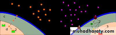
The left diagram shows a steroid (lipid) hormone (1) entering a cell and (2) binding to a receptor protein in the nucleus, causing (3) mRNA synthesis which is the first step of protein synthesis. The right side shows protein hormones (1) binding with receptors which (2) begins a transduction pathway. The transduction pathway ends (3) with transcription factors being activated in the nucleus, and protein synthesis beginning. In both diagrams, a is the hormone, b is the cell membrane, c is the cytoplasm, and d is the nucleus.
Regulation of hormones secretion
The quantities of hormone was regulated by feed-back inhibition which combine the :Hypothalamus + pituitary glands + Target tissues
• Hypothalamus
• Releasing hormones• Pituitary glands
• Stimulating hormones(Tropic hormones)
• Target organ
• Target hormone
Regulation
The rate of hormone biosynthesis and secretion is often regulated by negative feedback control mechanism. Such a mechanism depends on factors that influence the metabolism and excretion of hormones. Thus, higher hormone concentration alone cannot trigger the negative feedback mechanism. Negative feedback must be triggered by overproduction of an "effect" of the hormone.Hormone secretion can be stimulated and inhibited by: Other hormones (stimulating- or releasing -hormones)

Negative feedback means that when enough hormone is in the body, the body stops producing the hormone until it is needed again.

You eat. Glucose (sugar)
in the blood increases.
Increased glucose is detected by receptors
that notify the brain. It sends a message
to the pancreas to produce insulin.

Insulin tells muscle and liver to take up
glucose from the bloodstream and use it for energyor store it for later. Brain reduces appetite.

Blood glucose level drops as
it is removed by the cells.
Pancreas stops making insulin.

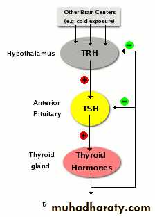
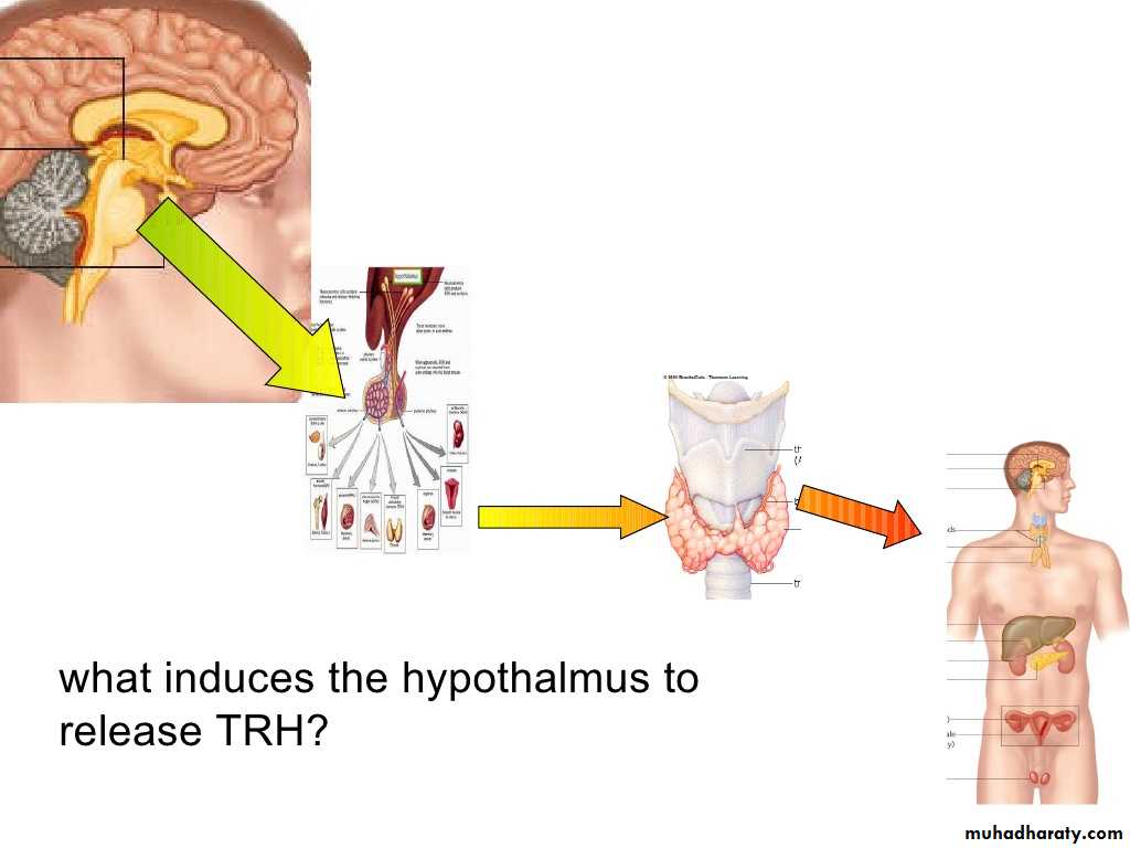
One special group of hormones is the tropic hormones that stimulate the hormone production of other endocrine glands. For example, thyroid-stimulating hormone (TSH) causes growth and increased activity of another endocrine gland, the thyroid, which increases output of thyroid hormones.
To release active hormones quickly into the circulation, hormone biosynthetic cells may produce and store biologically inactive hormones in the form of pre- or prohormones. These can then be quickly converted into their active hormone form in response to a particular stimulus.
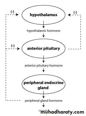
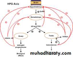
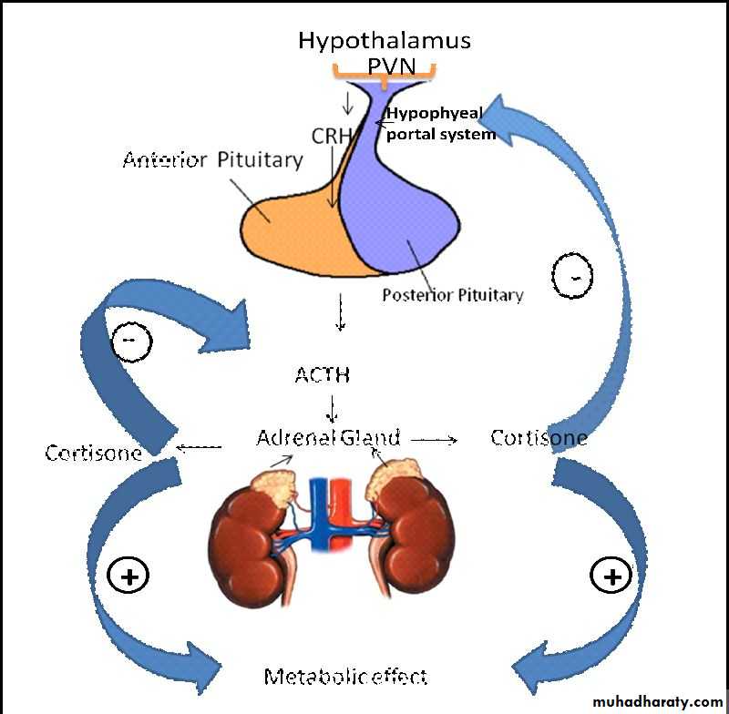
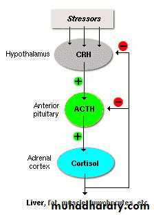
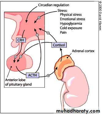
The feed-back inhibition takes these ways:
1- If the quantity of hormone was increased in target tissue, the hypothalamus hormone was inhibition, then the feed-back take the long loop.2- The target hormone was inhibit the tropic hormone, then the feed-back take the short loop.
3- The tropic hormone was inhibit the hypothalamus hormone, then the feed-back take the short loop.4- If the quantity of target hormone was decreased, the hypothalamus hormone was activation and regulate the secretion of pituitary glands. These glands are stimulated to secrete their specific hormones, which are carried by the blood to the target tissues to secrete the third hormone that is the organ tissue was needed.
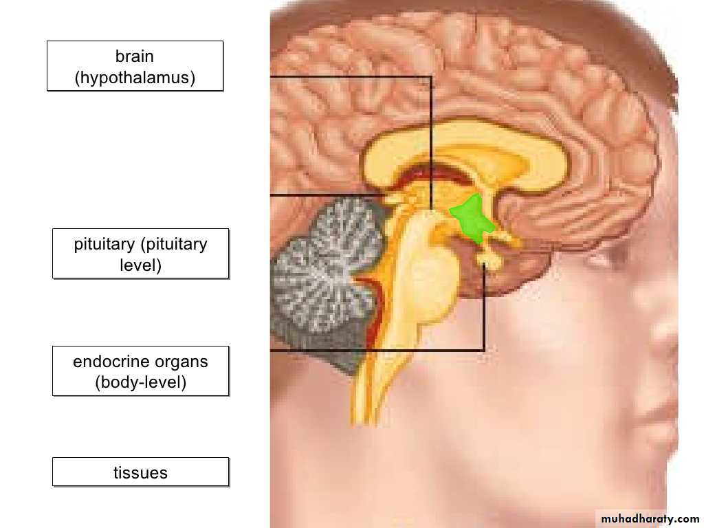
The hypothalamus gland :Small structure at the base of the brain .Regulates many body functions, including:
1- Appetite.
2- Body temperature.3- Regulates the pituitary glands.4- It function in stress situation . 5- Mood regulation.
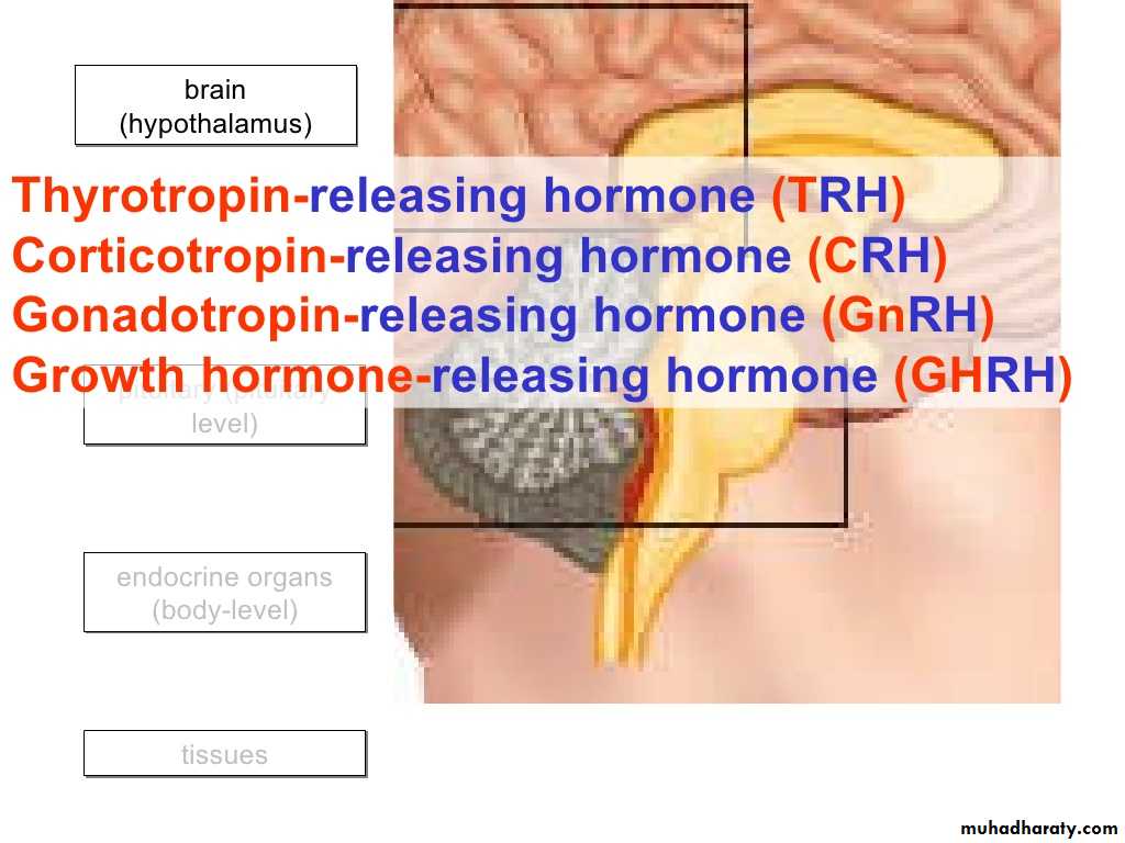
secrete hormones which are called releasing hormones (or releasing factors) , which are send to the pituitary gland , located just below the hypothalamus. Hypo means: underThalamus is from Greek word meaning : a bed
Hypothalamus hormones
Releasing hormones (releasing factors) of hypothalamusSecreted like neurotransmitters from neuronal axons into capillaries and veins to anterior pituitary :
1- TRH (thyroid releasing hormone) -----turns on* TSH
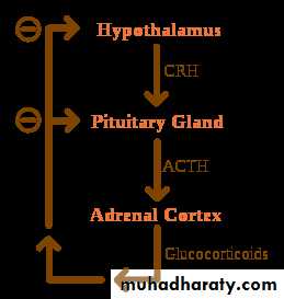
2- CRH (corticotropin releasing hormone) -----turns on ACTH
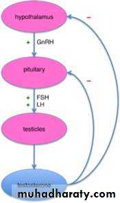
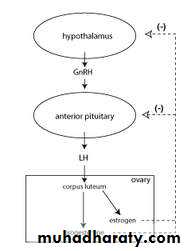
3- GnRH (gonadotropin releasing hormone) ---turns on FSH and LH
4- PRH (prolactin releasing hormone) -----turns on PRL
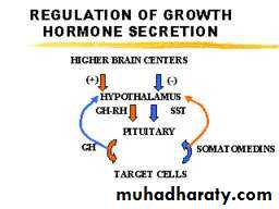
5- GHRH (growth hormone releasing hormone) ----turns on GH
The hypothalamus controls secretion of hormones which in their turn control the secretion of hormones by the thyroid gland, the adrenal cortex and gonads: in this way the brain controls these endocrine glands
64
*Note: “turns on” means causes to be released
The hypothalamus gland secrete hormones which are called releasing hormone(or releasing factors)which are send to the pituitary gland, located just below hypothalamus.
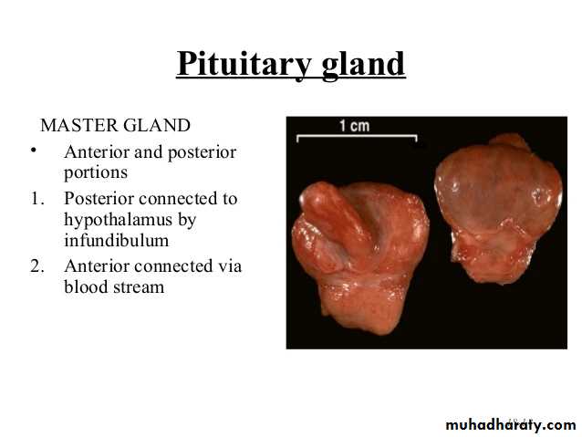
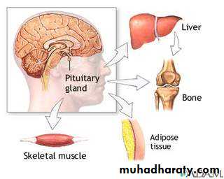
What is the pituitary gland?
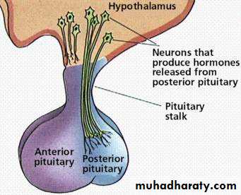
The pituitary is an important gland in the body and it is often referred to as the 'master gland', because it controls several of the other hormone glands (e.g. adrenals, thyroid).It is usually about the size of a pea and consists of two parts (often called lobes) - a front part, called the anterior pituitary and a back part, called the posterior pituitary.
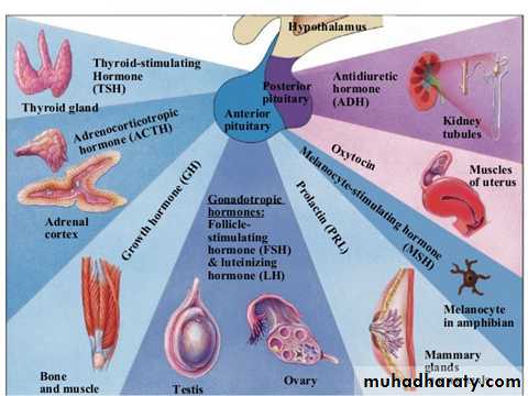
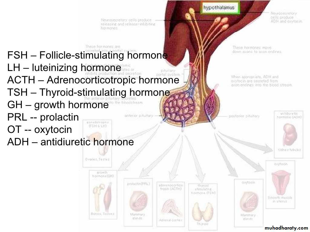
The anterior pituitary produce 7 hormones called tropic (tropin) hormones:
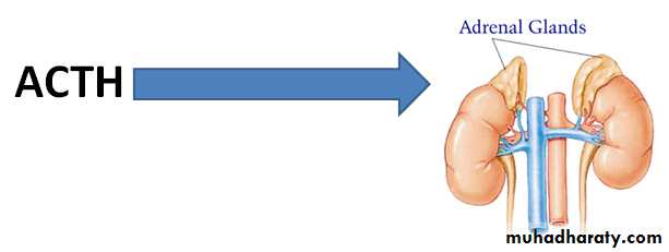
GH ( somatrotropic hormone) stimulates growth of skeletal epiphyseal plates and body to synthesize proteinACTH stimulates the adrenal cortex to produce corticosteroids: aldosterone and cortisol
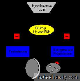
FSH stimulates follicle growth and ovarian estrogen production; stimulates sperm production
LH has a role in ovulation and the growth of the corpus luteum; stimulates androgen secretion in testes ;stimulates secretion of testosterone in male
71
The others from the anterior pituitary…
TSH stimulates the thyroid to produce thyroid hormone(thyroxine)PRL stimulates mammary glands in breast to make milk
MSH stimulates melanocytes; may increase mental alertness72
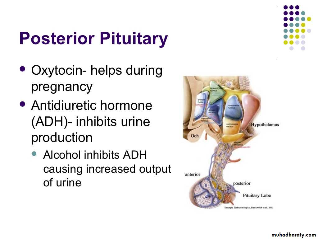
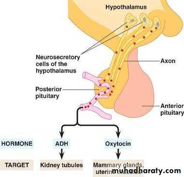
From the posterior pituitary
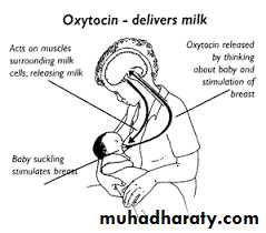
ADH (antidiuretic hormone , vasopressin) stimulates the kidneys to reclaim more water from the urine, raises blood pressureOxytocin prompts contraction of smooth muscle in reproductive tracts, in females initiating labor and ejection of milk from breasts
73
75
TSH: thyroid-stimulating hormone
ACTH: adrenocorticotropic hormone
FSH: follicle-stimulating hormone
LH: luteinizing hormone
GH: growth hormone
PRL: prolactin
MSH: melanocyte-stimulating hormone
ADH: antidiuretic hormone
Oxytocin
TRH (thyroid releasing hormone)
turns on TSH
CRH (corticotropin releasing hormone)
turns on ACTH
GnRH (gonadotropin releasing hormone) turns on FSH and LH
PRF (prolactin releasing hormone)
turns on PRL
GHRH (growth hormone releasing hm)
turns on GH
TSH stimulates the thyroid to produce thyroid hormone
ACTH stimulates the adrenal cortex to produce corticosteroids: aldosterone and cortisolFSH stimulates follicle growth and ovarian estrogen production; stimulates sperm production and androgen-binding protein
LH has a role in ovulation and the growth of the corpus luteum; stimulates androgen secretion by interstitial cells in testes
GH ( somatrotropic hormone) stimulates growth of skeletal epiphyseal plates and body to synthesize protein
PRL stimulates mammary glands in breast to make milk
MSH stimulates melanocytes; may increase mental alertness
ADH (antidiuretic hormone or vasopressin) stimulates the kidneys to reclaim more water from the urine, raises blood pressure
Oxytocin prompts contraction of smooth muscle in reproductive tracts, in females initiating labor and ejection of milk from breasts
Can we put it all together?
Red is from hypothalamus
Black is from pituitary
Anterior pituitary hormones are:
1-Growth hormone (GH) ,Somatotropin :
is a protein hormone secreted by somatotrope cells which are part of anterior pituitary gland.
The secretion is stimulated by sleep, stress, exerise, fasting, certain drugs.
GHR GH Target organ
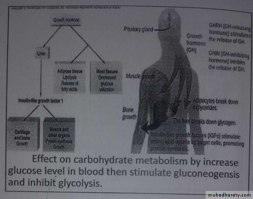
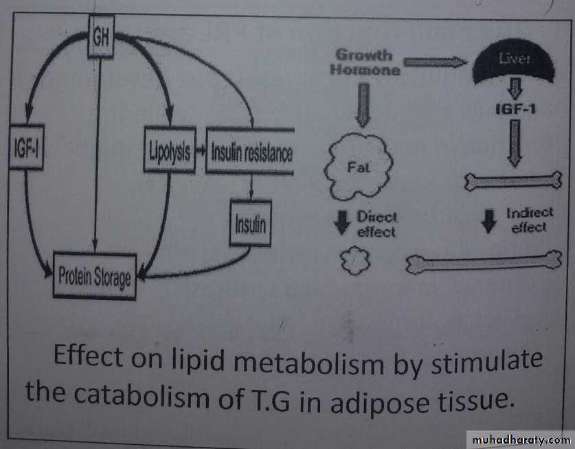
Hypothalamus pituitaryThe main function of GH: Effect on protein metabolism by increase amino acids transport to muscle cells for build protein. Effect on carbohydrate metabolism by increase glucose level in blood then stimulate gluconegenolysis and inhibit glycolysis. Effect on lipid metabolism by stimulate catabolism of T.G in adipose tissue. Retention certain minerals them its stimulate bone growth.
GH (Acromegaly) GH (Dwarfism)
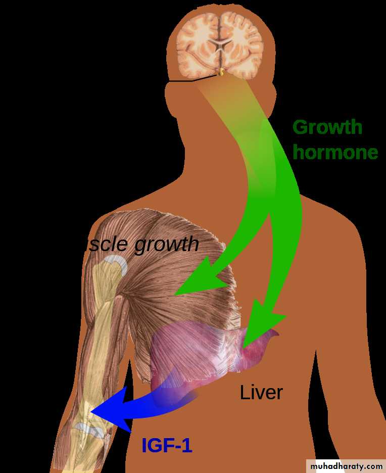


Effect on protein metabolism by increase amino acids transport to muscle cells for build protein.
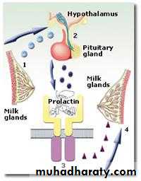
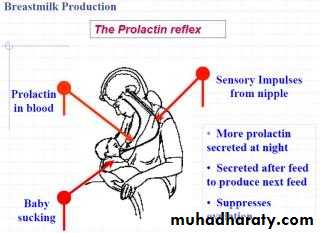
2 – Prolactin (PRL) Lactogenic hormone (Lactotropin)Is a protein hormone . Number and size of these cells increased in pregnancy.
The main function of PRL are:
Stimulate the breast tissue development and milk production in female, while the function in male is un cleare, however might have some role in the fertilization process.Synthesis two proteins:
- Diphospho galactosyl transferase which is responsible for lactose synthesis.
- α lact albumin which is develop the first enzyme to covert:
Glucose Galactose
PRH PRL Target organ
3- Gonadotrophic hormones
1- Follicle-Stimulating Hormone(FSH):-FSH produced by gonadotropine cells which are part of anterior pituitary gland.
It is a glycoprotein.
The main function of FSH:
In female:-
Stimulate the growth of ovarian follicles.
In male:-
Stimulate the growth of spermatogenesis.
2- Luteinizing Hormone(LH):-LH produced by gonadotrop cells which are part of another pitutary gland.The main function of LH:In female:- Stimulate the ova synthesis and corpus luteum synthesis . Stimulate estrogen and progesterone synthesis.In mail:- Stimulate testosterone synthesis.LHRH LH and FSH
4- Thyotropin (Thyroid stimulating hormone) (TSH)
TSH produced by thyrotrop cell which are part of anterrior pitutary gland.TSH is glycoprotein .It is combine with plasma membrane receptor by activation adenylate cyclase to convert:ATP c-AMP (second messenger)which response of TSH action.
The main function of TSH:- Stimulate the thyroid hormone (T3,T4).- Control of iodide concentration and converted it to organic compound by binding with thyroglobulin then it was hydrolyze.- Increase the concentration and synthesis of protein, phospholipid and nuclic acid.- Lipolysis the nutrient lipid in adipose tissues. TRH TSH T3,T4hypothalamus anterial pituitary target hormone
5- Adrencorticotropic hormone (ACTH):-
The ACTH is poly peptide consist of 39 amino acids.
The main function of ACTH :-1- Increased steroid hormone from adrinal cortix by conversion cholesterol to pregnenolone.( the precursor for all steroid) hormones).
2-Regulate production and secretion all adrinal cortix hormones.3-Transfer the unsaturated fatty acid from adipose tissues to blood plasma.4-Increase synthesis of ketone bodies. 5-Decrease synthesis of urea from amino acid.6-Delay the loss of adrinal cortix hormones in liver.
6-Melanocyte-stimulating hormone (MSH):
Also called melanotropin hormone. Are a family of peptide hormones produced by the skin, pituitary gland and hypothalamus in response to ultraviolet (UV) radiation that are produced by cells in the intermediate lobe of the pituitary gland.The main function:
1- MSH stimulate the production and release of melanin in skin ,hair and eyes. It does this by inducing skin cells called melanocytes to produce a pigment called melanin.2- MSH actions in the brain have effects on appetite and sexual arousal.
An increase in MSH will cause darker skin in humans. MSH increases in humans during pregnancy. This, along with increased estrogens, causes increased pigmentation in pregnant women.
The MSH include α-MSH, β-MSH, and γ-MSH. They are distinguished from one other by their preferential binding to different melanocortin receptors .
Different levels of MSH are not the major MSH of racialعنصري variation in skin colour. However, in many red-headed people, and other people who do not tan well, there are variations in their hormone receptors, causing them to not respond to MSH in the blood.
What happens if I have too much melanocyte-stimulating hormone?
A direct consequence of high levels of MSH is increased production of melanin. This can occur as a result of prolonged exposure to the sun or skin tanning. However, people with a high blood level of MSH do not necessarily tan very well or have even skin pigmentation. Very fair skinned people tend to produce less melanin due to variations in their melanocyte-stimulating hormone receptors, which means they do not respond to MSH levels in the blood.
Hyperpigmentation or abnormal darkening of the skin is found in patients with primary adrenal insufficiency, the adrenal glands do not produce enough hormones (including cortisol). This leads to a positive feedback loop where the hypothalamus stimulates the pituitary gland to release more ACTH to try and stimulate the adrenal glands to produce more cortisol. ACTH can be broken down to produce MSH leading to hyperpigmentation of the skin.
What happens if I have too little melanocyte-stimulating hormone?
A deficiency in MSH results in a lack of skin pigmentation and subsequent loss of natural protection from UV rays of the sun. In secondary adrenal insufficiency, damage to the pituitary gland prevents release of ACTH and MSH and there is reduced pigmentation of the skin.MSH deficiency can cause increased inflammation, pain, and sleeping problems as well as a reduction in the levels of anti-diuretic hormone(ADH) which causes thirst and frequent urination. MSH deficiency may also result in increased food intake and obesity.
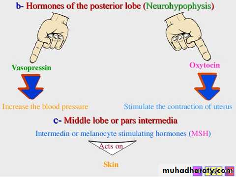
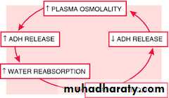
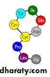
Vsopressin
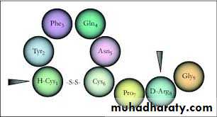
Vasopressin, also known as antidiuretic hormone (ADH), is a neurohypophysial hormone found in most mammals.
Vasopressin is a peptide hormone.
Most of it is stored in the posterior pituitary.Functions
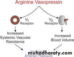
One of the most important roles of ADH is to regulate the body's retention of water in the body; it is released when the body is dehydrated and causes the kidneys to conserve water, thus concentrating the urine and reducing urine volume. At high concentrations, it also raises blood pressure by inducing moderate vasoconstriction.It also increases peripheral vascular resistance, which in turn increases arterial blood pressure. It plays a key role in homeostasis, by the regulation of water, glucose, and salts in the blood.
Increasing the water permeability of distal convoluted tubule and collecting duct cells in the kidney, thus allowing water reabsorption and excretion of more concentrated urine, i.e., antidiuresis
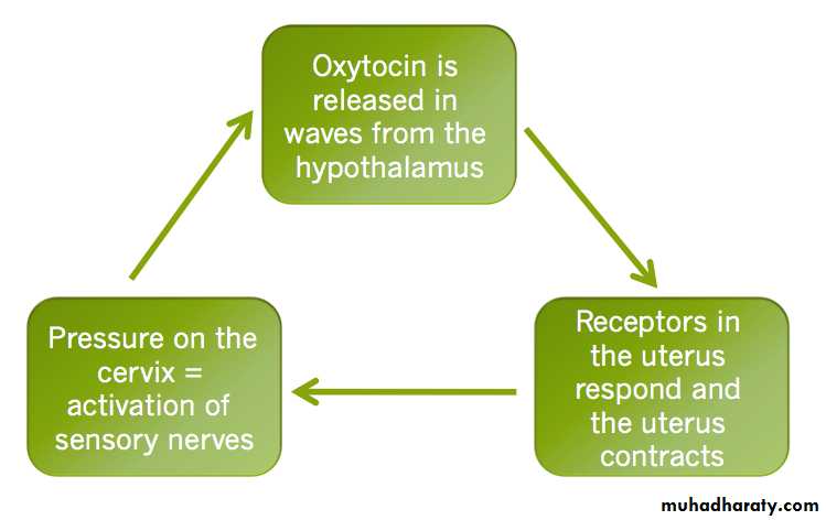
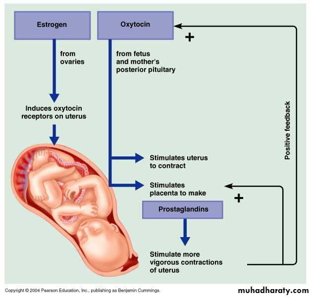
oxytocin
Oxytocin is used to cause contraction of the uterus in order to start labor or increase the speed of labor, and to stop bleeding following delivery. For this purpose, it is given either by injection into a muscle or into a vein.
The use of oxytocin as excessive contraction of the uterus that can cause distress in an unborn baby. Common side effects in the mother include nausea and a slow heart rate. Serious side effects include water intoxication with an excessive dose and uterus rupture. Allergic reactions may also occur.
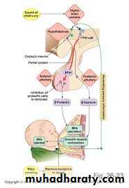
Oxytocin is normally produced in the hypothalamus.It plays a role in social bonding, sexual reproduction in both sexes, and during and after childbirth. Oxytocin is released into the bloodstream as a hormone in response to stretching of the cervix and uterus during labor and with stimulation of the nipples from breastfeeding. This helps with birth, bonding with the baby, and milk production
In lactating (breastfeeding) mothers, oxytocin acts at the mammary glands, causing milk to be 'let down' into subareolar sinuses, from where it can be excreted via the nipple.

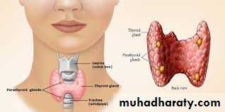
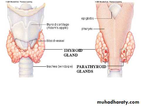
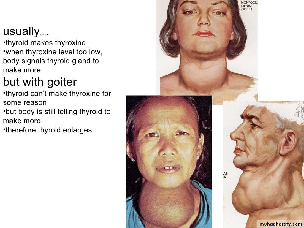
Thyroid gland
The thyroid gland is a butterfly-shaped organ located in the base of the neck. It releases hormones that control metabolism.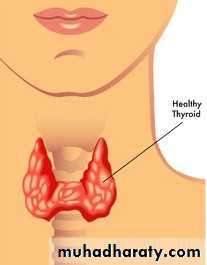
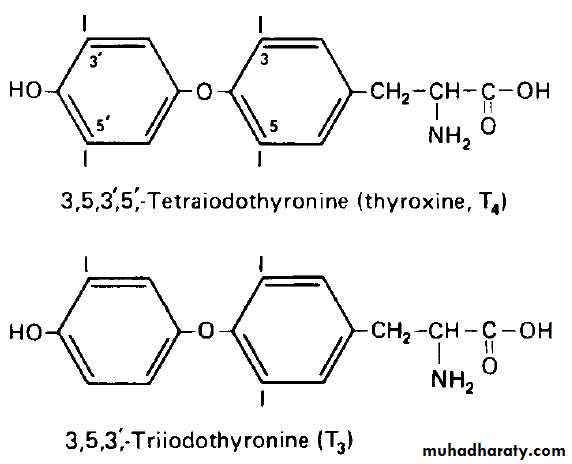
The thyroid hormones, triiodothyronine (T3) and its prohormone, thyroxine (T4), are tyrosine-based hormones produced by the thyroid gland that are primarily responsible for regulation of metabolism.
T3 and T4 are partially composed of iodine. A deficiency of iodine leads to decreased production of T3 and T4, enlarges the thyroid tissue and will cause the disease known as simple goitre.
The major form of thyroid hormone in the blood is thyroxine (T4), which has a longer half-life than T3.
T4 is converted to the active T3 within cells by deiodinases (5'-iodinase). These are further processed by decarboxylation and deiodination. All three isoforms of the deiodinases are selenium-containing enzymes, thus dietary selenium is essential for T3 production.
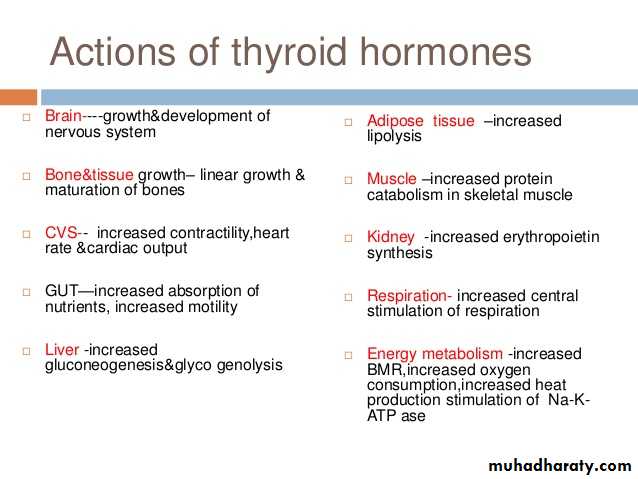
The main functions:
The thyroid's hormones regulate vital body functions, including:Breathing
Heart rateCentral and peripheral nervous systems
Body weight
Muscle strength
Menstrual cycles
Body temperature
Cholesterol levels
How the Thyroid Gland Works
The thyroid is part of the endocrine system, which is made up of glands that produce, store, and release hormones into the bloodstream so the hormones can reach the body's cells. The thyroid gland uses iodine from the foods you eat to make two main hormones:
Triiodothyronine (T3)
Thyroxine (T4)It is important that T3 and T4 levels are neither too high nor too low. Two glands in the brain—the hypothalamus and the pituitary communicate to maintain T3 and T4 balance.
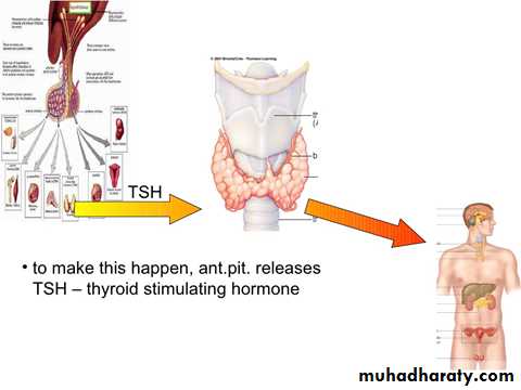
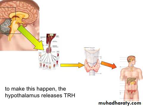
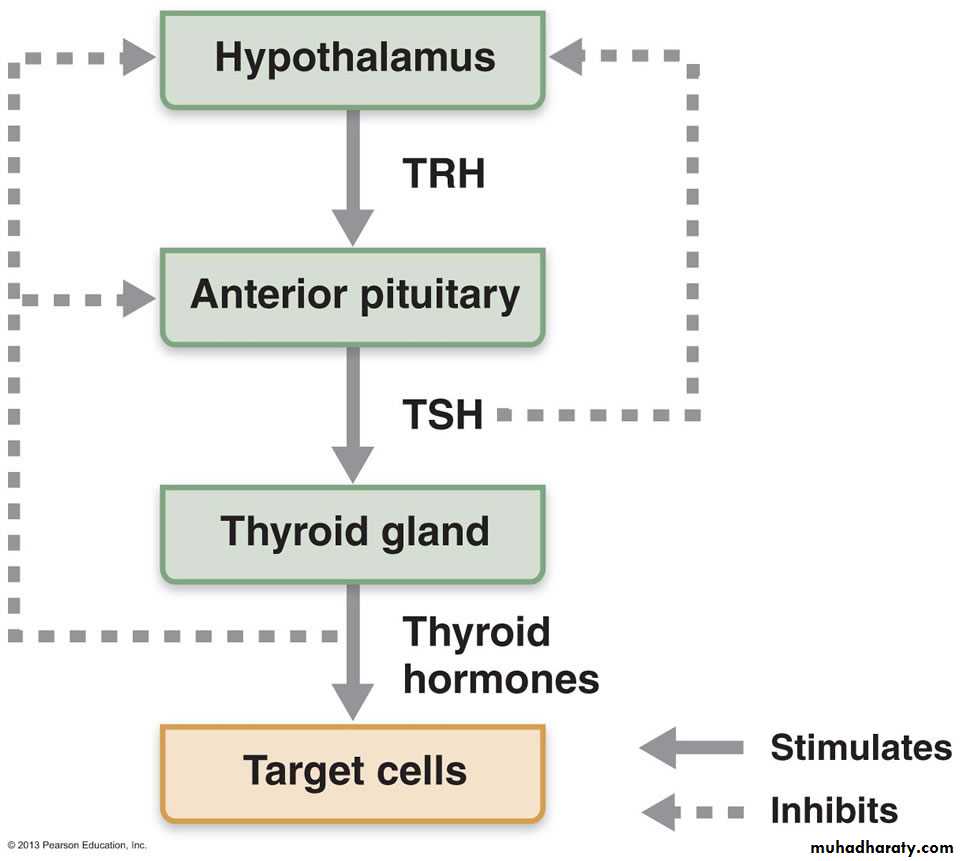
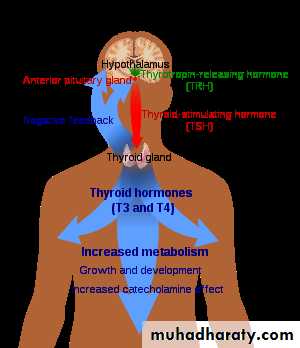
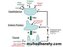
The hypothalamus produces Thyroid Releasing Hormone (TRH) that signals the pituitary to tell the thyroid gland to produce more or less of T3 and T4 by either increasing or decreasing the release of a hormone called thyroid stimulating hormone (TSH).
When T3 and T4 levels are low in the blood, the pituitary gland releases more TSH to tell the thyroid gland to produce more thyroid hormones.
If T3 and T4 levels are high, the pituitary gland releases less TSH to the thyroid gland to slow production of these hormones.
Why You Need a Thyroid Gland??
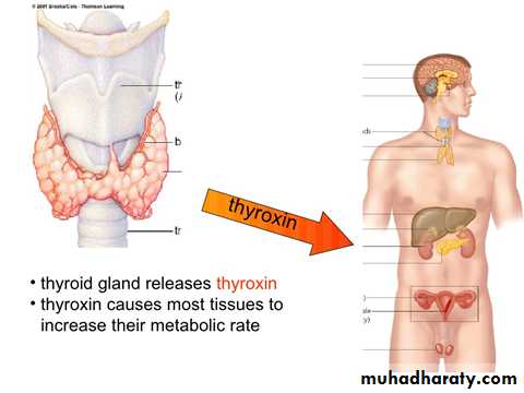
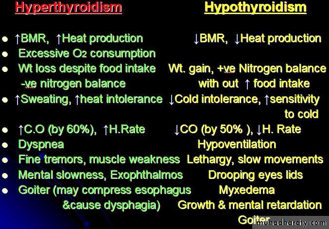
T3 and T4 travel in the bloodstream to reach almost every cell in the body. The hormones regulate the speed with which the cells/metabolism work. For example, T3 and T4 regulate heart rate and how fast your intestines process food. So if T3 and T4 levels are low, your heart rate may be slower than normal, and you may have constipation/weight gain. If T3 and T4 levels are high, you may have a rapid heart rate and diarrhea/weight loss.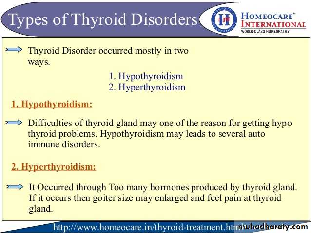
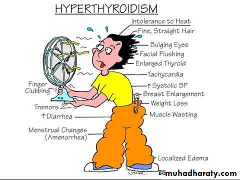
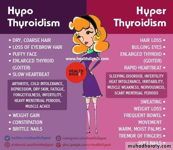
Listed below are other symptoms of too much T3 and T4 in your body (hyperthyroidism):
AnxietyIrritability or moodiness
Nervousness, hyperactivity
Sweating or sensitivity to high temperatures
Hand trembling (shaking)
Hair loss
Missed or light menstrual periods
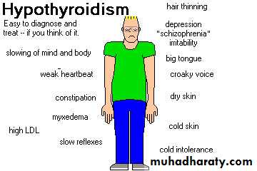
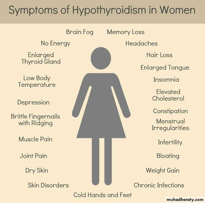
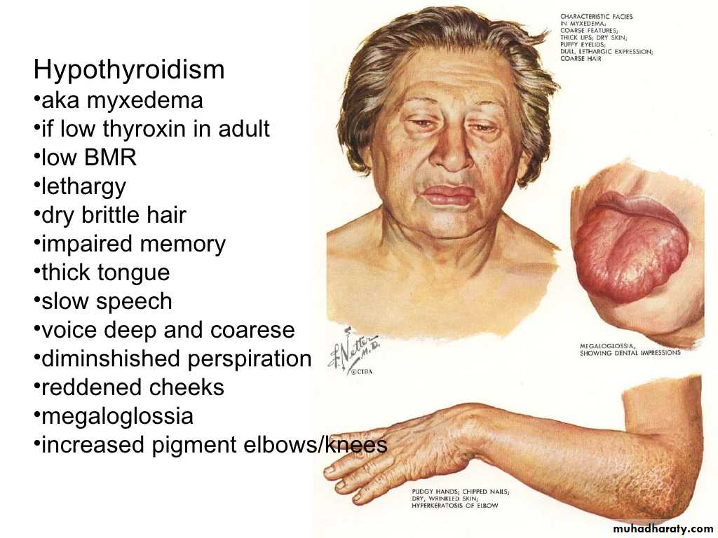
The following is other symptoms of too little T3 and T4 in your body (hypothyroidism):
Trouble sleepingTiredness and fatigue
Difficulty concentrating
Dry skin and hair
Depression
Sensitivity to cold temperature
Frequent, heavy periods
Joint and muscle pain
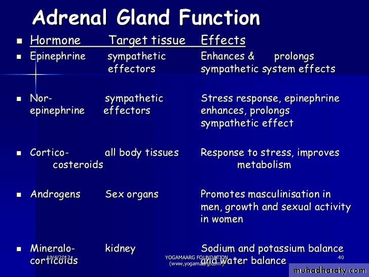
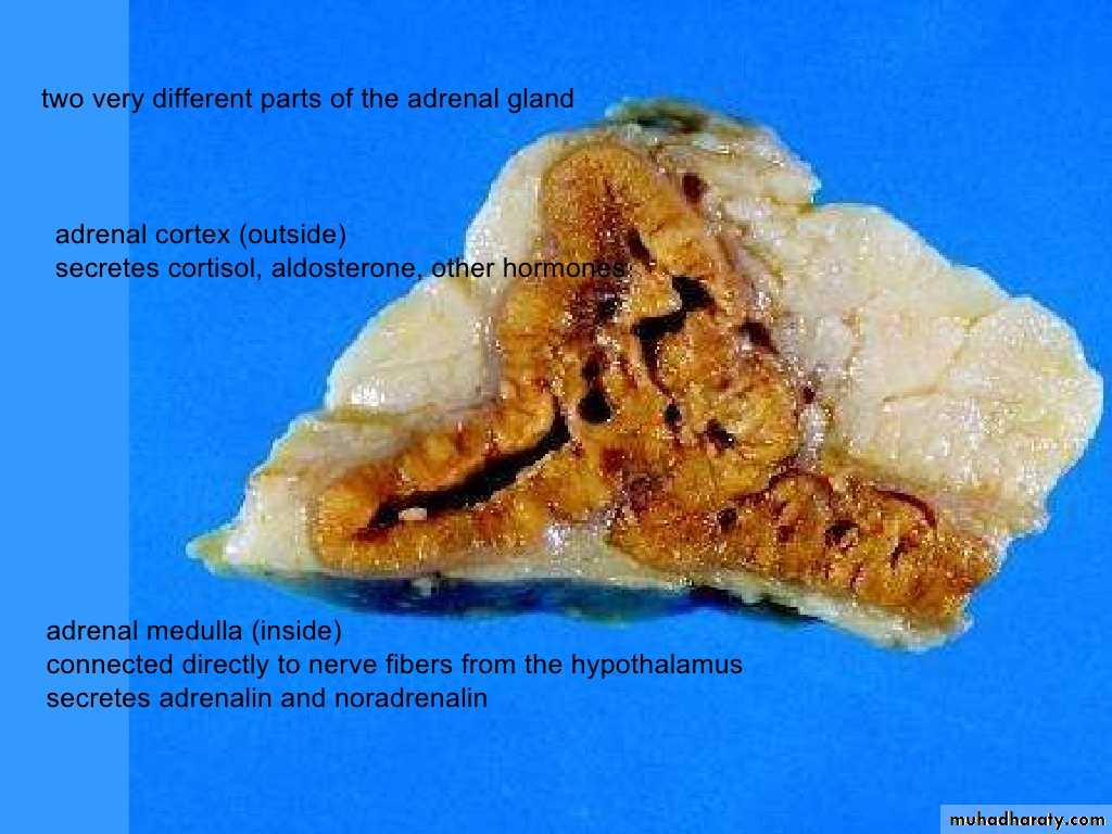
Adrenal gland:-1- Adrenal medulla.2- Adrenal cortex.
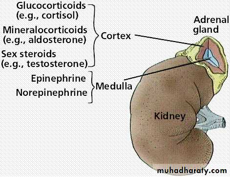
Adrenal medulla hormones
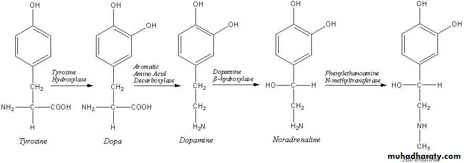
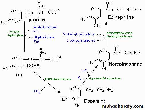
The adrenal medulla: is part of the adrenal gland. It is located at the center of the gland, being surrounded by the adrenal cortex. It is consisting of cells that secrete epinephrine (adrenaline), norepinephrine (noradrenaline), and a small amount of dopamine.The adrenal medulla is the principal site of the conversion of the amino acid tyrosine into the catecholamines epinephrine, norepinephrine, and dopamine.
In response to stressors such as exercise or imminent danger, medullary cells release the catecholamines adrenaline and noradrenaline into the blood. Adrenaline composes about 85% of the released catecholamines, and noradrenaline the other 15%.
Adrenaline and noradrenaline include increased heart rate and blood pressure, blood vessel constriction in the skin and gastrointestinal tract, smooth muscle, dilation, and increased metabolism, all of which are characteristic of the fight-or-flight response. Release of catecholamines is stimulated by nerve impulses, and receptors for catecholamines are widely distributed throughout the body.
Effects:
Catecholamines cause general physiological changes that prepare the body for physical activity (fight-or-flight response). Some typical effects are increases in heart rate, blood pressure, blood glucose levels, and a general reaction of the sympathetic nervous system. Some drugs, raise the levels of all the catecholamines.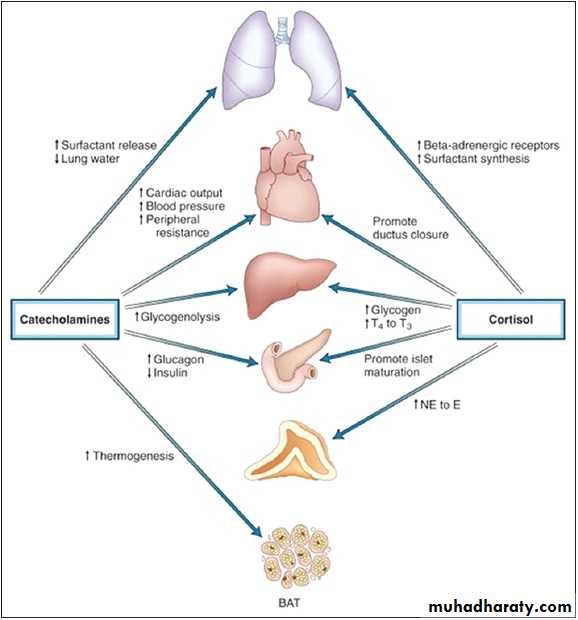
Adrenal cortex hormones
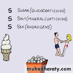
adrenal cortex mediates the stress response through the production of mineralocorticoids and glucocorticoids, such as aldosterone and cortisol, respectively. It is also a secondary site of androgen synthesis. Recent data suggest that adrenocortical cells under pathological as well as under physiological conditions show neuroendocrine properties; within the normal adrenal, this neuroendocrine differentiation seems to be restricted to cells and might be important for an autocrine regulation of adrenocortical functionThe adrenal cortex produces a number of different corticosteroid hormones.
Mineralocorticoids
The primary mineralocorticoid, aldosterone, is produced in the adrenocortical zona glomerulosa by the action of the enzyme aldosterone synthase . Aldosterone is largely responsible for the long-term regulation of blood pressure. Aldosterone effects on the distal convoluted tubule and collecting duct of the kidney where it causes increased reabsorption of sodium and increased excretion of both potassium (by principal cells) and hydrogen ions (by intercalated cells of the collecting duct).
Glucocorticoids:
The primary glucocorticoid released by the adrenal gland is cortisol in humans and corticosterone in many other animals. Its secretion is regulated by the hormone ACTH from the anterior pituitary. Upon binding to its target, cortisol enhances metabolism in several ways:It stimulates the release of amino acids from the body
It stimulates lipolysis, the breakdown of fatIt stimulates gluconeogenesis, the production of glucose from newly released amino acids and lipids
It increases blood glucose levels in response to stress, by inhibiting glucose uptake into muscle and fat cells
It strengthens cardiac muscle contractions
It increases water retention
Androgens:
Testosterone: a hormone with a wide variety of effects, ranging from enhancing muscle mass and stimulation of cell growth to the development of the secondary sex characteristics.Dihydrotestosterone (DHT): a metabolite of testosterone, and a more potent androgen than testosterone in that it binds more strongly to androgen receptors.
Androstenedione (Andro): an androgenic steroid produced by the testes, adrenal cortex, and ovaries. While androstenediones are converted metabolically to testosterone and other androgens, they are also the parent structure of estrone.