
Infectious Lecture 4th Year 27-12-2016 Dr.Osamah Muwafk
1
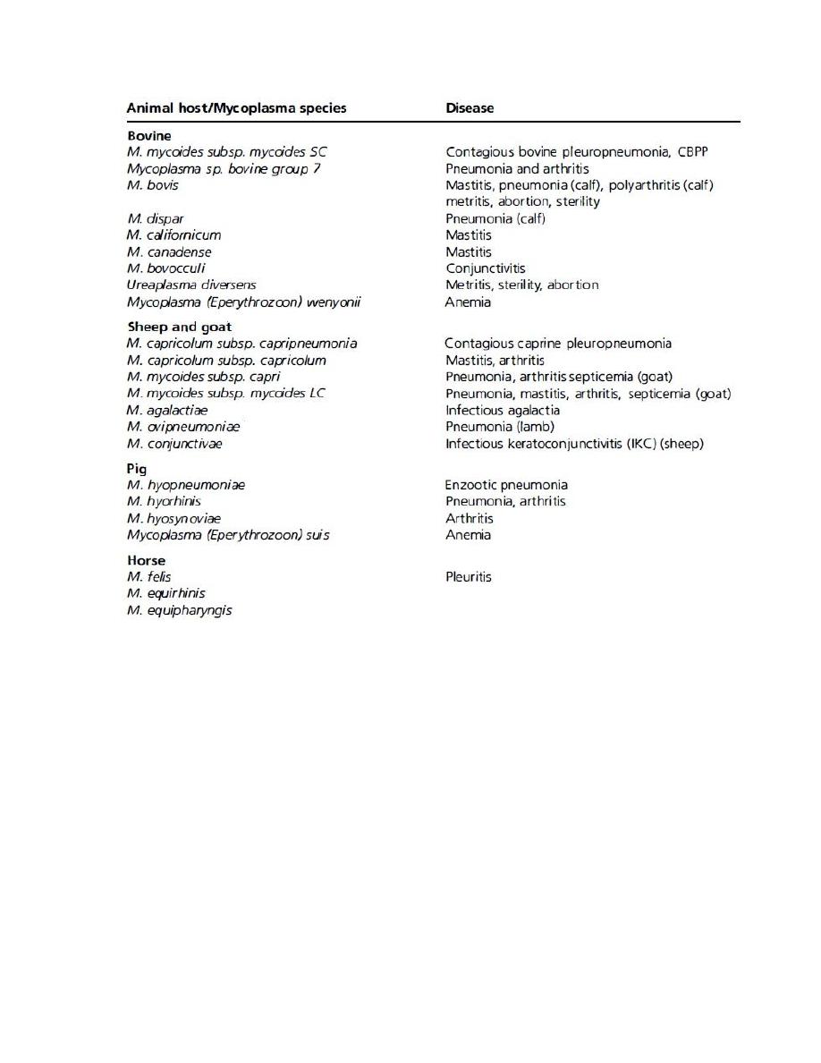
Diseases associated with Mycoplasma spp.
1. A class of Gram-negative bacteria
2. consisting of cells bounded by plasma membrane
3. Its organisms differ from other bacteria in that they are
deficient in cell walls
4. Mycoplasmas are the smallest prokaryotes with autonomous
replication.
5. They are extracellular parasites with an affinity for mucous
membranes, where they exist as commensals or pathogens
6. Pathogenic mycoplasma have a predilection for the
respiratory system, urogenital tract mammary gland and
serous membrane
7. In general, mycoplasmas are not highly virulent but rather
induce chronic diseases
8. Clinically normal cattle harbor M. bovis in the upper
respiratory tract with no apparent adverse effect
9. may shed the organism through the nasal discharge for
months to years
10. direct contact between infected and susceptible animals is the
primary mode of transmission
11. Calves fed discarded milk from cows with mycoplasma
mastitis may develop pneumonia and otitis media
12. Calves fed discarded milk from cows with mycoplasma
mastitis may develop pneumonia and otitis media
Infectious Lecture 4th Year 27-12-2016 Dr.Osamah Muwafk
2
Infectious Lecture 4th Year 27-12-2016 Dr.Osamah Muwafk
3

Infectious Lecture 4th Year 27-12-2016 Dr.Osamah Muwafk
4
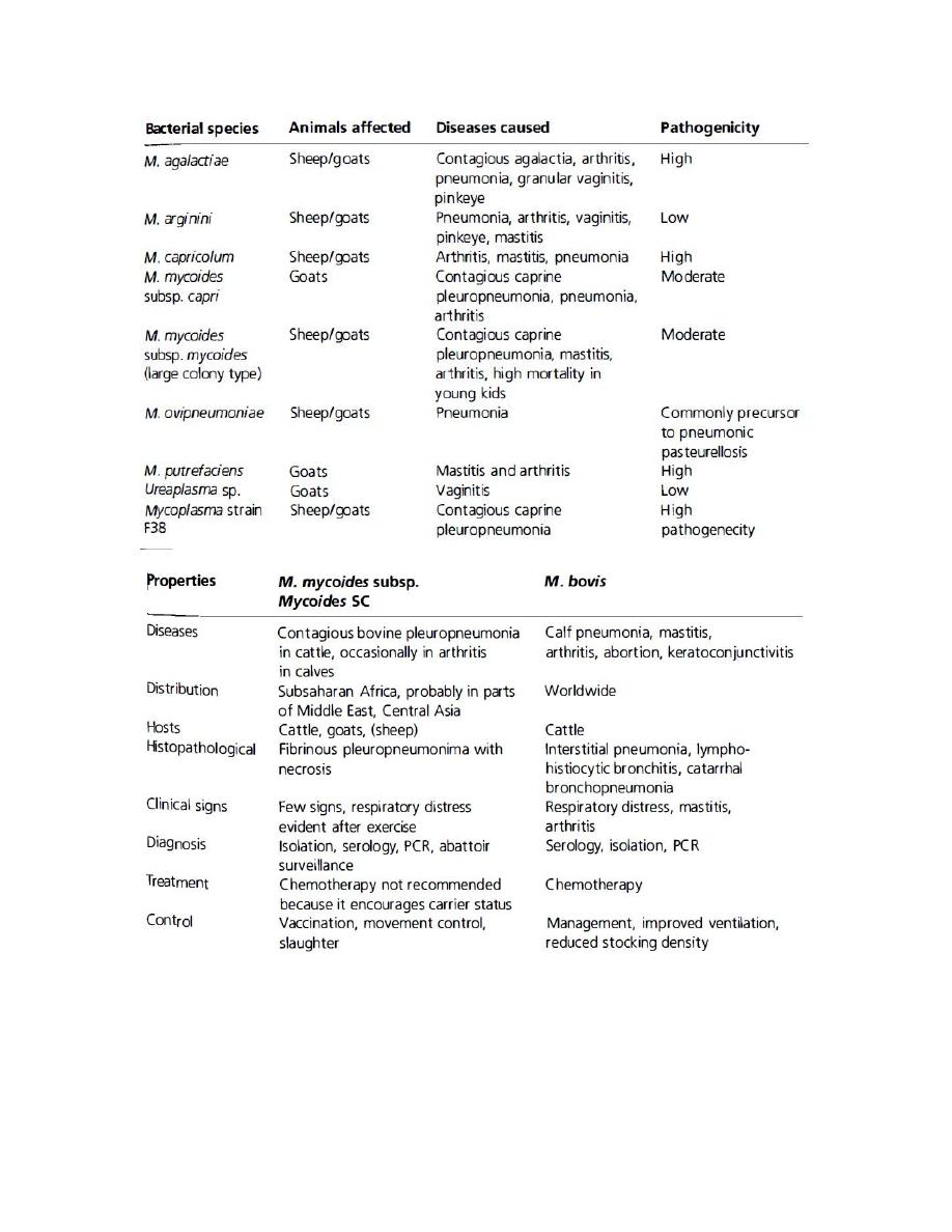
CONTAGIOUS BOVINE PLEUROPNEUMONIA
Etiology and Epidemiology:
1. Mycoplasma mycoides subsp. mycoides (Small colony) (M
mmSC)
2. occurs in cattle of the species Bos and allied animals
including buffalo, yak, bison and even reindeer
3. widespread in Africa and occurs in some countries of Asia
and Europe
4. Sporadic outbreaks have been recognized in the Middle East,
probably derived from importation of cattle from Africa
5. morbidity approaches 90%, the case mortality may be as
high as 50% and 25 % of the infected cattle remain as
recovered carriers with or without clinical signs
6. The focus of infection is often provided by recovered
'carrier' animals
7. pulmonary sequestrum preserves a potential source of
organisms for periods as long as 3 years
8. Transmission occurs from direct and repeated contacts
between sick and healthy animals
9. The principal route of infection is by the inhalation of
infective droplets from active or carrier cases of the disease.
10. It is sensitive to all environmental influences, including
disinfectants, heat and drying, and do not ordinarily survive
outside the animal body for more than a few hours
PATHOGENESIS
1. The organism invades the lungs of cattle and causes a
mycoplasmemia

Infectious Lecture 4th Year 27-12-2016 Dr.Osamah Muwafk
5
2. this results in localization in numerous other sites including
the kidneys and brain
3. resulting in high morbidity and mortality
4. thrombosis in the pulmonary vessels, probably prior to the
development of pneumonic lesions
5. Death results from anoxia and presumably from toxemia
CLINICAL FINDINGS:
A. Acute form
1. After an incubation period of 3-6 weeks
2. sudden onset of high fever (40°C; 105°F)
3. fall in milk yield, anorexia and cessation of
rumination
4. Coughing, at first only on exercise, and thoracic pain
are evident
5. affected animals are disinclined to move, standing
with the elbows out, the back arched and head
extended
6. Respirations are shallow, rapid and accompanied by
expiratory grunting
7. Pain is evidenced on percussion of the chest.
8. Auscultation reveals pleuritic friction sounds in the
early stages of acute inflammation
9. dullness, fluid sounds and moist gurgling crackles in
the later stages of effusion
10. Dullness of areas of the lung may be detectable on
percussion.
11. Edematous swellings of the throat and dewlap may
occur
12. swelling of the large 'movable joints may be present.

Infectious Lecture 4th Year 27-12-2016 Dr.Osamah Muwafk
6
13. In calves, valvular endocarditis and myocarditis may
occur
14. In fatal cases death occurs after a variable course of
from several days to 3 weeks
15. In the hyperacute form, affected cattle may die
within 1 week after the onset of respiratory distress
B. Chronic and subacute forms
1. Toxemia and unthriftiness due to inactive sequestrum
in the lung, with a necrotic center of sufficient size
2. a chronic cough, and mild respiratory distress on
exercise
3. These sequestra commonly break down when the
animal is exposed to environmental stress and cause
an acute attack of the disease
CLINICAL PATHOLOGY
1. Isolation or detection of organism
2. The polymerase chain reaction (PCR) has been used to
identify the specific organism and differentiate it from other
members of the cluster
3. Latex agglutination test
4. Serological tests: The complement fixation test (CFT),
ELISA
NECROPSY FINDINGS
1. Lesions are confined to the thoracic cavity and lungs and the
lesions are usually unilateral
2. The pleural cavity may contain large quantities of clear,
yellow-brown fluid containing pieces of fibrin

Infectious Lecture 4th Year 27-12-2016 Dr.Osamah Muwafk
7
3. Caseous fibrinous deposits are present on the parietal and
visceral surfaces of the lungs
4. The interlobular septae are prominently distended with
ambercolored fluid surrounding distended lymphatics.
5. lobules vary in color with red, gray, or yellow hepatization
6. Consolidation of the lungs with a typically marbled
appearance is characteristic
7. In chronic or advanced cases, a sequestrum of necrotic lung
varying size from 1-10 cm in diameter is surrounded by a
fibrous capsule
8. In affected calves, exudative peritonitis, arthritis, bursitis and
fibrinous arthritis of carpal and tarsal joints may be present
DIFFERENTIAL DIAGNOSIS:
1. Rinderpest erosive: stomatitis, dysentery, and erosions
throughout the alimentary tract
2. Foot and mouth disease: Salivation, lameness, fever, and
vesicular stomatitis
3. Hemorrhagic septicemia: Acute disease with death in 6 to 72
hours. Edema of the neck and brisket, lung lesions similar to
CBPP. Culture of Pasteurella spp.
4. Theileriosis (East Coast fever): Coughing, nasal and ocular
discharge, diarrhea, enlargement of peripheral lymph nodes,
ulceration of abomasum . No lung lesions
5. Ephemeral fever: Ocular discharge, drooling saliva,
lameness, enlarged joints, self-limiting disease of short
duration; most affected cattle recover quickly; fluctuating
fever; secondary pneumonia may occur

Infectious Lecture 4th Year 27-12-2016 Dr.Osamah Muwafk
8
6. Pulmonary abscesses: Large abscesses containing foul-
smelling purulent material; may have total destruction of
lung
7. Tuberculosis: Tubercular nodules may resemble CBPP
sequestra but they are degenerative cheese-like lesions, often
calcified
8. Farcy: Abscesses of lungs containing foul smelling material
and enlarged local lymph nodes
9. Actinobacillosis: Generalized lesions of lung and other
adjacent tissues
10. Echinococcal (hydatid cysts): Pulmonary cysts with a double
wall and containing clear fluid, often calcified when old
TREATM ENT
1. No therapeutic treatment is effective
2. Antibiotics can alleviate the clinical course of the disease
enabling some improvement in condition
3. Tilmicosin and danafloxacin were effective both in terms of
mycoplasmastatic, and mycoplasmacidal activity
4. Florofenicol and tetracycline were intermediate
5. spectinomycin was ineffective against some strains
CONTROL
The major obstacles to the control and eradication of the disease
are:
1. Difficulty in controlling the movements of cattle, especially
in sub-Sahara Africa
2. Complications of applying quarantine and slaughter policies
3. Lack of rapid pen-Side diagnostic tests
4. Ineffective vaccines

Infectious Lecture 4th Year 27-12-2016 Dr.Osamah Muwafk
9
5. Insufficient funds to implement control policies
6. Civil strife and drought, which have an effect on the spread
of the disease in Africa.
The possible strategies used for control in affected countries or
regions are:
1. Slaughter of all sick and in-contact cattle
2. Slaughter of all sick cattle and vaccination of in-contact cattle
3. Vaccination of healthy cattle with slaughter of sick cattle in
an epidemic and revaccination of cattle at risk
On a herd basis:
1. Removal of sources of infection
2. Hygiene: Any procedure which brings the animals together
should be avoided
3. Vaccination : Current vaccine strains (T144 and T1SR) for
CBPP are made from freeze dried broth cultures of live
attenuated Mycoplasma mycoides subsp. mycoides SC

Infectious Lecture 4th Year 27-12-2016 Dr.Osamah Muwafk
10
CONTAGIOUS CAPRINE PLEUROPN EUMONIA
Etiology and Epidemiology
1. Mycoplasma capricolum subsp. Capripneumoniae
2. CCPP is one of the most serious fatal diseases of goats
3. The disease is called Abu Nini in the Sudan
4. Infectivity is high with a morbidity of 100%
5. the illness is acute and severe with a case mortality rate of
60-100 %
6. transmission by inhalation
7. carrier or infected animal is the source of infection
CLINICAL FINDINGS:
1. Cough, Dyspnea
2. Lagging, lying down a lot (but the animal can stand and
walk)
3. fever (40.5-41Se; 104.5-106°F)
4. in the terminal stages, mouth-breathing, tongue protrusion
and frothy salivation
5. death in two or more days
NECROPSY FINDINGS:
1. hepatization of parts of the lung
2. an increase in pleural fluid with a fibrinous pleuritis
TREATMENT
1. Tylosin tartrate 10 mg/kg BW or oxytetracycline (15
mg/kg/d) is highly successful in limiting the severity of
disease

Infectious Lecture 4th Year 27-12-2016 Dr.Osamah Muwafk
11
2. The severity of the disease is reduced but treated animals are
still sources of infection
Control :
1. Herd biosecurity to prevent contact with infected animals is
important
2. Vaccination with an inactivated mycoplasma F38
3. A booster dose 1 month after the first vaccination provides
additional protection