Avian Influenza (AI)
CauseAvian Influenza is caused by an Orthomyxovirus; there are several serotypes.
Currently we know there are 16 H- types and 9 N-types and they can show up in all kinds of combinations. For poultry the most important ones are H5, H7 and H9. Pathogenicity varies with the strains HPAI and LPAI (high or low pathogenic AI).
Transmission
AI virus is excreted from nares, mouth, conjunctiva and cloaca.Airborne virus particles from the respiratory tract, droppings, and people carrying virus on their clothing and equipment are the main routes of transmission. Migratory water fowl and other wild birds infected with AI virus may be a source of infection.
Species affected
Avian Influenza viruses have been shown to naturally infect a wide variety of wild and domestic birds. In poultry production main problems are in chickens, turkeys and ducks.Clinical signs
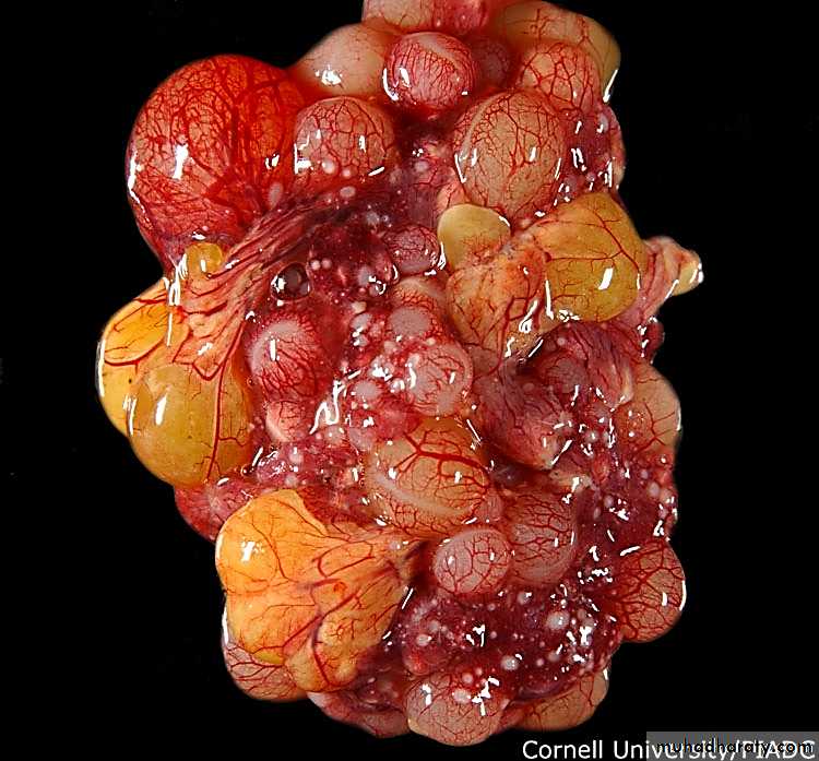
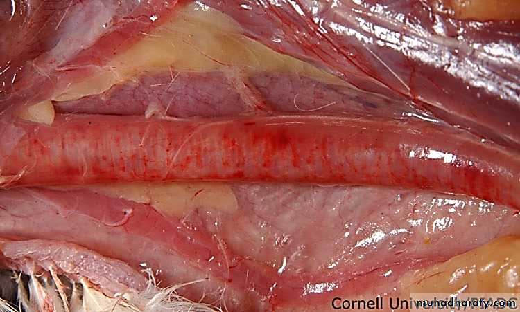
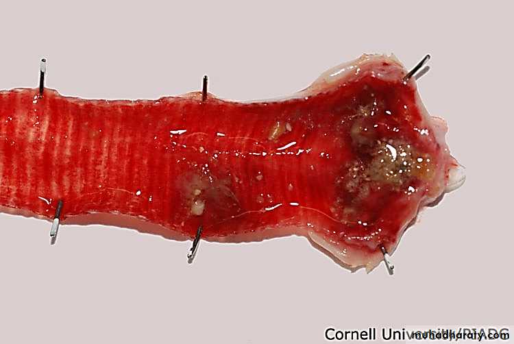
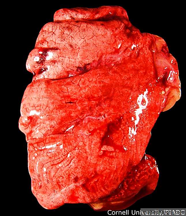
Clinical signs will vary, depending on the pathogenicity (HPAI and LPAI) of AI virus involved and other factors as host species, sex, concurrent infections, acquired immunity and environmental factors.LPAI shows generally mild symptoms: respiratory coughing sneezing, wet eyes, nasal discharge, depression, lethargy, limited reduction of feed intake and limited drop in egg production; low mortality rate. HPAI shows fast onset with increased mortality even before clinical signs are seen, depression, drop in feed and water intake, severe drop in egg production and mortality can vary between 50-90%.
Diagnosis
Clinical signs are indicative for AI; final confirmation by laboratory testing:- Direct detection of AI proteins or Nucleic acids(RNA) using PCR.
- Virus isolation from infected organs, tracheal or cloacal swabs.
- Serology from blood samples after infection and for routine monitoring showing specific AI antibodies.
Treatment
There is no treatment for Avian Influenza. Antibiotics will help to control secondary bacterial infections.
Prevention and control
Vaccination is generally done with inactivated AI vaccines based on the strain H-type causing the outbreaks.Picture 1
Picture 2Picture 3
Picture 4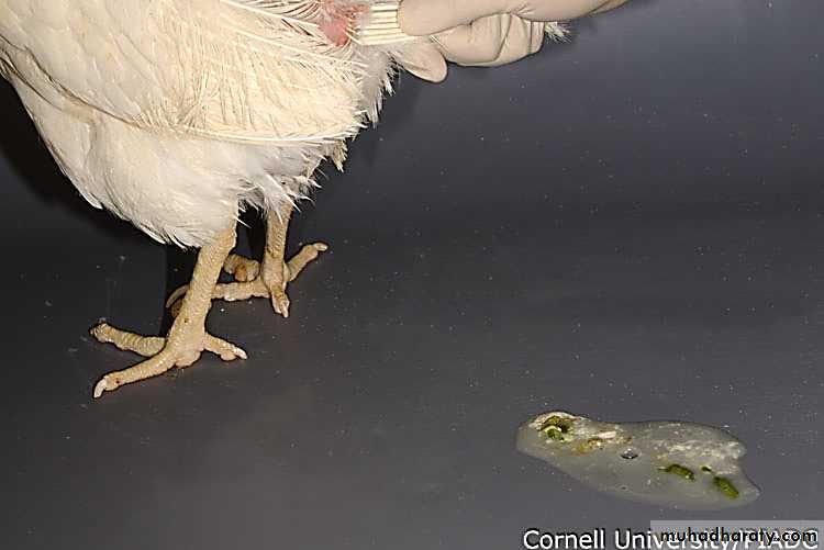
Picture 5
Picture 6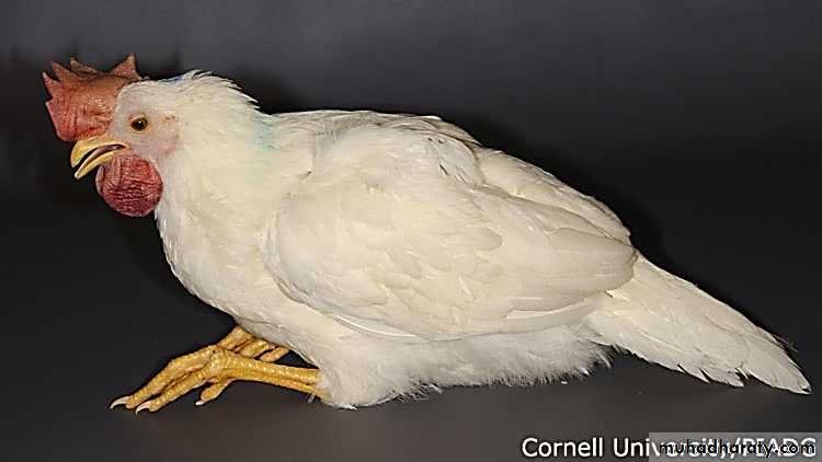
Picture 7
Picture 8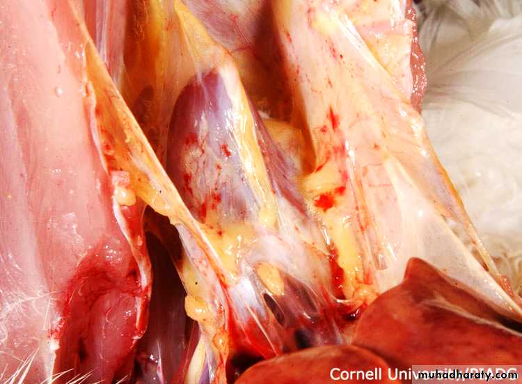
Picture 9
Picture 10
Picture 11
Picture 12
Picture 13
Picture 14
Infectious Bursal Disease(Gumboro disease, IBD)Cause
The disease is caused by a Birnavirus of serotype 1. Virus strains can be divided in classical and variant strains. The virus is very stable and is difficult to eradicate from an infected farm.Transmission
IBD virus is very infectious and spreads easily from bird to bird by way of droppings. Infected clothing and equipment are means of transmission between farms.Species affected
Chickens and turkeys appear to be natural hosts.
Clinical signs
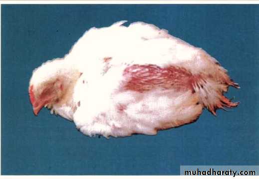
Clinical IBD occurs usually between 3 and 8 weeks of age depending on maternal antibody levels. Affected birds are listless and depressed, pale, huddling producing watery white diarrhea. Mortality varies. Usually new cases of IBD have a mortality rate of about 5 to10% but can be as high as 60% depending on the pathogenicity of the strain involved. Highly pathogenic strains are called “very virulent” IBD (vvIBD) resulting in high mortality.Clinical signs
Subclinical IBD occurs with infections before 3 weeks of age. Early IBD infection result in permanent immunosuppression without mortality.Immunosuppression is economically important due to increased susceptibility to secondary infections especially in the respiratory tract.
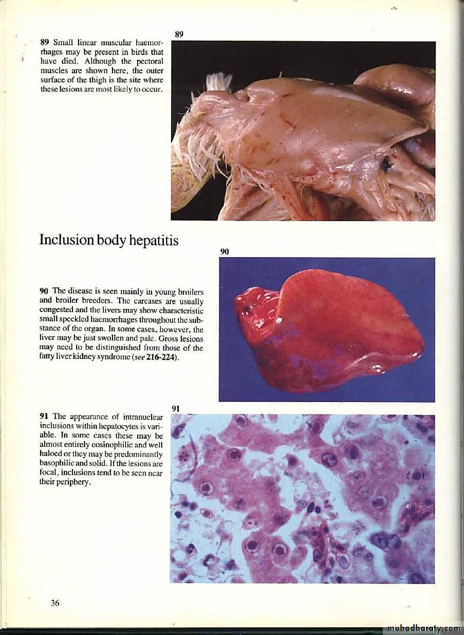
Gumboro disease related diseases such as inclusion body hepatitis are also more frequent in these birds. In broilers this form of the disease results in bad performance with lower weight gains and higher feed conversion ratios.
Diagnosis
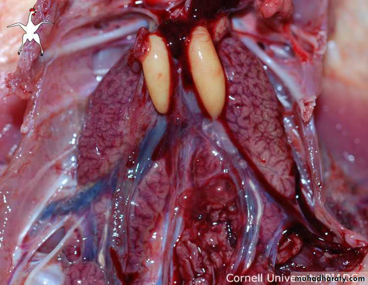
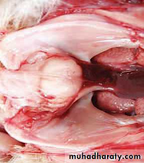
Typical clinical signs and post mortem lesions are found after IBD infection. Post mortem lesions; in acute cases the bursa of Fabricius is enlarged and gelatinous, sometimes even bloody. Muscle haemorrhages and pale kidneys can be seen. Infection by variant strains is usually accompanied by a fast bursal atrophy (in 24 - 48 hours) without the typical signs of Gumboro disease. Also in chronic cases the bursa is smaller than normal (atrophy). The bursa destruction is apparent on histologic examination. The lack of white blood cells (lymphocytes) results in a reduction in the development of immunity and decreased resistance of the birds to other infections.Histopathological examination, serology, virus isolation and PCR are confirming tools. IBD can be confused with sulfonamide poisoning, aflatoxicosis, and pale bird syndrome (Vitamin E deficiency).
Treatment
No treatment is available for IBD.Control
Vaccination of breeders and young chicks is the best means of control.The induction of a high maternal immunity in the progeny of vaccinated breeders, together with the vaccination of the offspring is the most effective approach to successful IBD control. A variety of live and inactivated vaccines have been developed to enhance the control of classical, variant and vvIBD challenges.
Recently a new generation of recombinant vector vaccines based on HVT-vector carrying an insert of the VP2 part of the IBD-virus entered the market for the control of IBD.
Picture 1
Picture 2Picture 3
Picture 4

Picture 5
Picture 6