Fowl Cholera
(Avian Cholera, Pasteurellosis, Avain hemorrhagic septicaemia)Cause
Fowl cholera is caused by a bacterium: Pasteurella multocida (several serotypes).Transmission
Transmission of fowl cholera is mainly from bird to bird by water or feed contamination. There is no evidence for egg transmission. Vectors like flies and red mite can be carriers and can add to the spread. Rodents (rats and mice) also appear to play a role in contamination of water and feed with Pasteurella multocida.Species affected
Turkeys, chickens, ducks and geese, and other bird species are susceptible.Clinical signs and lesions
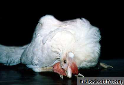
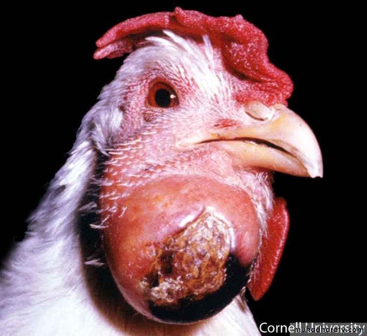
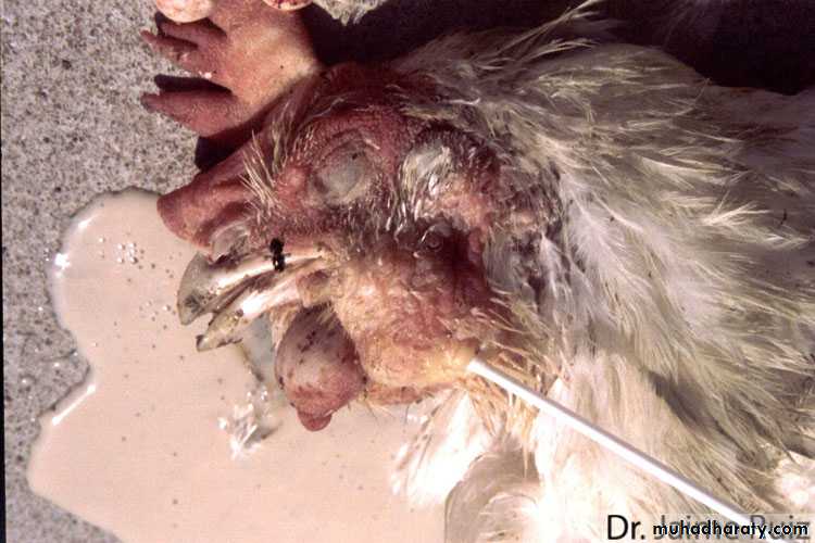
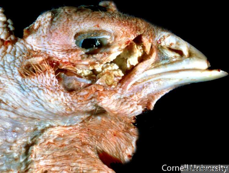
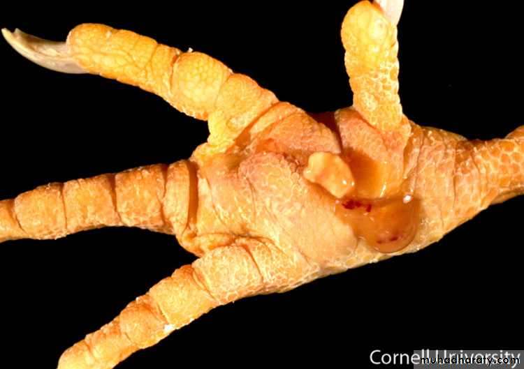
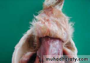
Affected birds are depressed and have decreased appetite. Egg production will drop 5-15 % and mortality will be high in acute fowl cholera. Birds that die from acute fowl cholera frequently have bluish combs and wattles. Chronic fowl cholera will not cause high mortality, although there will be an increase in deaths. Swollen wattles is a feature of chronic fowl cholera.Clinical signs and lesions
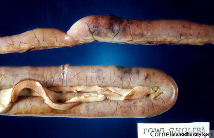
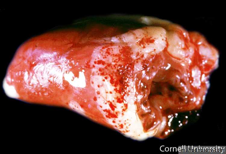
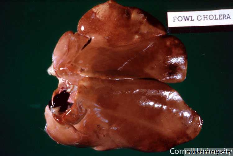
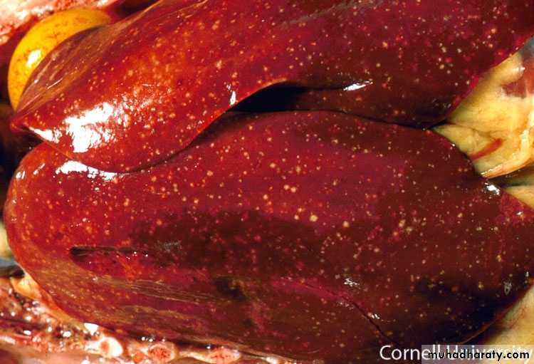
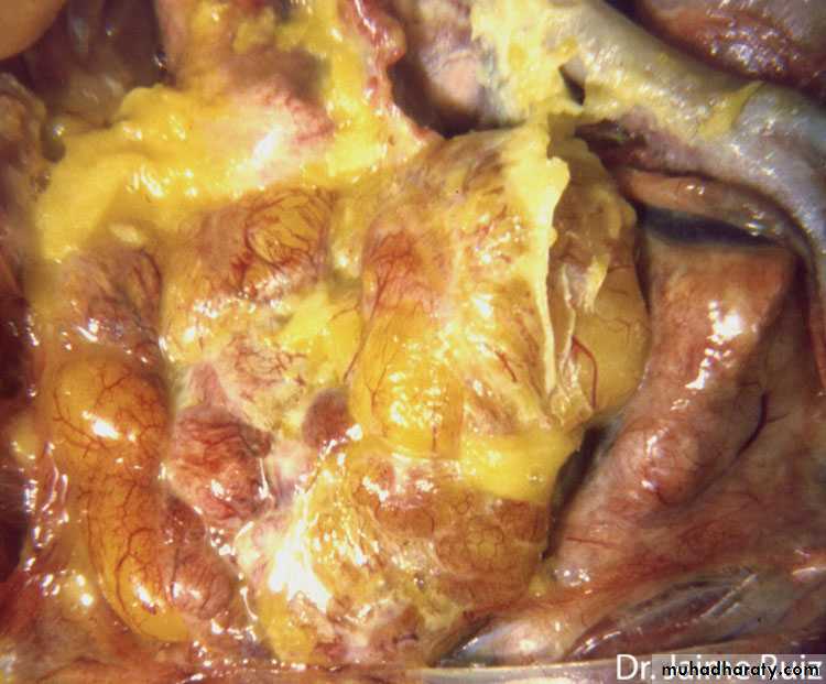
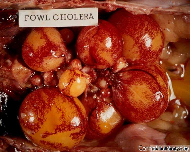
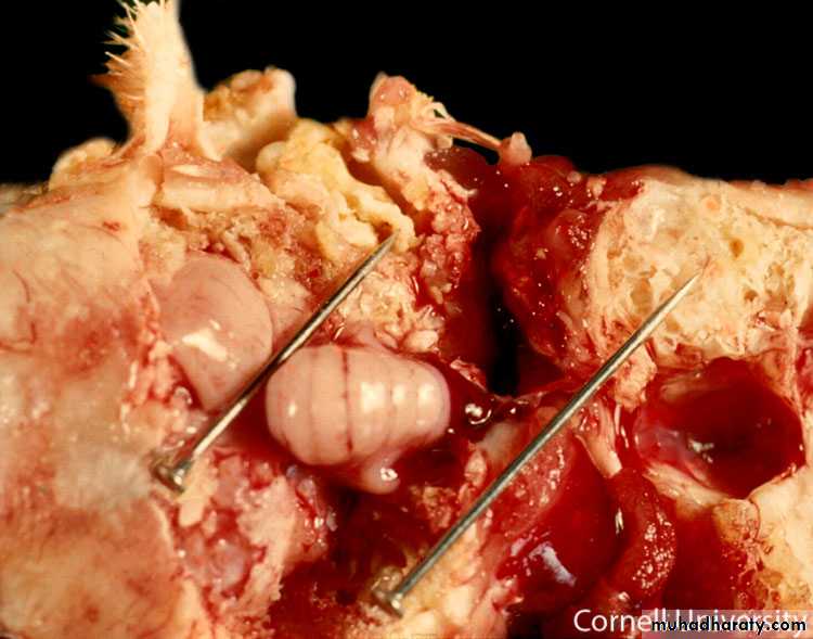
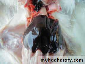
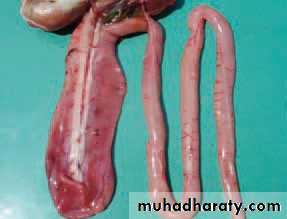
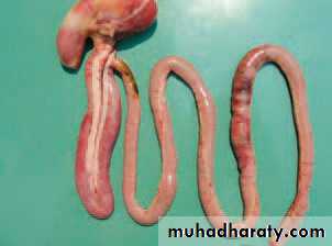
Lesions;acute phase septicaemia, vascular changes in abdominal viscera, hemorrhages, liver swelling with focal necrosis, ovaries appear flaccid and hemorrhagic and show ruptured yolks,
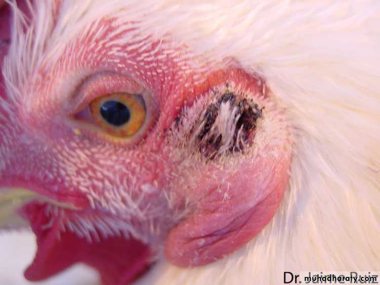
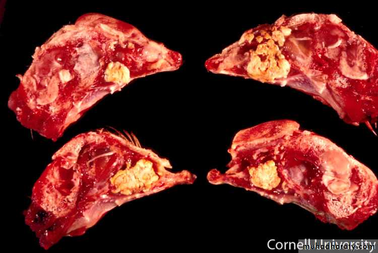
Chronic phase; localized infections in conjunctiva, fecial edema, middle ear infection resulting in torticollis, meningeal infection.
Diagnosis
Clinical signs in combination with isolation and identification from samples from birds that died of acute Fowl cholera. (Fresh death birds)
Treatment
Antibiotics based on antibiotic sensitivity test, the earlier the diagnosis the better change of a positive effect of an antibiotic treatment.Control
Hygiene management and rodent control to eliminate possible sources of Pasteurella multocida.Vaccination can be considered in areas where Pasteurella multocida is prevalent. Both live and inactivated vaccines are available.
Picture 1
Picture 2
Picture 3
Picture 4
Picture 5
Picture 6
Picture 7
Picture 8
Picture 9
Picture 10
Picture 11
Picture 12
Picture 13
Picture 14
Picture 15
Necrotic Enteritis(NE, Clostridial enteritis, enterotoxemia)
Cause
Bacterial infection with Clostridium perfringens (Gram+, toxin forming, spore forming, anaerobe).Clostridia including Clostridium perfringens are normal inhabitants of the gut. When the microflora balance in the gut is disturbed, potentially pathogenic clostridia begin to produce toxins and proteolytic enzymes. The toxins will induce cell damage known as Necrotic Enteritis (NE). Factors involved in microflora disturbance include: intestinal infections (eg coccidiosis), nutritional factors (protein source, grain source, diet changes), management: type of litter, timing of feed changes, antibiotic treatments.
Transmission
Clostridium perfringens is an ubiquitous organism that can be found in faeces, soil, dust contaminated feed and litter.
Species affected
Chickens, turkeys and quail. Natural outbreaks have reported in chickens from 2 weeks to 6 monthsof age. Majority of NE problems are identified in broilers raised on litter (2 -5 weeks of age).
Clinical signs and lesions
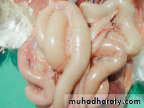
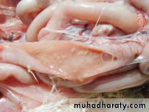
There are 2 forms of Necrotic Enteritis (NE), acute clinical form and the subclinical form.Although it can be seen at any age, the acute clinical form is primarily a disease in young chickens, showing severe depression, reluctance to move, diarrhea, ruffled feathers and sudden death and increased mortality. The subclinical form produces not outward signs but has big impact on performance (weight loss, reduced weight gain and impaired FCR).
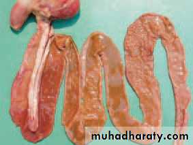
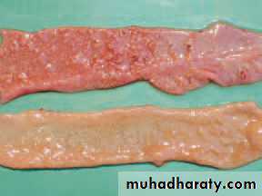
Clinical signs and lesions
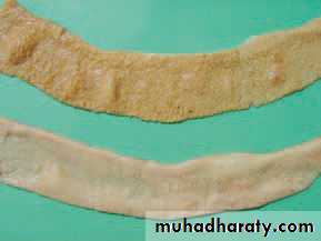
Lesions: necrosis of the mucosa of the small intestine. In the clinical form the necrosis might progress into a fibrinonectoric enteritis forming a diphtheritic membrane.In the mild form, focal areas of intestinal mucosal necrosis without further clinical signs can be found.
Diagnosis
Clinical signs in combination with typical gross and microscopic lesions and isolation of the causative agent will confirm the clostridial infection.Treatment
antibioticControl
Vaccination of breeders with inactivated vaccines based on toxins inducing active and passive immunity have shown to offer good protection.
Maintain microflora balance with management of all related factors; management, coccidiosis control and nutritional factors.
Picture 1
Picture 2
Picture 3
Picture 4
Picture 5
Picture 6
Picture 7
Picture 8