Lymph Drainage of the Head and Neck
Lymph Drainage of the Head and NeckThe lymph nodes of the head and neck are arranged as a regional collar that extends from below the chin to the back of the head and as a deep vertical terminal group that is embedded in the carotid sheath in the neck.
Regional Nodes :
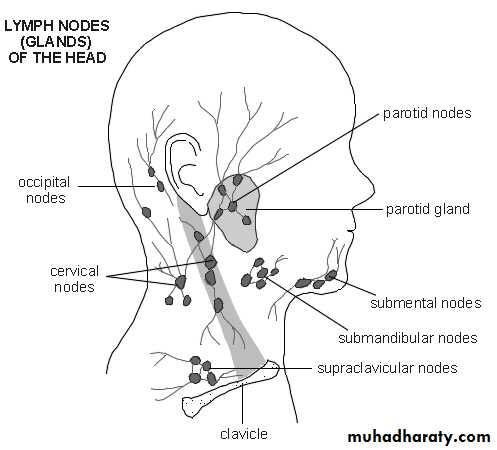
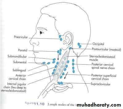
The regional nodes are arranged as follows:Occipital nodes: These are situated over the occipital bone on the back of the skull. They receive lymph from the back of the scalp.
Retroauricular (mastoid) nodes: These lie behind the ear over the mastoid process. They receive lymph from the scalp above the ear, the auricle, and the external auditory meatus.
Parotid nodes: These are situated on or within the parotid salivary gland. They receive lymph from the scalp above the parotid gland, the eyelids, the parotid gland, the auricle, and the external auditory meatus.
•Buccal (facial) nodes: One or two nodes lie in the cheek over the buccinators muscle. They drain lymph that ultimately passes into the submandibular nodes. •
Submandibular nodes: These lie superficial to the submandibular salivary gland just below the lower margin of the jaw. They receive lymph from the front of the scalp; the nose; the cheek; the upper lip and the lower lip (except the central part); the frontal, maxillary, and ethmoid sinuses, the upper and lower teeth (except the lower incisors); the anterior two thirds of the tongue (except the tip); the floor of the mouth and vestibule; and the gums.
Submental nodes: These lie in the submental triangle just below the chin. They drain lymph from the tip the tongue, the floor of the anterior part of the mouth, the incisor teeth, the center part of the lower lip, and the skin over the chin.
Anterior cervical nodes: These lie along the course of the anterior jugular veins in the front of the neck. They receive lymph from the skin and superficial tissues of the front of the neck.
Superficial cervical nodes: These lie along the course of the external jugular vein on the side of the neck. They drain lymph from the skin over the angle of the jaw, the skin over the lower part of the parotid gland, and the lobe of the ear.
•Retropharyngeal nodes: These lie behind the pharynx and in front of the vertebral column. They receive lymph from the naso pharynx, the auditory tube, and the vertebral column.
• Laryngeal nodes: These lie in front of the larynx. They receive lymph from the larynx.
• Tracheal (paratracheal) nodes: These lie alongside the trachea. They receive lymph from neighboring structures, including the thyroid gland.
Deep Cervical Nodes
The deep cervical nodes form a vertical chain along the course of the internal jugular vein within the carotid sheath. They receive lymph from all the groups of regional nodes. The jugulodigastric node, which is located below and behind the angle of the jaw, is mainly concerned with drainage of the tonsil and the tongue. The juguloomohyoid node, which is situated close to the omohyoid muscle, is mainly associated with drainage of the tongue. The efferent lymph vessels from the deep cervical lymph nodes join to form the jugular trunk, which drains into the thoracic duct or the right lymphatic duct.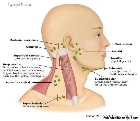
Pharynx
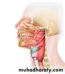
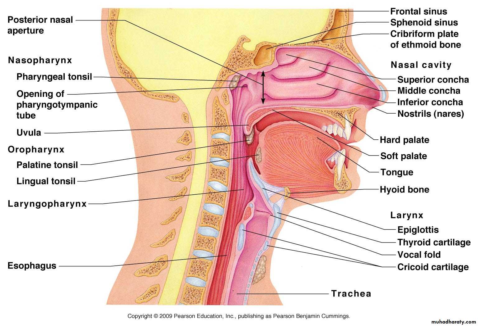
The pharynx is situated behind the nasal cavities, the mouth, and the larynx and may be divided into nasal, oral, and laryngeal parts. The pharynx is funnel shaped, its upper, wider end lying under the skull and its lower, narrow end becoming continuous with the esophagus opposite the 6th cervical vertebra. The pharynx has a musculomembranous wall, which is deficient anteriorly. Here, it is replaced by the posterior openings into the nose (choanae), the opening into the mouth, and the inlet of the larynx. By means of the auditory tube, the mucous membrane is also continuous with that of the tympanic cavity.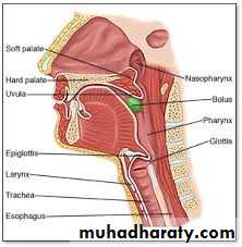
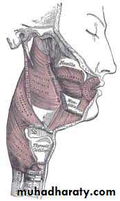
Muscles of the Pharynx
The muscles in the wall of the pharynx consist of the superior, middle, and inferior constrictor muscles, whose fibers run in a somewhat circular direction, and the stylopharyngeus and salpingopharyngeus muscles, whose fibers run in a somewhat longitudinal direction.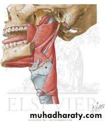
The three constrictor muscles extend around the pharyngeal wall to be inserted into a fibrous band or raphe that extends from the pharyngeal tubercle on the basilar part of the occipital bone of the skull down to the esophagus. The three constrictor muscles overlap each other so that the middle constrictor lies on the outside of the lower part of the superior constrictor and the inferior constrictor lies outside the lower part of the middle constrictor.
The lower part of the inferior constrictor, which arises from the cricoid cartilage, is called the cricopharyngeus muscle. The fibers of the cricopharyngeus pass horizontally around the lowest and narrowest part of the pharynx and act as a sphincter.
Interior of the Pharynx
The pharynx is divided into three parts: the nasal pharynx, the oral pharynx, and the laryngeal pharynx.Nasal Pharynx
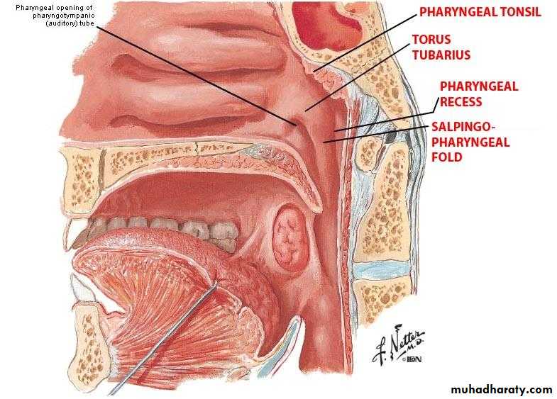
This lies above the soft palate and behind the nasal cavities. In the submucosa of the roof is a collection of lymphoid tissue called the pharyngeal tonsil. The pharyngeal isthmus is the opening in the floor between the soft palate and the posterior pharyngeal wall. On the lateral wall is the opening of the auditory tube, the elevated ridge of which is called the tubal elevation. The pharyngeal recess is a depression in the pharyngeal wall behind the tubal elevation. The salpingopharyngeal fold is a vertical fold of mucous membrane covering the salpingopharyngeus muscle.
Oral Pharynx
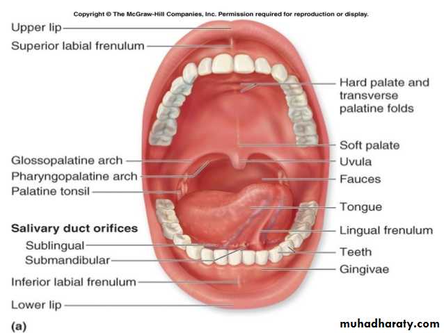
This lies behind the oral cavity. The floor is formed by the posterior one third of the tongue and the interval between the tongue and epiglottis. In the midline is the median glossoepiglottic fold, and on each side the lateral glossoepiglottic fold. The depression on each side of the median glossoepiglottic fold is called the vallecula.On the lateral wall on each side are the palatoglossal and the palatopharyngeal arches or folds and the palatine tonsils between them. The palatoglossal arch is a fold of mucous membrane covering the palatoglossus muscle. The interval between the two palatoglossal arches is called the oropharyngeal isthmus and marks the boundary between the mouth and pharynx. The palatopharyngeal arch is a fold of mucous membrane covering the PalatoPharyngus muscle .The recess between the palatoglossal and palatopharyngeal arches is occupied by the palatine tonsil.
Laryngeal Pharynx
This lies behind the opening into the larynx. The lateral wall is formed by the thyroid cartilage and the thyrohyoid membrane. The piriform fossa is a depression in the mucous membrane on each side of the laryngeal inlet.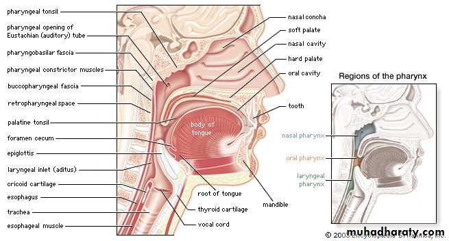
Sensory Nerve Supply of the Pharyngeal Mucous Membrane
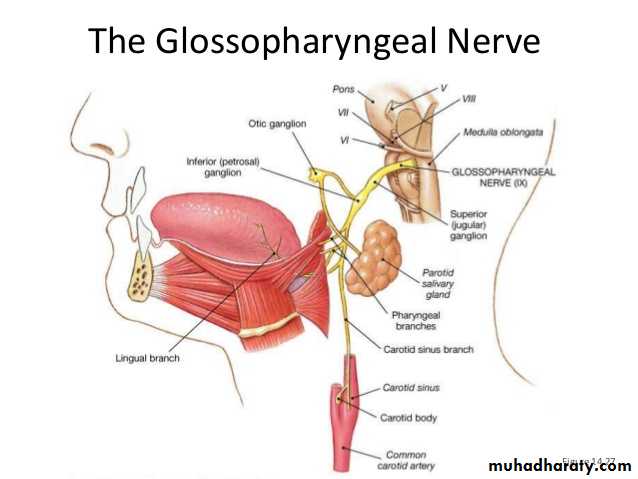
Nasal pharynx: The maxillary nerve (V2)Oral pharynx: The glossopharyngeal nerve
Laryngeal pharynx (around the entrance into the larynx): The internal laryngeal branch of the vagus nerve
Blood Supply of the Pharynx
Ascending pharyngeal, tonsillar branches of facial arteries, and branches of maxillary and lingual arteriesLymph Drainage of the Pharynx
Directly into the deep cervical lymph nodes or indirectly via the retropharyngeal or paratracheal nodes into the deep cervical nodesThe Process of Swallowing (Deglutition)
Masticated food is formed into a ball or bolus on the dorsum of the tongue and voluntarily pushed upward and backward against the undersurface of the hard palate. This is brought about by the contraction of the styloglossus muscles on both sides, which pull the root of the tongue upward and backward. The palatoglossus muscles then squeeze the bolus backward into the pharynx. From this point inward, the process of swallowing becomes an involuntary act.The nasal part of the pharynx is now shut off from the oral part of the pharynx by the elevation of the soft palate, the pulling forward of the posterior wall of the pharynx by the upper fibers of the superior constrictor muscle, and the contraction of the palatopharyngeus muscles. This passage of food and drink into the nasal cavities.
The larynx and the laryngeal part of the pharynx are pulled upward by the contraction of the stylopharyngeus, salpingopharyngeus, thyrohyoid, and palatopharyngeus muscles. The main part of the larynx is thus elevated to the posterior surface of the epiglottis , and the entrance into the larynx is closed. The bolus moves downward over the epiglottis, the closed entrance into the larynx, and reaches the lower part of the pharynx as the result of the successive contraction of the food slides down the groove on either side of the entrance into the larynx, that is, down through the piriform fossae. Finally, the lower part of the pharyngeal wall (the cricopharyngeus muscle) relaxes and the bolus enters the esophagus.
Palatine Tonsils
The palatine tonsils are two masses of lymphoid tissue, each located in the depression on the lateral wall of the oral part of the pharynx between the palatoglossal and palatopharyngea arches. Each tonsil is covered by mucous membrane, and its free medial surface projects into the pharynx. The surface is pitted by numerous small openings that lead into the tonsillar crypts. The tonsil is covered on its lateral surface by a fibrous capsule. The capsule is separated from the superior constrictor muscle by loose areolar tissue, and the external palatine vein descends from the soft palate‘ in 'this tissue to join the pharyngeal venous plexus. Lateral to the superior constrictor muscle lie the styloglossus muscle, the loop of the facial artery, and the internal carotid artery.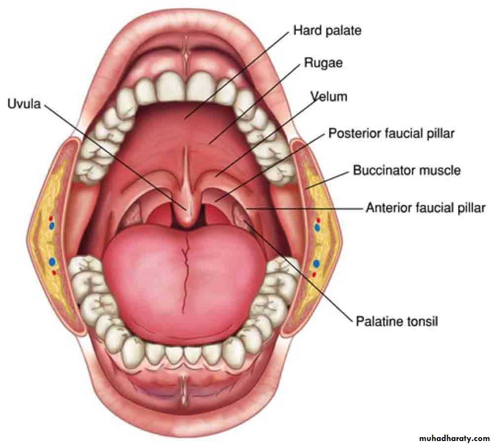
Blood Supply
The tonsillar branch of the facial artery.The veins pierce the superior constrictor muscle and join the pharyngeal, or the facial veins.
Lymph Drainage of the Tonsil
The upper deep cervical lymph nodes, just below and behind the angle of the mandible.
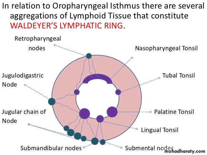
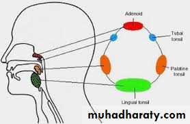
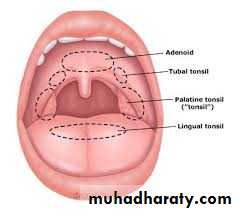
Waldeyer's Ring of Lymphoid Tissue :
The lymphoid tissue that surrounds the opening into the respiratory and digestive systems forms a ring. The lateral part of the ring is fanned by the palatine tonsils and tubal tonsils (lymphoid tissue around the opening of the auditory tube in the lateral wall of the nasopharynx).The pharyngeal tonsil in the roof of the nasopharynx forms the upper part, and the lingual tonsil on the posterior third of the tongue forms the lower part.
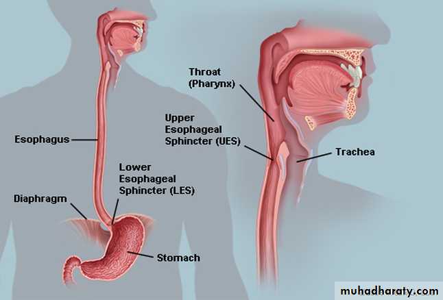
The Esophagus
The esophagus is a muscular tube about 10 in. (25 cm) long, extending from the pharynx to the stomach It begins at the level of the cricoid cartilage, opposite the body of the sixth cervical vertebra. It commences in the midline, but as it descends through the neck, it inclines to the left side.Relations in the Neck
•Anteriorly: The trachea; the recurrent laryngeal nerves ascend one on each side, in the groove between the trachea and the esophagus.
• Posteriorly: The prevertebral layer of deep cervical fascia, the longus colli , and the vertebral column
• Laterally : On each side lie the lobe of the thyroid gland and the carotid sheath.
Blood Supply in the Neck
The arteries of the esophagus in the neck are derived from the inferior thyroid arteries.The veins drain into the inferior thyroid veins.
Lymph Drainage in the Neck
The lymph vessels drain into the deep cervical lymph nodes.
Nerve Supply in the Neck
The nerves are derived from the recurrent laryngeal nerves and from the sympathetic trunks.
Thank
you
For
listening