
1
Fifth stage
Ortho Surgery
Lec
د.يقضان
12/3/2017
Slipped capital femoral epiphysis
Introduction :
It is also called epiphysiolysis .
It is uncommon .
It occur in children going through the pubertal growth spurt .
It affect boys more than girls .
Age incidence 14 -16 years old .
Left hip affected more than the right .
Aetiology
The slip occur through the growth plate .The aetiological factors are :
1- hormonal imbalance .
2- trauma .
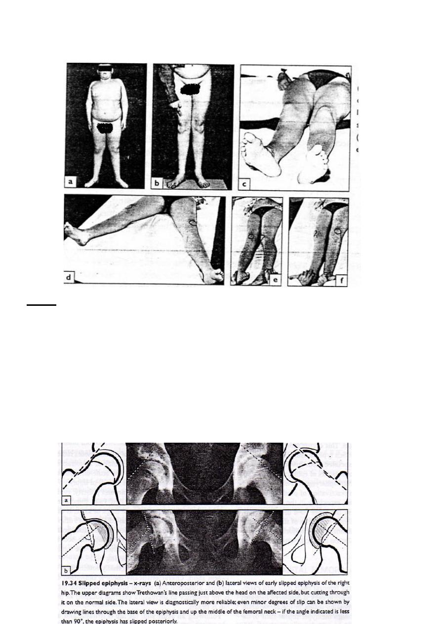
Clinically
In 50% of the cases there is history of trauma .The patient usually a child around puberty
,typically over weight , or very tall and thin.
Symptoms :
1. pain is the presenting symptom , usually it is in the groin or the thigh or even at the
knee . It come’s in attacks exaggerated by exercise
2. limping , it is more constant .
Examination:
1 – the leg is externally rotated .
2 – shortening 1 – 2 cm .
3 – limitation of flexion , abduction and internal rotation .
2
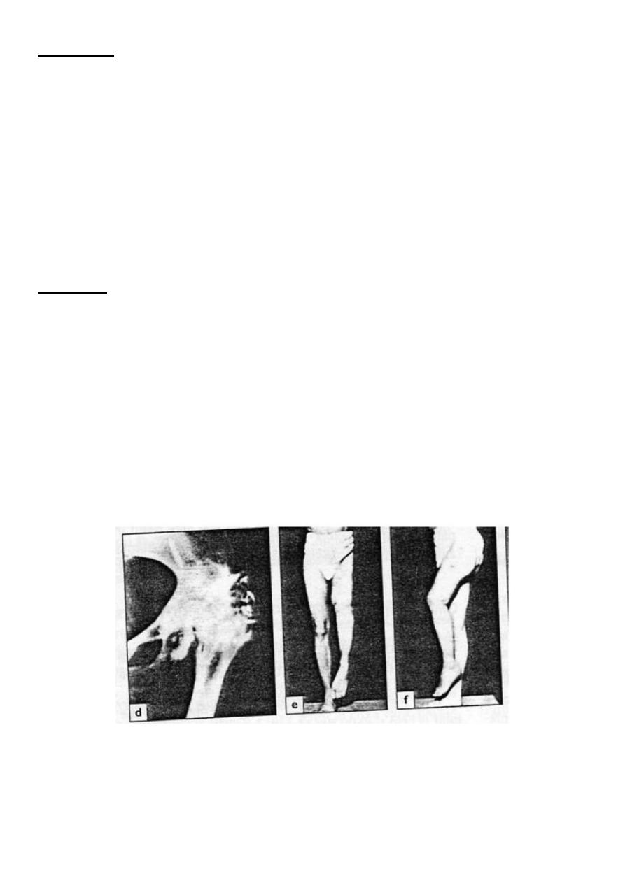
4 – the classical sign is increase external rotation as the hip is flexed .
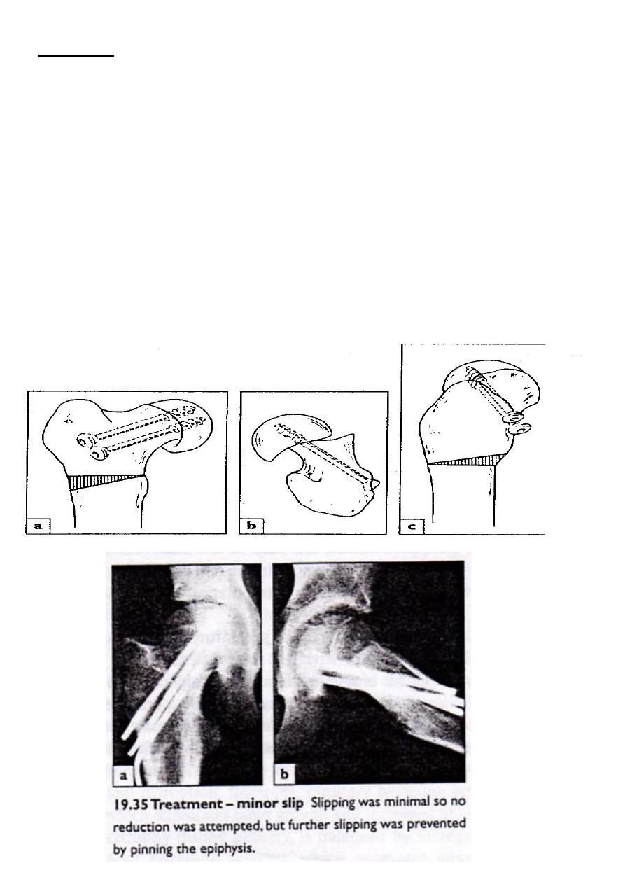
X-ray
1 – in early stage it is normal and the patient should be followed up later on by x – ray if the
symptoms persist .
2 – in anteroposterior view a line drawn along the superior surface of the neck remain
superior to the head instead of passing through it (trethowan’s) sign
3 – in lateral view the femoral epiphysis is tilted back ward and this can be detected by
measuring the angle between the axis of the femoral neck and the epiphyseal base , this
normally 90* (right angle) , if it is less than 87* it mean the epiphysis is tilted posteriorly .
3
Treatment
The aim of the treatment :
1 – preserve the epiphyseal blood supply .
2 – to stabilize the physis .
3 – to correct any residual deformity .
4 – to prevent further slipping .
Treatment :
1 - Manipulation under anesthesia is very dangerous because it may lead to avascular
necrosis of the head of the femur .
2 - surgical treatment for fixation of the epiphysis by pins or screws carry better prognosis .

4
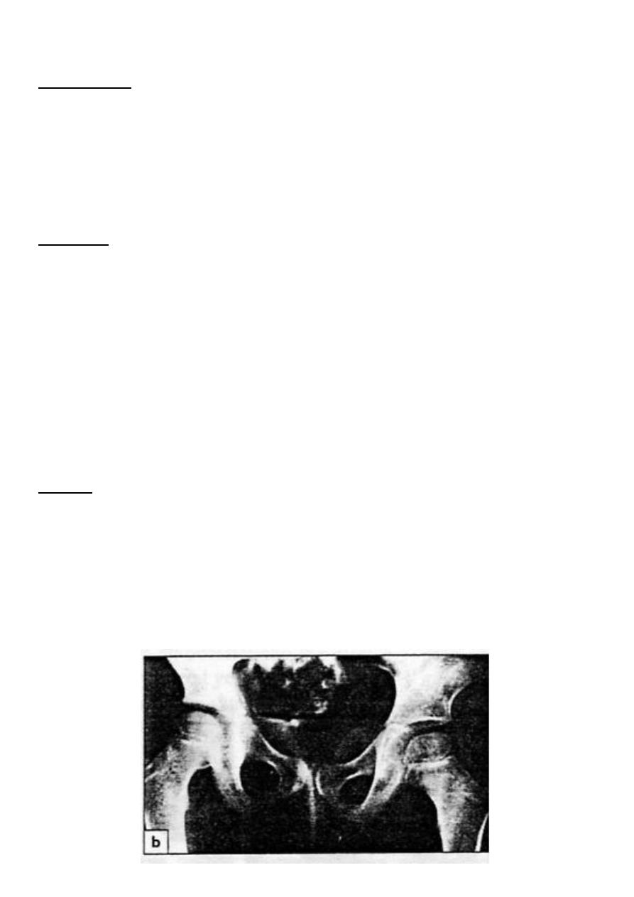
Pyogenic arthritis of the hip
Introduction
It can occur in any age even in newly born baby , and usually seen in children under two
years old .The causative micro organism is staph . aures , and it reach the joint either
directly from distant focus by blood or by local spread from osteomyelitis of the femur .
The head of the femur at this age is cartilaginous , so the treatment should be start early ,
other wise it will be completely destroyed by the proteolytic enzymes of the bacteria and pus
Adult also may develop pyogenic hip infection either as primary events e.g. debilitation or
more often secondary to invasive procedure around the hip .
Clinically
The child look acutely ill , feverish, dehydrated and he is in pain , but the site of the pain is
not clearly marked .
The patient resist any movement of the joint , but with careful examination we can detect the
site of maximum tenderness over the hip joint .
In newly born baby the infection come usually from infected umbilical vein ; the baby look
ill some time no fever and no cause is apparent , so in this condition we should think for
deep sepsis
investigation
1 – x –ray : It is of little value:
some time it show soft tissue swelling .
show displacement of the femoral head laterally .
2 – ultrasound : it is helpful in detection joint effusion .
3 – blood invest.
increase ESR .
increase WBC count mainly polymorph .
increase C REACTIVE protein .
4 - The diagnosis is difficult , and it can be confirmed by aspiration of pus or fluid from the
joint and send it to culture and sensitivity
5
Treatment
1 – intravenous antibiotic should be given as soon as the diagnosis is settled (anti staph.) .
2 – when pus come out during aspiration , anterior arthrotomy should be done , washing of
the joint , local antibiotic , then close the wound without drain .
3 – skin traction is applied until the activity of the disease is subside .
Complication
1 – destruction of the head and neck of the femur and this lead some time to pathological
dislocation of the hip , bony ankylosis .
2 – secondary osteoarthritis .
6
TUBERCULOSIS OF THE HIP
Introduction
It can started as synovitis or osteomyelitis of the femur, then start the arthritis and this cause
destruction of the joint and some time pathological dislocation .
Healing of this lesion leaves a fibrous ankylosis with considerable limb shortening and
deformity
Clinically
The condition start insidiously : pain in the groin and thigh , limping .
Later on the pain become more sever and may awake the patient from sleep .
In early stage of the disease (synovitis or osteomyelitis) the patient keep his limb flexed and
abducted , the extreme of all movements are restricted and painful .
Until x ray changes appear the diagnosis is very difficult and the hip remain irritable .
If arthritis supervenes the hip become flexed , adducted and internally rotated , muscle
wasting become obvious and all movements are grossly limited by pain and spasm .
X – ray
1 – earliest changes is general rarefaction with normal joint space and outline .
2 – in stage of arthritis in addition to rarefaction there is destruction of the acetabular roof
(wondering acetabulum) ; or there is destruction of the femoral head or usually both and all
these will lead to pathological dislocation and fibrous ankylosis .
TB. LT . Hip (marked osteoporosis)
7
Treatment
1 – anti tuberculous drugs are essentials .
2 – skin traction is applied .
3 – if there is absces it should be evacuated.
4 – if the joint has been destroyed then : arthrodesis may be necessary ; but after all signs
of the activity of the disease are subside and not before 14 years old .
5 - In older patient with residual pain and deformity , and if the disease activity is
subside for long time then total hip replacement is advisable .
prognosis
1 – in early disease , if properly treated there will be complete healing and normal hip .
2 – if articular surface is damaged ; this lead to fibrous joint .
In untreated cases there will be :
A – shortening .
B – fixed flexion deformity .
C – adduction deformity
Outcome of untreated TB hip
R