Objectives
After studying this chapter you should know the following:■Describe the various parts of the eye and list the functions of each. ■ Describe the organization of the retina. ■ Explain how light rays in the environment are brought to a focus on the retina and the role of accommodation in this process. ■ Define hyperopia, myopia, astigmatism, presbyopia, and strabismus. ■ Describe the electrical responses produced by rods and cones, and explain how these responses are produced. ■ Trace the neural pathways that transmit visual information from the rods and cones to the visual cortex.
VISION
The eye is present within protective casing (orbit), each eye has a layer of photoreceptors that respond to light, a lens system that focuses the light on these receptors, and a system of nerves that conducts impulses from the receptors to the brain.
Anatomy of the eye (figure-1)
The eye consists of 3 layers. The outer protective layer of the eyeball is the sclera or the “white of the eye” through which no light can pass. It is modified anteriorly to form the transparent cornea, through which light rays enter the eye.
Just inside the sclera, the second layer which is the choroid, a vascular layer that provides oxygen and nutrients to the structures in the eye. Lining the posterior two thirds of the choroid is the retina, the innermost layer where the neural tissue containing the photoreceptors.
The iris (coloured portion of the eye) contains circular muscle fibers that constrict and radial fibers that dilate the pupil (a hole in the center of the iris that allows light to enter the posterior part of the eye). Variations in the diameter of the pupil can produce up to a fivefold change in the amount of light reaching the retina.
The aqueous humor is a clear protein-free liquid that nourishes the cornea and iris; it is produced in the ciliary body by diffusion and active transport from plasma. It is normally reabsorbed through a network of trabeculae into the canal of Schlemm, which is a venous channel at the junction between the iris and the cornea (anterior chamber angle). Obstruction of this outlet leads to increased intraocular pressure, a critical medical condition known glaucoma.
The vitreous chamber is the space between the lens and the retina that is filled primarily with a clear gelatinous material called the vitreous humor.
The eye is well protected from injury by the bony walls of the orbit. The cornea is moistened and kept clear by tears that course from the lacrimal gland in the upper portion of each orbit across the surface of the eye to empty via the lacrimal duct into the nose. Blinking helps keep the cornea moist.
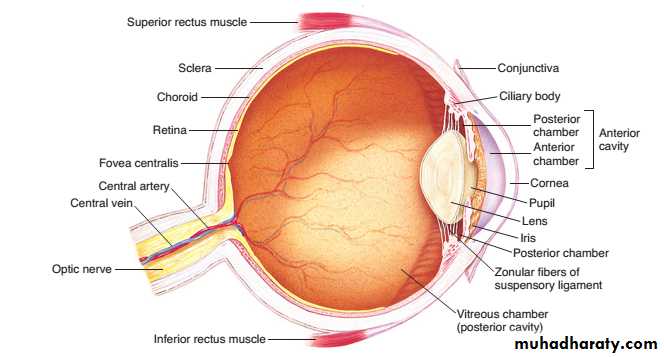
Figure 1: anatomy of the eye
Retinais organized into layers containing different types of cells and neural processes (axons and dendrites). The outer nuclear layer contains the photoreceptors, the rods and cones. The rods and cones layer of the retina rests on the pigment epithelium next to the choroid. The pigment epithelium contains melanin black pigment absorbs light rays, preventing the reflection of rays back through the retina. Such reflection would otherwise produce blurring of the visual images. Also pigment epithelium contains large quantities of vitamin A that is important in the synthesis of photosensitive chemicals of rods and cones (see figure 2 below).
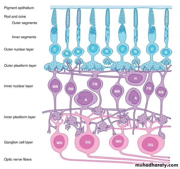
Figure 2: layers of the retina
The optic nerve leaves the eye at a point 3 mm medial to and slightly above the posterior pole of the globe. This region is visible through the ophthalmoscope as the optic disk. Since there are no visual receptors over the disk, this area of the retina does not respond to light and is known as the blind spot (figure 3).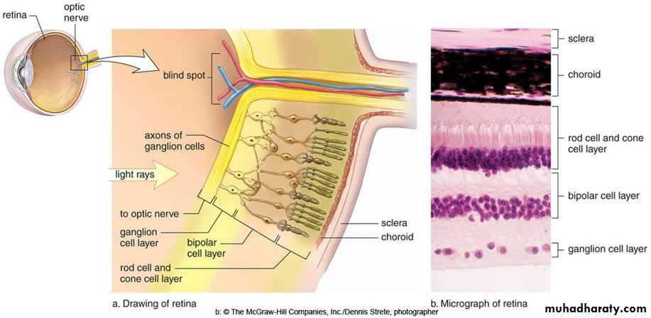
Figure 3: blind spot
Near the posterior pole of the eye, there is a yellowish pigmented spot called the macula .The fovea (figure 4) is in the center of the macula; it is a thinned-out, rod-free portion of the retina. In it, the cones are densely packed, and each synapses on a single bipolar cell, which, in turn, synapses on a single ganglion cell, providing a direct pathway to the brain. There are very few overlying cells (various layers of the retina are pulled aside) and no blood vessels allowing light to pass unimpeded to the cones. Consequently, the fovea is the point where visual acuity is greatest. When attention is attracted to or fixed on an object, the eyes are normally moved so that light rays coming from the object fall on the fovea.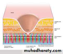
Figure 4: fovea centralis
Photoreceptors (rods and cones) (figure 5)The rods are extremely sensitive to light and are the receptors for night vision (scotopic vision). The scotopic visual apparatus is incapable of resolving the details and boundaries of objects or determining their colour. The cones have a much higher threshold, but the cone system has a much greater acuity and is the system responsible for vision in bright light (photopic vision) and for colour vision. Accordingly, there are thus two kinds of inputs to the central nervous system (CNS) from the eye: input from the rods and input from the cones.
The distribution of rods and cones in the retina is different with the rods predominate in the extrafoveal region, while the cones are present exclusively in the fovea.
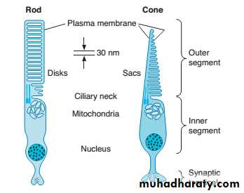
Figure 5: rods and cones
Photosensitive chemicalsWhen light is absorbed by Photosensitive chemicals, their structure changes, and this triggers a sequence of events that initiates neural activity through the initiation of action potential.
RHODOPSIN (figure 6)
The photosensitive pigment (rhodopsin) is comprised of retinal, an aldehyde of vitamin A, and a protein called opsin. Because of the importance of vitamin A in the synthesis of retinal, it is not surprising that a deficiency in this vitamin produces visual abnormalities.
Action potential formed in the photoreceptors is then transmitted to the optic nerve and visual cortex.
Regarding cone photoreceptors (colour pigments) there are 3 types of colour pigments red, green and blue each is activated by its own wavelength.
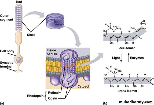
Figure 6: rhodopsin and its role in vision
The lens (figure 7)Light rays are bent when they pass from a medium of one density into a medium of a different density. The bending of light rays is called refraction and is the mechanism that allows one to focus an accurate image onto the retina.
Light rays from an object that strike a lens more than 6 m (20 ft) away are considered to be parallel. Refractive power is greatest when the curvature of a lens is greatest. The refractive power of a lens is conveniently measured in diopters.
The lens is composed of elastic capsule filled with viscous proteinaceous but transparent fluid. When the lens is in a relaxed state with no tension on its capsule, it assumes an almost spherical shape, owing mainly to the elastic retraction of the lens capsule. However, suspensory ligaments that hold the lens in place by attachment to the ciliary muscles are constantly tensed. The tension on the ligaments causes the lens to remain relatively flat under normal conditions of the eye.
Also the ciliary muscle has two separate sets of smooth muscle fibers and supplied by parasympathetic nerve fibers through the 3rd cranial nerve. When the eyes look to near object, these muscle fibers contract, releasing the ligaments' tension on the lens, making the lens more spherical and increasing its refractive power. The reverse occur when the eye look to far object. This process is called accommodation.
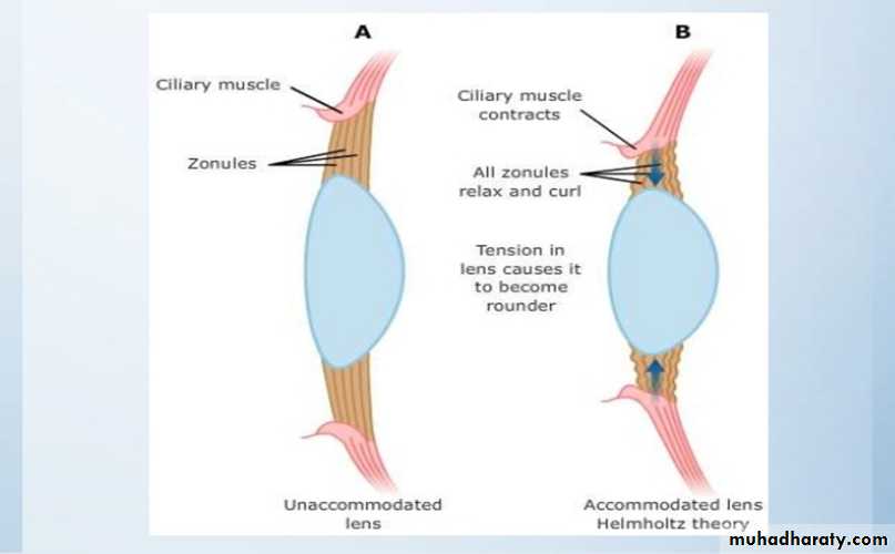
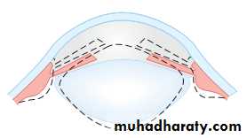
Figure 7: the lens and accommodation mechanism
Common defects of the image forming mechanism (figure 8)
In some individuals, the eyeball is shorter than normal and the parallel rays of light are brought to a focus behind the retina. This abnormality is called hyperopia or farsightedness. Sustained accommodation, even when viewing distant objects, can partially compensate for the defect, but the prolonged muscular effort is tiring and may cause headaches and blurring of vision. The defect can be corrected by using glasses with convex lenses.
In myopia (nearsightedness), the anteroposterior diameter of the eyeball is too long. Myopia is said to be genetic in origin. However, there is a positive correlation between sleeping in a lighted room before the age of 2 and the subsequent development of myopia. In young adult humans the extensive close work involved in activities such as studying accelerates the development of myopia. This defect can be corrected by glasses with biconcave lenses.
Astigmatism is a common condition in which the curvature of the cornea is not uniform. When the curvature in one meridian is different from that in others, light rays in that meridian are refracted to a different focus, so that part of the retinal image is blurred. Astigmatism can usually be corrected with cylindrical lenses.
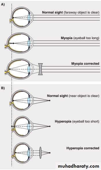
Figure 8: defects of refraction
Responses in the visual pathways & cortexNeural pathways
The axons of the ganglion cells pass caudally in the optic nerve and optic tract to end in the lateral geniculate body in the thalamus. The fibers from each nasal hemiretina decussate in the optic chiasm. In the lateral geniculate body (LGN), the fibers from the nasal half of one retina and the temporal half of the other synapse on the cells whose axons form the geniculocalcarine tract (optic radiation). This tract passes to the occipital lobe of the cerebral cortex. The effects of lesions in these pathways on visual function are discussed in the practical part and is required here. Some ganglion cell axons bypass the lateral geniculate nucleus (LGN) to project directly to the pretectal area; this pathway mediates the pupillary light reflex and eye movements. The frontal cortex is also concerned with eye movement, and especially its refinement.
Lateral geniculate body is part of the thalamus and represent a relay center for the visual pathway between the retina and occipital cortex.
Qx 1: according to this figure, define the visual field defects labelled as A, B, C and D.
Qx 1: according to this figure, define the visual field defects labelled as A, B, C and D.
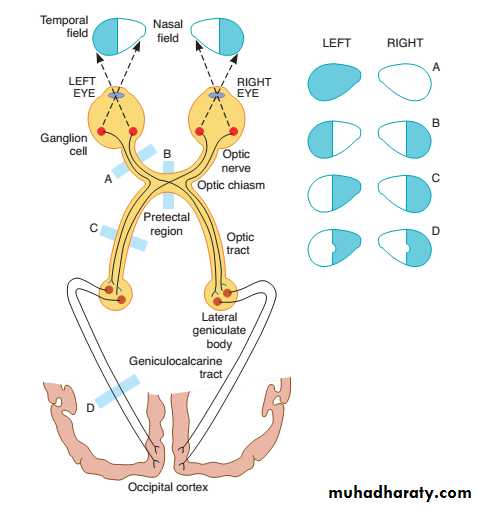
Figure 9: optic nerve pathway
Primary Visual cortex (figure 10)It is located principally on the sides of the calcarine fissure in the occipital lobe. Optic radiation fibers arising from LGN terminate in this area; and like LGN it has detailed spatial representation with point-point representation of the retina. The macula has a large area of representation in the primary visual cortex. The fovea is responsible for the highest degree of visual acuity. Based on retinal area, the fovea has several hundred times as much representation in the primary visual cortex as do the most peripheral portions of the retina.
Secondary Visual Areas of the Cortex (figure 11)
There are about dozen of Brodmann areas concerned with vision called secondary visual areas or secondary association areas. They lie around the primary visual cortex. They receive their input from the primary visual cortex and somatosensory cortex for further analysis and dissection of the visual image.
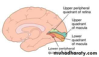
Figure 10: occipital cortex
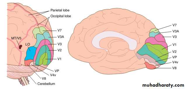
Figure 11: visual association areas
Qx 2: what are Bradmann areas?Qx 3: what are the lesions resulting from damage to superior colliculus
Qx 4: functions of: choroid layer of the eye, ciliary muscles, pigmented epithelium layer of the retina, lateral geniculate body,
Qx 5: what is presbyopia?
Qx 6: what happen when parasympathetic supply to the eye is injuried?
MULTIPLE CHOICE QUESTIONS
For all questions, select the single best answer unless otherwise directed.
1. A visual exam in an 80-year-old man shows he has a reduced ability to see objects in the upper and lower quadrants of the left visual fields of both eyes but some vision remains in the central regions of the visual field. The diagnosis is
A. central scotoma.
B. heteronymous hemianopia with macular sparing.
C. lesion of the optic chiasm.
D. homonymous hemianopia with macular sparing.
E. retinopathy.
2. A 45-year-old female who had never needed to wear glasses experienced difficulty reading a menu in a dimly-lit restaurant. She then recalled that as of late she needed to have the newspaper closer to her eyes in order to read it. A friend recommended she purchase reading glasses. Visual accommodation involves
A. increased tension on the lens ligaments.
B. a decrease in the curvature of the lens.
C. relaxation of the sphincter muscle of the iris.
D. contraction of the ciliary muscle.
E. increased intraocular pressure.
3. A 28-year-old male with severe myopia made an appointment to see his ophthalmologist when he began to notice flashing lights and floaters in his visual field. He was diagnosed with a retinal detachment. The retina
A. is epithelial tissue that contains photoreceptors.
B. lines the anterior one-third of the choroid.
C. has an inner nuclear layer that contains bipolar cells, horizontal cells, and amacrine cells.
D. contains ganglion cells whose axons form the oculomotor nerve.
E. contains an optic disk where visual acuity is greatest.
4. A 62-year-old Caucasian woman experienced a rapid onset of blurry vision along with loss of central vision. A comprehensive eye exam showed that she had wet age-related macular degeneration. The fovea of the eye
A. has the lowest light threshold.
B. is the region of highest visual acuity.
C. contains only red and green cones
D. contains only rods.
E. is situated over the head of the optic nerve.
5. Which of the following parts of the eye has the greatest concentration of rods?
A. Ciliary body
B. Iris
C. Optic disk
D. Fovea
E. Parafoveal region
6. Which of the following is not correctly paired?
A. Rhodopsin: retinal and opsin
B. Obstruction of the canal of Schlemm: elevated intraocular pressure
C. Myopia: convex lenses
D. Astigmatism: nonuniform curvature of the cornea
E. Inner segments of rods and cones: synthesis of the photosensitive compounds
7. The correct sequence of events involved in phototransduction in rods and cones in response to light is:
A. activation of transducin, decreased release of glutamate, structural changes in rhodopsin, closure of Na+ channels, and decrease in intracellular cGMP.
B. decreased release of glutamate, activation of transducin, closure of Na+ channels, decrease in intracellular cGMP, and structural changes in rhodopsin.
C. structural changes in rhodopsin, decrease in intracellular cGMP, decreased release of glutamate, closure of Na+ channels, and activation of transducin.
D. structural changes in rhodopsin, activation of transducin, decrease in intracellular cGMP, closure of Na+ channels, and decreased release of glutamate.
E. activation of transducin, structural changes in rhodopsin, closure of Na+ channels, decrease in intracellular cGMP, and decreased release of glutamate.
8. A 25-year-old medical student spent a summer volunteering in the sub-Saharan region of Africa. There he noted a high incidence of people reporting difficulty with night vision due to a lack of vitamin A in their diet. Vitamin A is a precursor for the synthesis of
A. rods and cones.
B. retinal.
C. rod transducin.
D. opsin.
E. cone transducin.
9. An 11-year-old male was having difficulty reading the graphs that his teacher was showing at the front of classroom. His teacher recommended he be seen by an ophthalmologist. Not only was he asked to look at a Snellen letter chart for visual acuity but he was also asked to identify numbers in an Ishihara chart. He responded that he merely saw a bunch of dots. Abnormal color vision is 20 times more common in men than women because most cases are caused by an abnormal
A. dominant gene on the Y chromosome.
B. recessive gene on the Y chromosome.
C. dominant gene on the X chromosome.
D. recessive gene on the X chromosome.
E. recessive gene on chromosome 22
10. Which of the following is not involved in color vision?
A. Activation of a pathway that signals differences between
S cone responses and the sum of L and M cone responses
B. Geniculate layers 3–6
C. P pathway
D. Area V3A of visual cortex
E. Area V8 of visual cortex
11. A 56-year-old female was diagnosed with a tumor near the base of the skull, impinging on her optic tract. Which of the following statements about the central visual pathway is correct?
A. The fibers from each temporal hemiretina decussate in the optic chiasm, so that the fibers in the optic tracts are those from the temporal half of one retina and the nasal half of the other.
B. In the geniculate body, the fibers from the nasal half of one retina and the temporal half of the other synapse on the cells whose axons form the geniculocalcarine tract.
C. Layers 2 and 3 of the visual cortex contain clusters of cells called globs that contain a high concentration of cytochrome oxidase.
D. Complex cells have a preferred orientation of a linear stimulus and, compared to simple cells, are more dependent on the location of the stimulus within the visual field.
E. The visual cortex is arranged in horizontal columns that are concerned with orientation.0