Mycoplasma &Chlamydia
Mycoplasmas (Mycoplasmas spp)Mycoplasmas are groups of small, wall-less organisms. They are the smallest free-living organisms (0.3μm in diameter).Important properties:1) Mycoplasmas stain poorly with Gram stain.2) The outer surface is a flexible three layer cell membrane; hence the organisms can assume a variety of shapes.3) It contain cholesterol in their bacterial membrane.4) The colony frequently has a characteristic (fried egg) shape, with a raised center & a thinner outer edge.
In human there are four important species:1) Mycoplasma pneumoniae 2) M. Hominis3) M. Genitalium 4) Ureaplasma urealyticum
Pathogenesis:Pathogenic Mycoplasmas have flask-like or filamentous shapes & have specialized polar tip structures that mediate the adherence to host cells (ciliated & non ciliated cells).
1) M. pneumoniae•Is transmitted from person to person by means of infected respiratory secretions & cause atypical pneumonia. May be ranged from asymptomatic infection to serious pneumonitis. Incubation period 1-3 weeks.
• Symptoms:Fever, headache, sore throat, & cough which is non-productive .Complications are uncommon, but sometimes hemolytic anemia, meningitis & pericarditis may occur
• Diagnosis: Culture: haert infusion peptone broth (with 2% agar & 30% human ascitic fluid or animal serum, pH 7.8). Complement fixation (cf test). Cold hemagglutination (at 4C°). ELISA.• Treatment: Tetracycline & erythromycin.
2) Mycoplasma hominis: causes infections of uterine tubes (salpingitis) & tubo-ovarian abscesses (in Women).3) Mycoplasma genitalium: Associated with some infections of chronic nongonococcal urethritis (in men).
4)Ureaplasma urealiticum: causes nongonococcal urethritis in some men (may play role in male infertility).Lung diseases in premature low birth-weight infants (acquired during birth
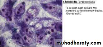
Chlamydiae (Chlamydia spp.)• Chlamydiae: are obligate intracellular bacteria, lack theability to produce sufficient energy to grow independently & therefore can grow only inside host cell. Chlamydiae have a replicative cycle different from that of all other bacteria. Within cells site of replication appears as an inclusion body, which can be stained & visualizedmicroscopically. These inclusions are useful in the identification of these organisms in the clinical laboratory.•They have rigid cell wall but they don’t have typicalpeptidoglycan. Their cell walls resemble those of G-negative but lack muramic acid.
Chlamydia spp., inclusion bodies within host cells
Pathogenesis:Chlamydiae infect primarily epithelial cells of the mucous membrane and the lungs.Diseases:1)Chlamydia psittaci: infects the lungs →humanpsittacosis, this disease may be asymptomatic or producehigh fever & pneumonia.2) C. pneumoniae →upper & lower respiratory tractinfections especially bronchitis & pneumonia.3) C. trachomitis types A, B&C →trachoma (chronicconjunctivitis endemic in Africa & Asia).
4) C. trachomatis types D-K→genital tract infections, which occasionally transmitted to the eyes or the respiratory tract.• In men: it common cause of non-gonococcal urethritis, which may progress to epididymitis, prostatitis or proctitis • In women: cervictitis develops & may progress tosalpingitis & pelvic inflammatory disease →this may result infertility or ectopic pregnancy.• Infants borne to infected mothers often develop mucopurulent eye infections (neonatal inclusionconjunctivitis) 7-12 days after delivery. Some develop chlamydial pneumonitis 2-12 weeks after birth.• 5) C. trachomitis L1-L3 immunotypes→lymphogranuloma venereum ,a sexually transmitted disease with lesions on genitalian & in lymph nodes .
Diagnosis:• Group-specific Ag. (lipopolysaccharide) → complement fixation test.• Species-specific & immunotype-specific Ag. (Protein)→ Immunofluorescence test .• Chlamydiae form cytoplasmic inclusions →stain with Giemsa →immunofluorescence test .Treatment:Tetracyclines such as doxycycline & macrolides,such as erythromycin & azithromycin .