
Hussien Mohammed Jumaah
CABMLecturer in internal medicine
Mosul College of Medicine
2016

learning-topics
Critical illness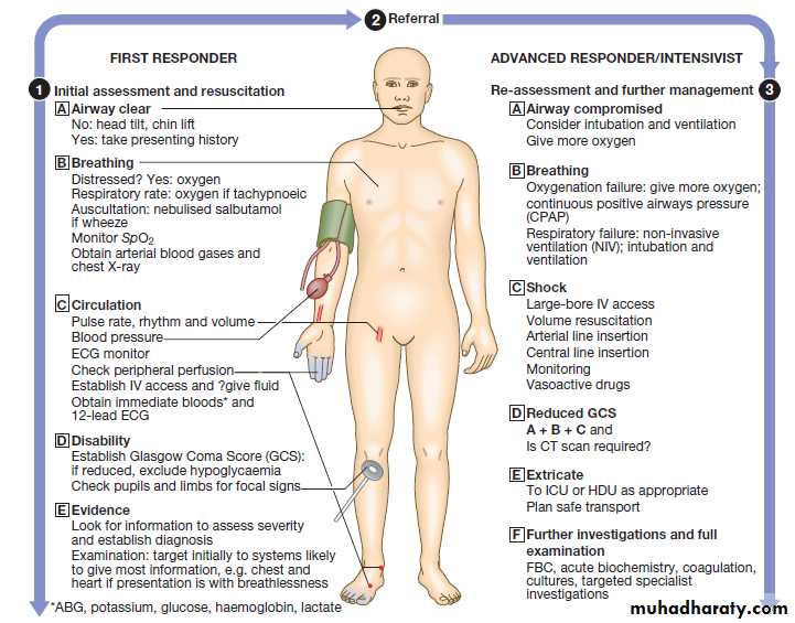


A critically ill patient is at imminent risk of death. Recognition, assessment and management of critical illness
are thus fundamental to clinical care in any area of
medicine. The principle underpinning intensive care is the simultaneous assessment of illness severity and stabilisation of life-threatening physiological abnormalities.
The goal is to prevent deterioration and effect improvements as the diagnosis is established, and treatment of the underlying definitive disease process(es)
is initiated. Blinkered attention to either resuscitation or diagnosis in isolation results in worse outcomes and increased mortality; the two processes are inextricably
interlinked. Appropriate physiological monitoring is required to allow continuing assessment and re-assessment of response to therapy, wherever the clinical environment.
PHYSIOLOGY OF CRITICAL ILLNESS
Oxygen transportThe principal function of the heart, lungs and circulation
is the provision of oxygen (and other nutrients) to the
various organs and tissues of the body. During this process, carbon dioxide and other metabolic waste products are removed. The rate of supply and removal should match the specific metabolic requirements of the individual tissues. This requires adequate oxygen uptake in the lungs, global matching of delivery and consumption, and regional control of the circulation. Failure to supply sufficient oxygen to meet the metabolic requirements of the tissues is the cardinal feature of circulatory failure or ‘shock’, and optimisation of tissue oxygen delivery and consumption is the goal of resuscitation.
Atmospheric oxygen moves down a partial pressure
gradient from air, through the respiratory tract, fromalveoli to arterial blood and then to the capillary beds
and cells, diffusing into the mitochondria, where it is
utilised at cytochrome a3 . The movement of
oxygen from the left ventricle to the systemic tissue
capillaries is known as oxygen delivery (DO2), and is
the product of cardiac output (flow) × arterial oxygen
content (CaO2) The latter is the product of haemoglobin
(Hb) × arterial oxygen saturation of haemoglobin (SaO2)
× 1.34.
By increasing cardiac output, arterial oxygen saturation or haemoglobin concentration, DO2 will be increased.
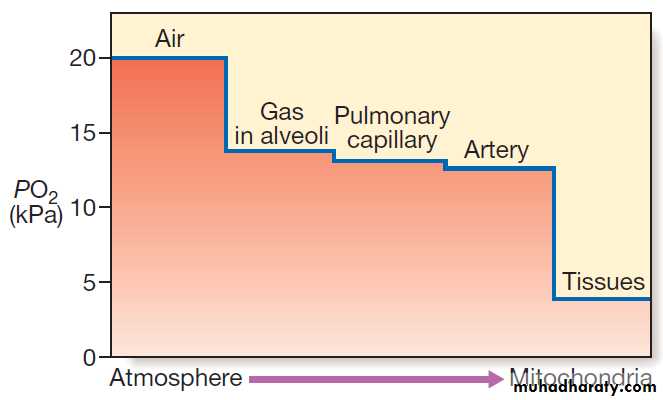
The oxygen cascade in the transport of oxygen from inspired gas to the cell. Gas containing oxygen is entrained into the lungs by inspiratory flow. The partial pressure of O2 falls as it moves from lungs to blood and then to the tissues. The partial pressure gradient
between capillary blood and the intracellular environment is pivotal to oxygen reaching the mitochondria.
(PO2 = oxygen partial pressure (kPa) 1 kPa = 7.5 mmHg).
The regional distribution of oxygen delivery is important.
If skin and muscle receive high blood flows but thesplanchnic bed does not, the gut will become hypoxic
even if overall DO2 is high.
The movement of oxygen from tissue capillary to cell
occurs by diffusion and depends on the gradient of
oxygen partial pressures, diffusion distance and the
ability of the target cell to take up and use oxygen.
Microcirculatory, tissue diffusion and cellular factors
thus also influence the oxygen status of the cell.
Cardiovascular component of oxygen delivery: flow
A key determinant of DO2 is cardiac output, which isdetermined by the ventricular ‘preload’ and ‘afterload’,
myocardial contractility and heart rate.
Preload
The atrial filling pressures, or preload, determine the
end-diastolic ventricular volume, which, according to
Starling’s Law and depending on myocardial contractility,
defines the force of cardiac contraction and the
stroke volume .The principal determinant of preload is venous return, determined by the intravascular volume, venous ‘tone’ and intrathoracic pressure. This can be measured as the central venous pressure (CVP).
When volume is lost (e.g. in major haemorrhage),
venous ‘tone’ increases and this helps to offset the consequent fall in atrial filling pressure and stroke volume.
If the equivalent volume is restored gradually by intravenous fluid administration, the right atrial pressure
will return to normal as the intravascular volume is normalized and the reflex increase in venous tone abates.
However, if fluid is infused too rapidly, there is insufficien
time for the venous and arteriolar tone to fall and pulmonary oedema may occur, even though the
intravascular volume has only been restored to the premorbid level.
If the preload is low, volume loading with intravenous
fluids is the priority and is the most appropriatemeans of improving cardiac output and tissue perfusion.
The choice of fluid for volume loading is controversial,
but as there is no clear advantage of colloid over crystalloid, sodium chloride is used. Red cells have traditionally been transfused to achieve and maintain a haemoglobin concentration of 100 g/L, but in the absence of significant heart disease, the target is 70–90 g/L .
When the preload is high due to excessive intravascular
volume or impaired myocardial contractility,
removing volume from the circulation by using diuretics
or haemofiltration, or increasing the capacity of the vascular bed by using venodilator therapy (glyceryl trinitrate, morphine) often improves stroke volume.
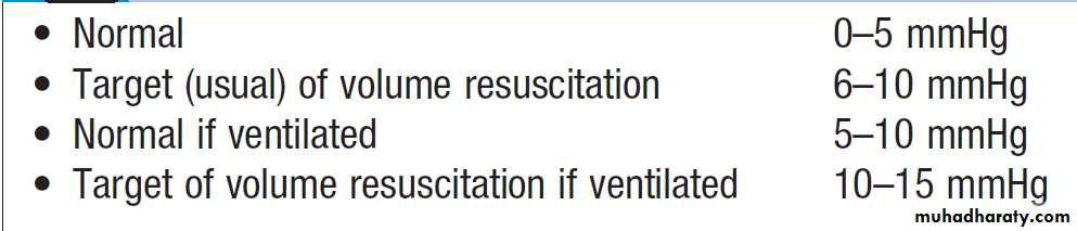
Central venous pressure measurements
Afterload
Afterload is the tension in the ventricular myocardiumduring systole, and is determined by the resistance to
ventricular outflow, which is a function of the peripheral
arteriolar resistance. High resistances produce lower flows at higher pressures for a given amount of ventricular work.
Therefore, a systemic vasodilator will allow the same cardiac output to be maintained for less ventricular work but with a reduced arterial BP. In hyperdynamic sepsis, the peripheral arteriolar tone and BP are low but the cardiac output is often high; therefore the vasoconstrictor noradrenaline (norepinephrine) is appropriate to restore BP, usually at the price of some reduction in cardiac output.
Myocardial contractility
This determines the stroke volume that the ventricle can generate against a given afterload for a particular preload. Myocardial contractility is frequently reduced in critically ill patients due to preexisting cardiac disease (usually ischaemic), drugs (e.g. β-blockers, verapamil) or to the disease process itself (particularly sepsis, as the associated low diastolic BP may compromise coronary arterial perfusion).Oxygenation component of oxygen delivery: content
The major determinants of the oxygen content of arterial
blood (CaO2) are the arterial oxygen saturation of haemoglobin (SaO2) and the haemoglobin.> 95% of oxygen carried in the blood is bound to haemoglobin.
The oxyhaemoglobin dissociation curve (Fig.)
describes the relationship between the saturation ofHaemoglobin (SO2) and the partial pressure (PO2) of
oxygen in the blood.
A shift in the curve will influence the uptake and release of oxygen by the haemoglobin molecule. If the curve moves to the right, the haemoglobin
saturation will be lower for any given oxygen
tension: less oxygen will be taken up in the lungs but
more will be released to the tissues. As capillary PCO2
rises, the curve moves to the right, increasing the unloading of oxygen in the tissues – a phenomenon known as the Bohr effect. Thus a shift to the right increases capillary PO2 and hence cellular oxygen supply.
Due to the shape of the curve, a small drop in arterial PO2 (PaO2) below 8 kPa (60 mmHg) will cause a marked fall in SaO2. The shape of the curve also means that increases in PaO2 beyond the level that ensures SaO2 is greater than 90% produce relatively small additional increases in CaO2 .Thus, in anaemic (Hb 60 g/L or 6 g/dL) and hypoxaemic (SaO275%) when breathing air (fractional inspired oxygen concentration (FiO2) 20%), supplementary oxygen at FiO2 40% will increase SaO2 to 93% and CaO2by 24%. However, further increases in FiO2, while raising PaO2, cannot produce any further useful increases in SaO2 or CaO2. However, increasing haemoglobin to 90 g/L (9 g/dL) by blood transfusion will result in a further 50% increase in CaO2.
Traditionally, the optimum haemoglobin concentration
for critically ill patients was considered to beapproximately 100 g/L (10 g/dL), representing a balance
between maximising the oxygen content of the blood and
avoiding regional microcirculatory problems due to
increased viscosity. However, improved outcomes have
been demonstrated when the haemoglobin is maintained
between 70 and 90 g/L (7–9 g/dL).
A target haemoglobin of 10 g/dL remains appropriate in the elderly and in patients with CAD, cardiogenic shock, significant aortic stenosis or acute brain trauma.
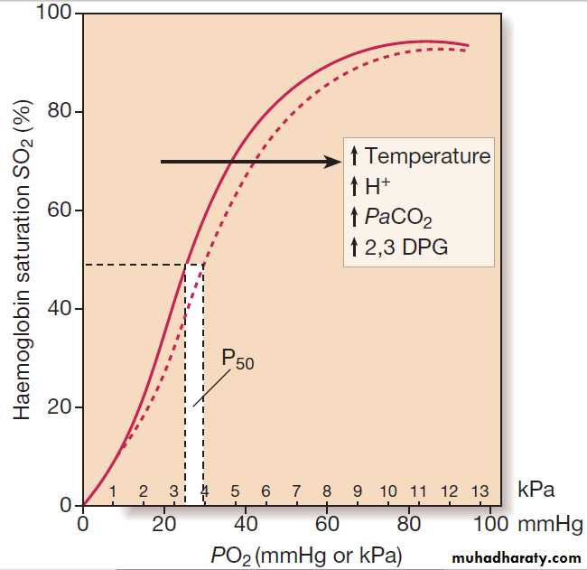
Oxyhaemoglobin dissociation curve: the relationship between oxygen tension (PO2) and percentage saturation of
haemoglobin with oxygen (SO2). The dotted line illustrates the rightward shift of the curve (i.e. P50 increases) caused by increases in temperature, PaCO2, metabolic a cidosis and 2,3 diphosphoglycerate (DPG).
Oxygen consumption
The sum of the oxygen consumed by the various organsrepresents the global oxygen consumption (VO2), and is
approximately 250 mL/min for an adult of 70 kg undertaking normal daily activities.
The oxygen saturation in the pulmonary artery, or
‘mixed venous oxygen saturation’ (SvO2), is a measure
of the oxygen not consumed by the tissues (DO2 − VO2).
The saturation of venous blood from different organs
varies considerably; the hepatic venous saturation
usually does not exceed 60% but the renal venous saturation may reach 90%, reflecting the difference in the
metabolic requirements of these organs, and the oxygen
content of the blood delivered to them.
Relationship between oxygen consumption and delivery
The tissue oxygen extraction ratio (OER) is 20–25% in a
normal individual at rest. The maximum OER is
approximately 60% for most tissues; at this point, no
further increase in extraction can occur and any further
increase in oxygen consumption or decline in oxygen
delivery will cause tissue hypoxia, anaerobic metabolism
and increased lactic acid production.
This ultimately results in multiple organ failure and an increased risk of death. In practice, if there is a metabolic acidosis, hyperlactataemia and/or oliguria that could be due to inadequate oxygen delivery, a therapeutic trial of increased oxygen delivery (while maintaining an adequate BP) may be helpful clinically.
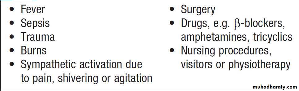
Pathophysiology of the inflammatory response
In critically ill patients, these have important consequences (Box).Fever, tachycardia with warm peripheries, tachypnoea
and a raised white cell count prompt a diagnosis
of sepsis, with the presentation caused by invading
microorganisms and their breakdown products. Other
conditions, such as pancreatitis, trauma, malignancy,
tissue necrosis (e.g. burns), aspiration syndromes, liver
failure, blood transfusion and drug reactions, can also
present in this way in the absence of infection.
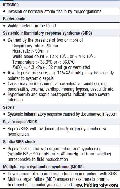
Terminology in the inflammatory state
Local inflammationThe body’s initial response to a noxious local insult is to
produce a local inflammatory response, with sequestration and activation of white blood cells and the release of a variety of mediators to overcome the primary ‘insult’ and prevent further damage locally or in distant organs. Normally, a delicate balance is achieved between pro- and anti-inflammatory mediators. However, if the response is excessive, a large array of pro-inflammatory mediators may be released into the circulation . The inflammatory and coagulation cascades are intimately linked, as the latter cause not only platelet activation and fibrin deposition, but also activation of
leucocytes and endothelial cells.
Conversely, leucocyte activation induces tissue factor expression and initiates coagulation pathways.
The natural anticoagulants, antithrombin (AT III), activated protein C (APC) and tissue factor pathway inhibitor (TFPI), inhibit proinflammatory cytokines.
Deficiency of AT III and APC (features of disseminated intravascular coagulation (DIC)) facilitates thrombin generation and promotes further endothelial cell dysfunction.
Systemic inflammation
In a severe inflammatory response, systemic release ofcytokines and other mediators triggers widespread
interaction between the coagulation pathways, platelets,
endothelial cells and monocytes, tissue macrophages,
and neutrophils. Activated neutrophils express adhesion
factors, which make them adhere to and initially
roll along the endothelium, before adhering firmly and
migrating through the damaged and disrupted endothelium
into the extravascular interstitial space (together with fluid and proteins), resulting in tissue oedema and inflammation. A vicious circle of endothelial injury, intravascular coagulation, microvascular occlusion, tissue damage and further release of inflammatory mediators ensues.
This can occur in all organs, manifesting in
the lungs as acute lung injury and in the kidneys as acute
tubular necrosis (ATN). Similar processes probably
account for damage to other organs, including the heart.
The endothelium itself produces mediators that
control local blood vessel tone. The profound vasodilatation
that characterises septic shock and some other acute systemic inflammatory states, such as pancreatitis, results from excessive production of nitric oxide (NO), due to activation of inducible NO synthase enzymes.
Systemic inflammatory processes also have important
effects on mitochondrial function, resulting inimpaired oxidative phosphorylation and aerobic energy
generation. This block to oxygen utilisation by cells is
sometimes called cytopathic hypoxia. Patients typically
have a reduced arteriovenous oxygen difference, a low
oxygen extraction ratio, a raised plasma lactate and a
paradoxically high mixed venous oxygen saturation
(SvO2), despite normal or supranormal oxygen delivery.
This is associated with the development of multiple
organ failure (MOF) and reduced survival.
MONITORING
Monitoring in intensive care includes a combination ofclinical and automated recordings. Electrocardiogram
(ECG), SpO2 (oxygen saturation), BP and usually CVP
recordings are taken at least hourly, using either a
24-hour chart or a computerised system. Urine output
measurement requires early catheterisation. All invasive haemodynamic monitoring should be referenced to the
mid-axillary line as ‘zero’. Clinical monitoring of physical
signs, such as respiratory rate, the appearance of the
patient, restlessness, conscious level and indices of
peripheral perfusion (pale, cold skin; delayed capillary
refill in the nailbed), is just as important as a set of blood
gases or monitor readings.
Monitoring the circulation
Electrocardiogram
Blood pressure
In critically ill patients, continuous intra-arterial monitoring
is necessary using a line placed in the radial artery
(or the femoral in vasoconstricted patients or where
access is difficult).
The brachial artery should be avoided, as it is an end artery of relatively small calibre and occlusion leads to ischaemia of the hand. When there is systemic vasoconstriction, the mean arterial pressure (MAP) may be normal or even high, although the cardiac output is low. Conversely, if there is peripheral vasodilatation, as in sepsis, MAP may be low, although cardiac output is high.
Central venous pressure
CVP or right atrial pressure (RAP) is monitored using acatheter inserted via either the internal jugular or the
subclavian vein, with the distal end sited in the upper
right atrium. The CVP may help in assessing the need
and the rate for intravascular fluid replacement. If the
CVP is low in the presence of a low MAP or cardiac
output, fluid resuscitation is necessary. However, a
raised level does not necessarily mean that the patient is
adequately volume-resuscitated. Right V-dysfunction, pulmonary hypertension, intrathoracic pressure and venous ‘tone’ also influence CVP, and may lead to a raised CVP even when the patient is hypovolaemic.
In addition, positive pressure ventilation raises
intrathoracic pressure and causes marked swings inatrial pressures and systemic BP in time with respiration.
Pressure measurements should be recorded at
end-expiration.
In severe hypovolaemia, the RAP may be sustained
by peripheral venoconstriction, and transfusion may
initially produce little or no change in the CVP.
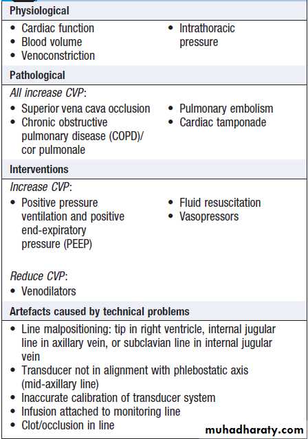
Factors affecting central venous pressure
Pulmonary artery catheterisation and pulmonary artery ‘wedge’ pressure (PAWP)The CVP is usually an adequate guide to the filling
pressures of both sides of the heart. However, certain
conditions, such as pulmonary hypertension or right
ventricular dysfunction, may lead to raised CVP levels
even in the presence of hypovolaemia. In these circumstances, it may be appropriate to insert a pulmonary artery flotation so that pulmonary artery pressure and PAWP which approximates to left atrial pressure (6 -12 mmHg ,measured from the mid-axillary line) can be measured. but in left heart failure it may be grossly elevated, >30 mmHg.
Provided the pulmonary capillary membranes are intact, the optimum PAWP when managing acute circulatory failure in the critically ill patient is generally 12–15 mmHg, because this will ensure good left ventricular filling without risking hydrostatic pulmonary oedema.
Pulmonary artery catheters also allow measurement
of cardiac output and sampling of blood from the pulmonary artery (‘mixed venous’ samples), permitting
continuous monitoring of the mixed venous oxygen
saturation (SvO2) by oximetry.
Measurement of SvO2 gives an indication of the adequacy of cardiac output (and hence DO2) in relation to the body’s metabolic requirements.
It is especially useful in low cardiac output states.
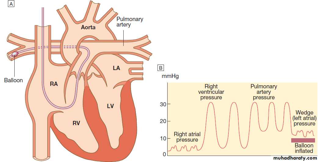
Fig. A pulmonary artery catheter.
A There is a small balloon at the tip of the catheter and pressure can be measured through the central lumen. The catheter is inserted via an internal jugular, subclavian or femoral vein and advanced through the right heart until the tip lies in the pulmonary artery. When the balloon is deflated, pulmonary artery pressure can be recorded.B Advancing the catheter with the balloon inflated will ‘wedge’ the catheter in the pulmonary artery. Blood cannot then flow past the balloon, so the tip of the catheter will now record the pressure transmitted from the pulmonary veins and left atrium (pulmonary artery wedge pressure), provides indirect measure of the left A pressure.
Cardiac output
Measurement is important, particularly when large doses of a vasopressor are administered, when there is cardiac disease and when volume resuscitation and vasoactive drug therapy. It is most accurately measured by indicator dilution methods. Most PA catheters incorporatea heating element, which raises blood temperature at frequent intervals, and the resultant temperature change is detected by a thermistor at the tip of the catheter.Oesophageal Doppler ultrasonography provides a rapid and useful assessment of volume status and cardiac performance to guide early fluid and vasoactive therapy.
A 6 mm probe is inserted into the distal oesophagus, allowing continuous monitoring of the aortic flow signal from the descending aorta .Using the stroke distance (area under the velocity/time waveform) an estimate of stroke volume and hence cardiac output can be made. Peak velocity is an indicator of left ventricular performance, while flow time is an indicator of left ventricular filling and peripheral resistance.
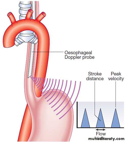
Oesophageal Doppler ultrasonography.
EchocardiographyIn many centres, echocardiography is increasingly being
used for rapid assessment of myocardial function and
volume status at the bedside. Continuous transoesophageal
echocardiography allows direct assessment of cardiac filling status and ventricular function in real time.
Urine output
This is a sensitive measure of renal perfusion, provided
that the kidneys are not damaged or affected by drugs such as diuretics or dopamine. Output is measured hourly and a lower limit of normal of 0.5 mL/hr/kg body weight is widely used. It reflects renal perfusion over the hours preceding measurement rather than in real time.
Peripheral skin temperature
In general, resuscitation is not complete until the
patient’s feet are warm and well perfused.
Blood lactate, hydrogen ion and base excess/deficit
Base excess or deficit is calculated as the difference between the patient’s bicarbonate and the normal bicarbonate after the PCO2 has been maintained in a blood gas machine at 5.33 kPa (40 mmHg). A metabolic acidosis with base deficit of > 5 mmol/L requires investigation. It often indicates increased lactic acid production in poorly perfused, hypoxic tissues, and impaired lactate metabolism and clearance due to poor hepatic perfusion. Serial lactate measurements may therefore be helpful in monitoring tissue perfusion and response to treatment.
Monitoring respiratory function
Oxygen saturation (SpO2)Measured by a probe attached to a finger or earlobe. Spectrophotometric analysis determines the relative proportions of saturated and desaturated haemoglobin. It is unreliable if peripheral perfusion is poor, in the presence of nail polish, excessive movement or high ambient light. It is not useful in carbon monoxide poisoning, as it does not detect carboxy-haemoglobin.
If this is suspected, PO2 must be measured in an arterial blood gas sample. In general, arterial oxygenation is safe if SpO2 is above 90%.
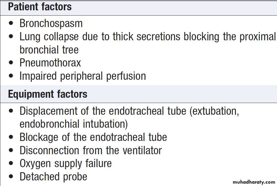
Causes of sudden changes in oxygen saturation
Arterial blood gasesArterial blood gases (ABGs) are measured several times
a day in a ventilated patient so that inspired oxygen
(FiO2) and minute volume can be adjusted to achieve the desired PaO2 and PaCO2 respectively. ABG results are
also used to monitor disturbances of acid–base balance.
Lung function
In ventilated patients, lung function is monitored by:
• arterial PO2 taken in relation to the fractional
inspired oxygen concentration (PO2/FiO2 ratio) and
level of end-expiratory pressure
• arterial and end-tidal CO2, reflecting alveolar
ventilation
• airway pressures and tidal volumes, reflecting lung
compliance and airways resistance.
Capnography
The CO2 concentration in inspired gas is zero, but during
expiration, after clearing the physiological dead space,
it rises progressively to reach a plateau that represents
the alveolar or end-tidal CO2 concentration. This cyclical
change in CO2 concentration, or capnogram, is measured
using an infrared sensor inserted between the ventilator
tubing and the endotracheal tube (Fig.). In
normal lungs, the end-tidal CO2 closely mirrors PaCO2,
and can be used to assess the adequacy of alveolar ventilation.
However, its use is limited as there may be
marked discrepancies in the presence of lung disease or
impaired pulmonary perfusion (e.g. due to hypovolaemia).
In combination with the gas flow and respiratory
cycle data from the ventilator, CO2 production andhence metabolic rate may be calculated. In clinical practice, end-tidal CO2 is used to confirm correct placement of an endotracheal tube, in the management of head injury, and during the transport of ventilated patients.
Continuous measurement of end-tidal CO2 is important
in the minute-to-minute monitoring of any patient ventilated
through an endotracheal tube or tracheostomy in
the acute setting.
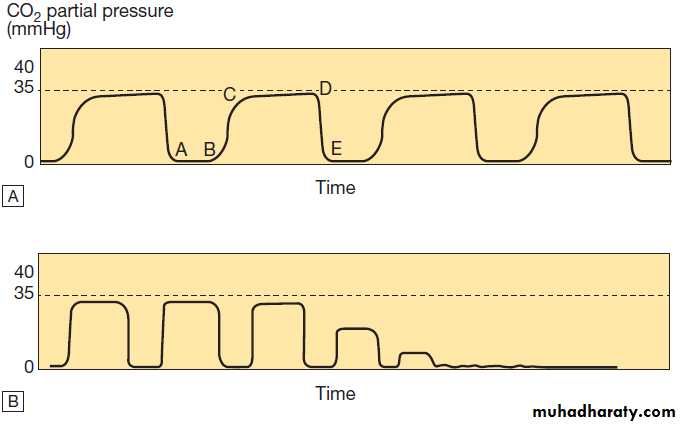
Capnography traces for normal and pathological states. A The normal
capnogram represents the varying CO2 level throughout the breath cycle. A–B = baseline, B–C = expiratory upstroke, C–D = expiratory plateau, D = end-tidal concentration, D–E = inspiration. B Loss of the capnograph can occur with loss of cardiac output, as well as airway displacement. (1 kPa = 7.5 mmHg)Transcutaneous PCO2
Monitors that measure transcutaneous PCO2 are nowavailable with an earlobe probe that incorporates a pulse
oximeter and CO2 electrode. The transcutaneous PCO2
closely approximates to PaCO2 and gives continuous
monitoring. This is useful in patients with no arterial
cannula but who require close monitoring: for example,
during ventilatory weaning (see below).
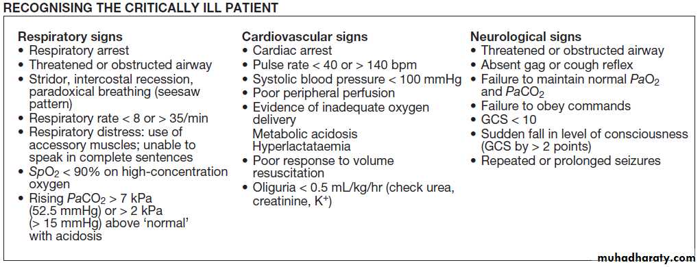
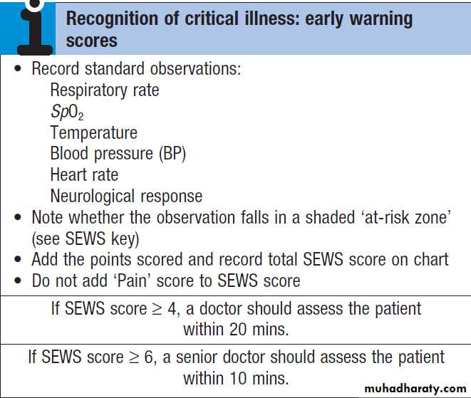
RECOGNITION OF CRITICAL ILLNESS
Assessment of:• the airway (for patency and noises, e.g. stridor, snoring, none)
• breathing (rate, symmetry, work of breathing,
accessory muscle use, paradoxical chest/ abdominal movement)
• peripheral circulation (temperature of the extremities)
• conscious level (the response of the patient).
Tachypnoea is often the earliest abnormality to
appear and the most sensitive sign of a worsening clinical state, but it is the least well documented.
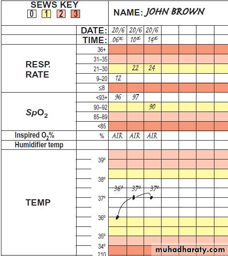
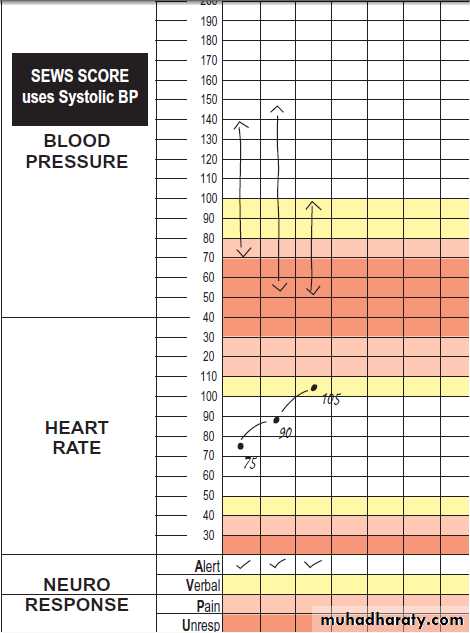
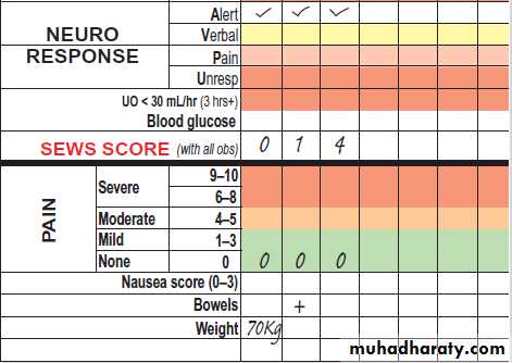
In the UK, the use of early warning scores, such as the Standard Early Warning System chart (SEWS), has been adopted to improve the recognition of critical illness. A patient with a SEWS score of 4 or more requires urgent review and appropriate interventions.
Assessment and initial resuscitation of the critically ill Airway and breathing
If the patient is talking, the airway is clear and breathing
is adequate. A rapid history should be obtained whilst
initial assessment is undertaken. Supplemental oxygen should be administered to patients who are breathless, tachypnoeic or bleeding, or who have chest pain or reduced conscious level. Critically ill should receive at least 60% oxygen initially. High-concentration oxygen is best given using a mask with a reservoir bag, which, at
15 L/min, can provide nearly 90% oxygen. ABGs should be checked early to assess oxygenation, ventilation (PaCO2) and metabolic state (pH or H+, HCO3 and base deficit). Oxygen therapy should be adjusted in light of the ABGs.
Early application of pulse oximeter monitoring is ideal, although this may not be reliable if the peripherally shut down. Intubation, while often essential, may be hazardous in a cardiorespiratory failure, and full monitoring and resuscitation facilities must be available.
Circulation
Carotid pulse should be sought in collapsed or unconscious, peripheral pulses checked in the conscious. Venous access is vital but often difficult in sick patients. Wide-bore cannulae are required. Ideally, two 16G or larger cannulae should be inserted, one in each arm, in the severely hypovolaemic patient. If the two cannulae are of different sizes, the pulse oximeter should be placed on the same side as the larger one, and the BP cuff on the same side as the smaller one.
This facilitates unimpeded volume resuscitation and uninterrupted oxygen saturation monitoring.
Pressure infusors and blood warmers should be utilised
for rapid, high-volume fluid resuscitation, particularly
of blood products. An 18G cannula is adequate for drug
administration.
Machine-derived cuff BP measurement is inaccurate
at extremes of BP and in tachycardia, especially atrial
fibrillation. Manual sphygmomanometer BP readings
tend to be more accurate in hypotension.
If severe hypotension is not readily corrected with fluid, early consideration should be given to arterial line insertion and vasoactive drug therapy.
Disability
Conscious level should be assessed using the GCS. Appropriate painful stimuli include supraorbital pressure and trapezius pinch. A score of 8 or less denotes coma with associated airway compromise and loss of airway protection, which necessitates intervention. Focal signs may indicate unilateral cerebral pathology. Abnormal pupil size, symmetry or reaction may indicate primary cerebral disease or cerebral global insults induced by drugs (e.g. opioids), hypoxia or hypoglycaemia.
Exposure, evidence and examination
‘Exposure’ indicates the need for targeted clinical examination, and ‘evidence’ may be gathered from any recent investigations, prescription or monitoring charts.
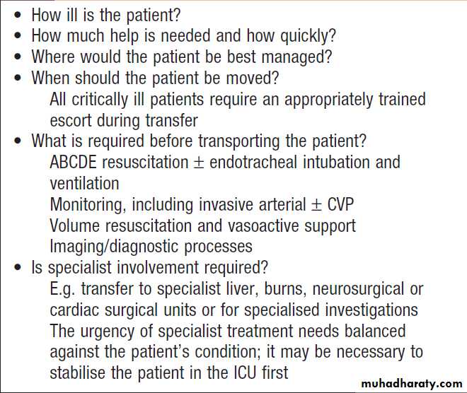
Clinical decision-making and referral to critical care
During the initial assessment and resuscitation, severaldecisions must be made (Box), but particularly
whether referral to the critical care service is necessary.
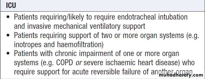
• Intensive care units allow management of the sickest
patients who require invasive ventilation, multimodal monitoring and multiple organ system support (Box).
• High-dependency care allows a greater degree of
monitoring, support and nursing/ medical input than the standard ward, for patients following major surgery, or for the septic patient requiring invasive haemodynamic monitoring and circulatory support alone, or for the patient with respiratory failure manageable with NIV or continuous positive airway pressure (CPAP).
Clinical decisions in the critically ill
HDU
• Patients requiring detailed observation or monitoring that
cannot be provided at ward level Direct arterial BP monitoring
CVP monitoring, Fluid balance, Neurological observations, regular GCS recording
• Patients requiring support for a single failing organ system,
excluding invasive ventilatory support:
CPAP or NIV , Moderate inotropic or vasopressor support
Renal replacement therapy in an otherwise stable patient
• Step down from intensive care
Admission criteria for intensive care (ICU)
and high-dependency units (HDU)

NIV = non-invasive (mask ) ventilation
PRESENTING PROBLEMS/MANAGEMENT OF MAJOR ORGAN FAILURECirculatory failure: ‘shock’
The defining feature of ‘shock’ is a level of oxygen delivery (DO2) that fails to meet the metabolic requirements of the tissues. ‘Shock’ is not synonymous with hypotension, which is often a late manifestation.
The cardiac output and oxygen delivery may be critically low, even though the BP remains normal. Objective markers of inadequate tissue oxygen delivery, such as increasing base deficit, elevated blood lactate and reduced urine output, can aid earlier identification of shock.
The causes of circulatory failure or ‘shock’ may be
categorised as either low flow or stroke volume, or lowperipheral arteriolar resistance (vasodilatation).
Low stroke volume
• Hypovolaemic: any condition provoking a major reduction in blood volume, e.g. haemorrhage, severe burns, salt and water depletion.
• Cardiogenic: severe cardiac impairment, e.g. myocardial infarction, acute mitral regurgitation. Subarachnoid haemorrhage may cause catecholamine-mediated myocardial stunning that can result in pulmonary oedema or cardiogenic shock.
• Obstructive: obstruction to blood flow, e.g. major pulmonary embolism, cardiac tamponade, tension pneumothorax.
Vasodilatation
• Sepsis/SIRS: infection or other causes of a systemic inflammatory response that produce widespread endothelial damage with vasodilatation, AV shunting, microvascular occlusion,capillary leak and tissue oedema.
• Anaphylactic: inappropriate vasodilatation triggered
by an allergen often associated with endothelial disruption and capillary leak.
• Neurogenic: major brain or spinal injury, which disrupts brainstem and neurogenic vasomotor control. High cervical cord trauma may result in disruption of the sympathetic outflow tracts, leading to inappropriate bradycardia due to a combination of loss of noradrenaline-mediated vasoconstriction and adrenaline-mediated chronotropy. Guillain–Barré syndrome involves the autonomic as well as the sensorimotor systems, which may result in periods of severe hypo- or hypertension.
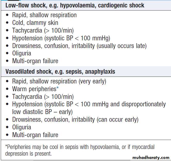
Clinical assessment and complications
Clinical features depend on the primary pathophysiology.Hypovolaemic, cardiogenic and obstructive causes of circulatory failure produce the ‘classical’ image
of shock with cold peripheries, reduced or absent
peripheral pulses, weak central pulses and evidence
of a low cardiac output. In early haemorrhagic shock, a
narrowed pulse pressure, i.e. a raised diastolic (DBP)
and reduced systolic (SBP) blood pressure, such as
105/95 mmHg, indicates the combination of hypovolaemia (reduced stroke volume, hence SBP) and activation of the sympathetic system, with noradrenaline inducing vasoconstriction and so raising the DBP.
In contrast, sepsis/SIRS and anaphylactic shock are
usually associated with warm peripheries, boundingpulses and features of a high cardiac output. The BP
pattern is again distinctive (e.g. 115/42 mmHg), with a
low DBP in the early stages due to peripheral vasodilatation, but a normal systolic BP, as the left ventricular afterload is reduced and stroke volume thus maintained, These patients are usually warm peripherally, but in more advanced septic or anaphylactic shock, SBP falls and the peripheries become cool. This is usually due to
the hypovolaemia associated with capillary leak and
will respond to fluid resuscitation.
If there is no improvement with this, myocardial depression may be present.
Neurogenic shock often results in vasodilated hypotension
with a paradoxically slow heart rate.
All forms of shock require early identification and
treatment because, if inadequate regional tissue perfusion
and cellular dysoxia persist, MOF will develop. Early institution of invasive haemodynamic monitoring is required.
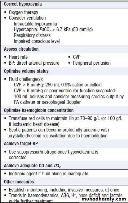
Immediate management of
circulatory collapseCirculatory support
The primary goals (Box) are to:• Restore global oxygen delivery (DO2) by ensuring
adequate cardiac output.
• Maintain an MAP that ensures adequate perfusion
of vital organs. The target pressure will be patient-specific,
depending on pre-morbid factors (e.g.
hypertension or coronary artery disease), and may
range from 60 to 90 mmHg.
The first objective is to ensure that an ‘appropriate’
ventricular preload is restored, initially by adequate
volume resuscitation.
Vasoactive drugs may then have to be considered.
Therapeutic options to optimize cardiac function
If the cardiac output is inadequate and contractility is poor, the available treatment options are to:
• Reduce afterload. achieved by using an arteriolar dilator (e.g. nitrates), but this may be limited by the consequent fall in systemic pressure. A counterpulsation intra-aortic balloon pump offers the ideal physiological treatment because it reduces LV afterload while increasing cardiac output, diastolic pressure and coronary perfusion. It is particularly valuable in myocardial ischaemia.
• Increase preload. If there is significant impairment of
contractility, giving fluids to increase filling pressures will only produce a small increase in stroke volume and cardiac output, and risks precipitating pulmonary oedema.
• Improve myocardial contractility. An inotrope may be
required to ensure adequate cardiac output andperipheral blood flow sufficient to secure adequate
oxygen delivery.
• Control heart rate and rhythm. The optimum heart
rate is usually between 90 and 110 beats per minute.
Correction of low serum potassium and magnesium
concentrations should be the first step in treating
tachyarrhythmias in the critically ill. Atrial fibrillation is common; IV amiodarone (300 mg over 30–60 minutes, followed by 900 mg over 24 hours) can be successful in controlling ventricular rate and in restoring and maintaining sinus rhythm.
Prognosis
If the precipitating cause and accompanying circulatoryfailure are dealt with promptly, before significant organ
failure occurs (‘early’ shock), the prognosis is good. If
not, there is progressive deterioration in organ function
and MOF ensues (‘late’ shock). The mortality of MOF is
high and increases with the number of organs that have
failed, the duration of organ failure and the patient’s
age. Failure of four or more organs is associated with a
mortality of more than 80%.
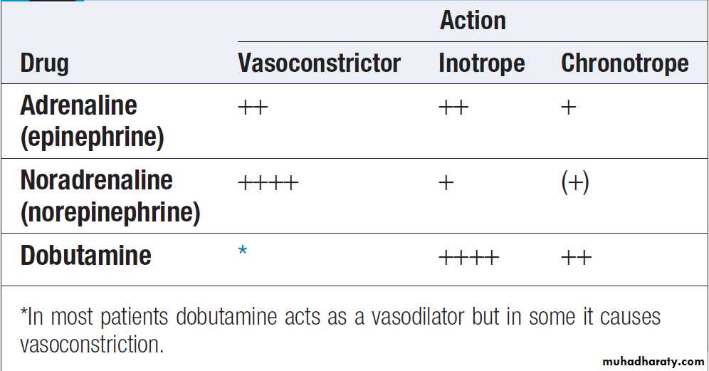
Actions of commonly used vasoactive agents
Respiratory failure and acute respiratory distress syndromeRespiratory failure may be the primary problem or
could constitute a secondary complication .The pattern of respiratory failure is classified using ABG analysis:
• type 1: hypoxaemia (PaO2 < 8 kPa (< 60 mmHg) when breathing air) without hypercapnia
• type 2: hypoxaemia with hypercapnia (PaCO2 > 6.5 kPa (> 49 mmHg) ) due to alveolar hypoventilation.
Acute hypoxaemia results from ventilation–perfusion mismatch within the lung and this can be caused by almost any pulmonary disease. The most extreme form of mismatch is pulmonary shunting, which occurs when an area of lung is not ventilated at all – for example, due to collapse or consolidation.
Acute or chronic hypercapnia usually results from
alveolar hypoventilation. Causes include:• central depression of respiratory drive
• impaired nerve transmission between the CNS and muscle (especially the diaphragm)
• reduced chest wall movements (including
diaphragmatic movements)
• reduced alveolar ventilation, pathology within the lungs. Critically ill patients may have both type 1 and 2 respiratory failure at some point, and the pattern and severity can change rapidly. The best method for assessing hypoxaemia is the ratio of the PaO2 to the fractional inspired oxygen delivered (PaO2/FiO2). This ‘PF’ ratio is lower, the more severe the disease.
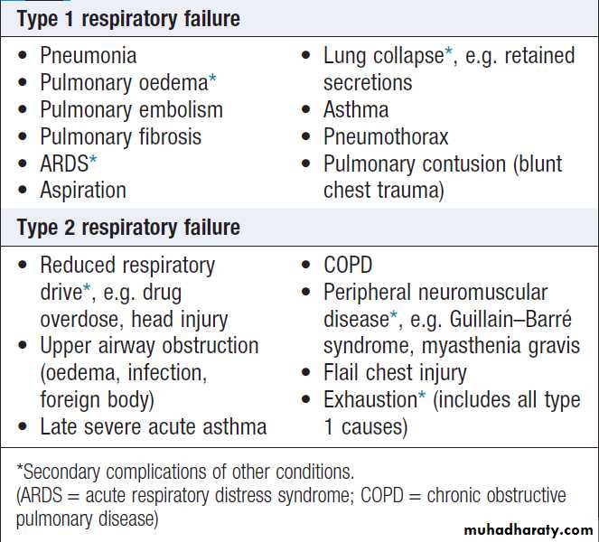
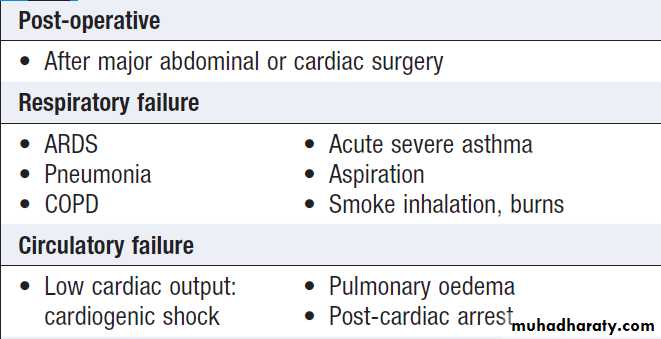
Common causes of respiratory failure in
critically ill patients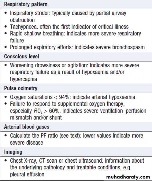
How to assess respiratory failure
Acute lung injury and the acute respiratory distress syndromeA range of conditions (Box) can result in a diffuse
acute inflammatory process in the lungs called acute
lung injury (ALI); when severe, this is termed the acute respiratory distress syndrome (ARDS). Inflammation occurs throughout the lungs, affecting both endothelial and epithelial surfaces. Activated neutrophils are sequestered into the lungs and capillary permeability is increased, with damage to type I and II alveolar cells.
This results in exudation and accumulation of protein-rich cellular fluid within alveoli and the formation of characteristic ‘hyaline membranes’.
Local release of cytokines and chemokines by activated macrophages and neutrophils results in progressive recruitment of inflammatory cells.
Secondary effects include loss of surfactant and impaired surfactant production.
The net effect is alveolar collapse and reduced lung compliance, which are most marked in dependent regions of the lung, where airspaces become fluid-filled (Fig.).
The combination of loss of surfactant and fluid
accumulation makes these areas difficult to ventilate,
which results in hypoxaemia due to ventilation–
perfusion mismatch and increased pulmonary shunt.
ALI and ARDS can be difficult to distinguish from fluid
overload or cardiac failure.
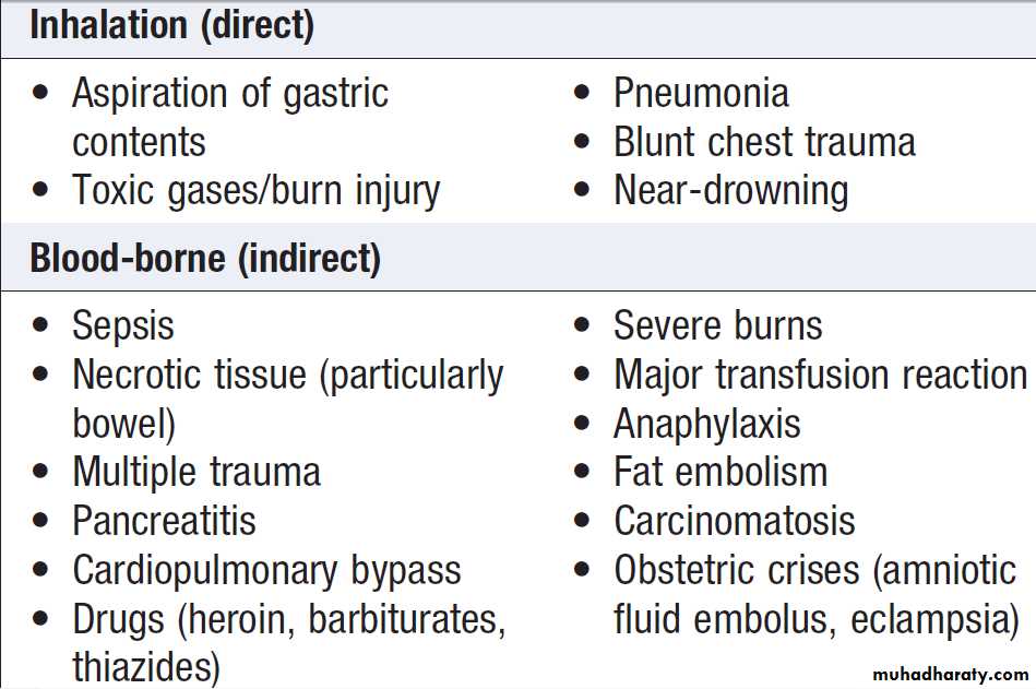
Conditions predisposing to ARDS
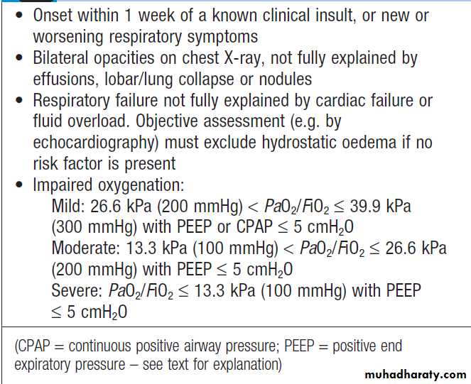
Berlin definition of ARDS
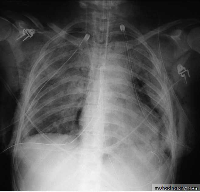
Chest X-ray in acute respiratory distress syndrome
(ARDS). Note bilateral lung infiltrates, pneumomediastinum,pneumothoraces with bilateral chest drains, surgical emphysema, and
fractures of the ribs, right clavicle and left scapula.
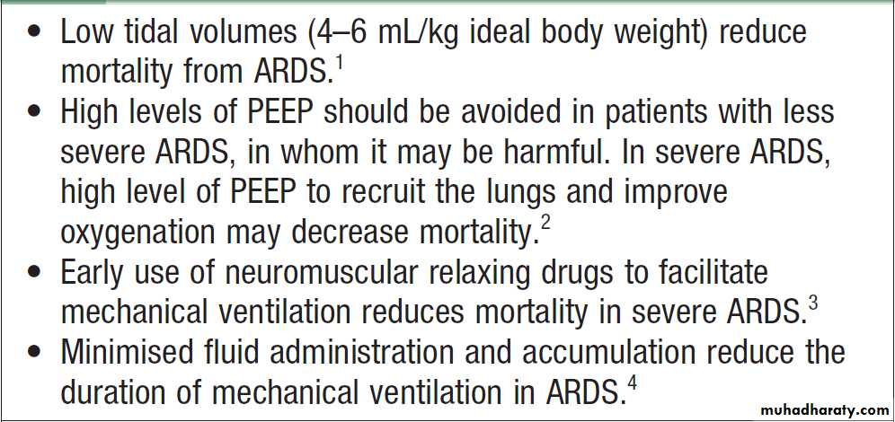
Mechanical ventilation in ARDS
Respiratory support
The aims are to maintain the patency of the airway, correct hypoxaemia, hypercapnia, reduce the work of breathing.Oxygen therapy
Oxygen is given to ensure arterial oxygenation (SpO2 > 90%), by facemask or nasal cannulae.The inspired oxygen concentration (FiO2) is adjusted, depending on pulse oximetry and ABG analysis. If this results in unacceptable hypercapnia, the patient requires some form of mechanical support. The risk of progressive hypercapnia in patients with COPD who are dependent on hypoxic drive has been overstated. Hypoxic cerebral damage is irreversible, so the theoretical risks of oxygen toxicity are not relevant if the patient is acutely hypoxaemic, as the maintenance of cerebral oxygenation takes precedence.
Non-invasive respiratory support
Non-invasive respiratory support includes techniquesthat do not require sedation or an endotracheal or tracheostomy tube. This helps preserve the patient’s respiratory muscle activity and reduces complications such
as nosocomial infection. It can be used to support
selected patients with type 1 or 2 respiratory failure, but
the patient’s conscious level must be adequate to ensure
airway protection from aspiration. Non-invasive respiratory
support is classified as continuous positive airway
pressure (CPAP) alone or CPAP plus additional support,
in the form of pressure applied to the breathing circuit
during inspiration (non-invasive ventilation, or NIV).
CPAP therapy
CPAP therapy involves the application of a continuouspositive airway pressure throughout the patient’s
breathing cycle, typically between 5 and 10 cmH2O.
CPAP recruits collapsed alveoli and can enhance clearance of alveolar fluid. It is particularly effective for treating pulmonary atelectasis (which may be post-operative) and pulmonary oedema, and helps correct hypoxaemia in some patients with pneumonia, especially the immunocompromised. CPAP therapy is most effective in correcting hypoxaemia in type 1 respiratory failure, but if it improves pulmonary compliance (by clearing fluid or improving lung volume), it can reduce the work of breathing and improve hypercapnia in type 2.
However, many patients with the latter require NIV or invasive ventilation. CPAP therapy can be delivered using tight-fitting facial masks, high-flow nasal cannulae, and hoods (Fig.). Usually, a CPAP mask is tried first, but different systems can be trialled until the most comfortable
for the patient is found. Patients must be cooperative,
able to protect their airway, and have the strength to
breathe spontaneously and cough effectively. Failure to
improve over 24–48 hours, or a further deterioration in
conscious level or blood gases, indicates that invasive
ventilation should be considered.
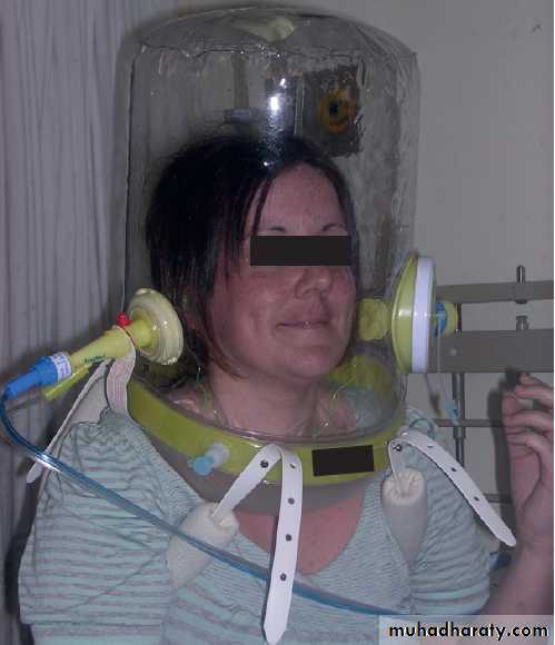
continuous positive airway
pressure(CPAP) delivery with a Castar hood.
Non-invasive ventilation (NIV)
Is ventilatory support by nasal or full facemask. It can be delivered by a simple bi-level (BiPAP) turbine ventilator, which delivers a higher pressure (approximately 15–25 cmH2O) for inspiration and a lower pressure (4–10 cmH2O) to allow expiration. If hypoxaemia is severe, a complex ICU ventilator is employed that allows higher oxygen concentrations to be administered. A simple breathing circuit with a leak rather than an expiratory valve is generally used, and ventilation can be spontaneous (triggered by the patient’s breaths) or timed (occurring at set intervals and/or frequency).Systems that synchronise with the patient’s
efforts are better tolerated and more effective.
NIV is the first-line therapy in patients with type 2 respiratory failure secondary to acute exacerbation of COPD because it reduces the work of breathing and offloads the diaphragm, allowing it to recover strength. It should be initiated early, especially when severe respiratory acidosis and/or decreased consciousness secondary to hypercapnia are present. Unless there is an improvement in acidosis within 4–6 hours, invasive ventilation is indicated.
NIV can also be used to support selected patients with hypercapnia secondary to pulmonary oedema or pneumonia, or during weaning from invasive ventilation,
but its effectiveness in these conditions is less certain. As with mask CPAP, NIV requires the patient to be conscious and cooperative.
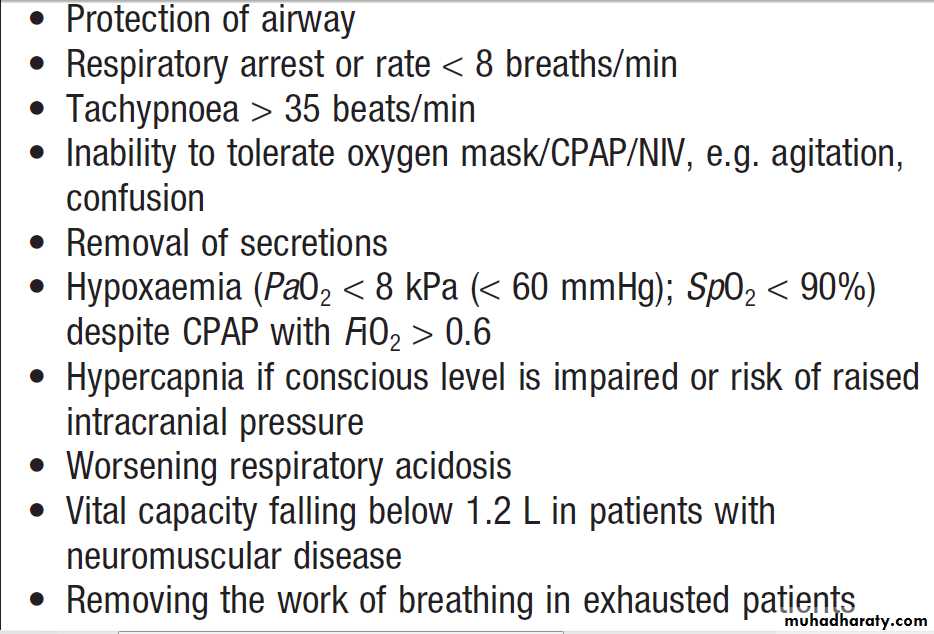
Emergency endotracheal intubation and mechanical ventilation
Many patients admitted to ICU require endotracheal
intubation and mechanical ventilation, mostly for respiratory failure (Boxes). The final decision to
undertake these is based on clinical judgement rather
than the results of ABGs in isolation. If possible, the
patient’s relatives should be given the chance to visit
prior to anaesthesia and intubation, as this may be the
last opportunity they have to speak together.
In the conscious patient, intubation requires induction
of anaesthesia and muscle relaxation, while in more obtunded patients, sedation alone may be adequate.
Intubation can be hazardous in the critically ill patient,
particularly if there is associated cardiovascular failure.Patients should be pre-oxygenated and cricoid pressure
applied, with continuous monitoring of heart rate, ECG and BP (preferably invasively), together with capnography (and subsequently a chest X-ray) to confirm correct endotracheal tube placement. Complications are common, and intubation is ideally performed in a critical care environment, or with expert assistance, resuscitation facilities and appropriate medication immediately available. Hypotension may follow sedation or anaesthesia due to the direct cardiovascular effects of the anaesthetic agent and loss of sympathetic drive. Positive pressure ventilation may compound this by increasing intrathoracic pressure, thereby reducing venous return and thus cardiac output.
Tracheostomy is usually performed electively when
endotracheal intubation is likely to be required for morethan 7–10 days (Box).
The timing is determined by individual patient factors and clinical judgement. Tracheostomy is usually carried out percutaneously in the ICU, to avoid transfer to an operating theatre. The passage of a smaller (4.5 mm internal diameter) ‘minitracheostomy’ tube through the cricothyroid membrane is a useful technique for clearing airway secretions in spontaneously breathing patients with a poor cough effort, particularly in the HDU and in post-operative patients.
Indications for tracheal intubation and mechanical ventilation
Neurological disease
• Coma of any cause • Status epilepticus • Drug overdose • Respiratory musclefailure • Head injury: to avoid hypoxaemia and hypercapnia, and reduce intracranial pressure
• Bulbar abnormalities causing risk of aspiration (e.g. stroke, myasthenia gravis)
Multiple trauma
Conditions requiring mechanical ventilation*
*Additional considerations:
Metabolic rate: ventilatory requirements rise as metabolic rate increases. Nutritional reserve: low potassium or phosphate reduces respiratory muscle power. Abdominal distension due to surgery or tense ascites: causes discomfort and splinting of the diaphragm, compromising spontaneous respiratory effort and promoting bilateral basal lung collapse.
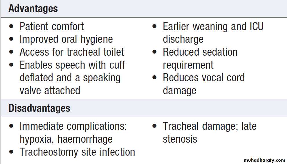
Advantages and disadvantages of tracheostomy
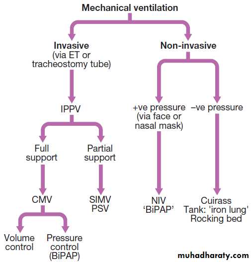
Types of invasive and non-invasive ventilatory
support. (BiPAP = bi-level positive airway pressure; CMV = controlledmandatory ventilation; ET = endotracheal; IPPV = intermittent positive
pressure ventilation; NIV = non-invasive ventilation; PSV = pressure
support ventilation; SIMV = synchronised intermittent mandatory ventilation)
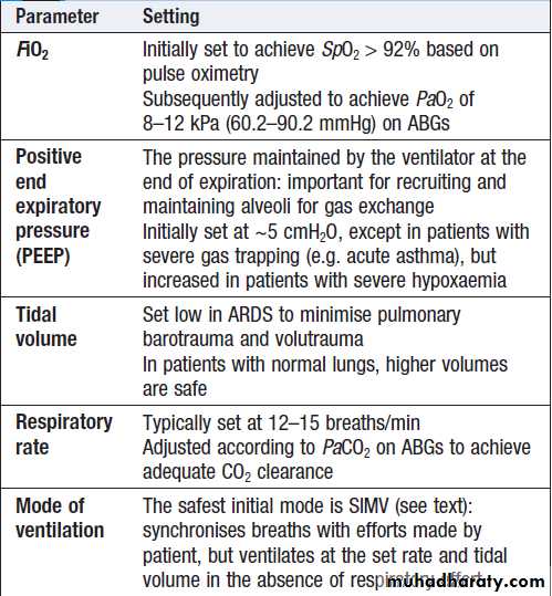
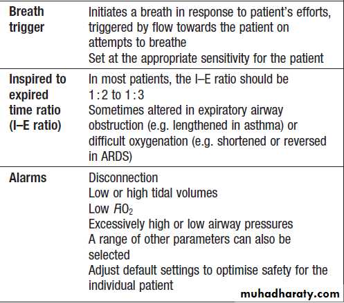
Ventilator parameter settings on initiating
mechanical ventilationGeneral considerations in the management of the ventilated/intubated patient
Modern ventilators allow enormous flexibility in theway ventilator support is provided. The terminology
used to describe ventilation modes can be confusing, because of subtle differences between modes, and the
use of different names by different manufacturers. Figure gives a classification of the different types of invasive ventilation support, and Box outlines the terminology used and the parameters that are set on any ventilator. The inspired gas should always be humidified and warmed, usually achieved with a heat and moisture exchanger, but occasionally with hot water humidification systems.
Initial settings
Following intubation, the ventilator is set to deliver a
safe mandatory mode of ventilation that will achieve
oxygenation and carbon dioxide clearance in the majority
of patients. Regular re-assessment of the patient’s
parameters will show if modification is required. Hyperoxia
is avoided, as it is associated with adverse outcomes.
Mandatory modes of ventilation
Volume-controlled modes. These are set to deliver a
preset tidal volume at a set frequency to guarantee a
specified minute ventilation. Synchronised intermittent
mandatory ventilation (SIMV) is the most widely used,
which also synchronises breaths with any efforts made
by the patient.
Volume-controlled modes will deliver the set volume, but the pressures generated in the patient’s lungs can be excessively high if the pulmonary compliance is low – for example, in ARDS – thus precipitating lung barotrauma or volutrauma, which could cause pneumothorax.
Pressure-controlled modes. These deliver a set pressure
for a specified duration. Pressure-controlled ventilation(PCV) and bi-level positive airway pressure ventilation
(BiPAP) are examples. The tidal and minute volumes
achieved are determined by the pulmonary compliance;
in patients with stiff lungs, only small tidal volumes may
be achieved, whereas in patients with normal compliance,
excessive volumes may result.
An advantage of these modes is that airway pressures are controlled, but the effect on blood gases needs to be regularly assessed to identify changes in pulmonary compliance.
Tidal volume is a useful safety alarm as it falls with increased resistance, such as with bronchospasm. In difficult-toventilate cases, several modes can be attempted sequentially to identify which is most effective.
Weaning or spontaneously breathing modes. Most
modern ventilators can detect whether a patient ismaking breathing efforts, and use a flow trigger to
augment each breath. Assisting breathing with additional
pressure is more comfortable than fixed tidal
volumes and allows sedation to be reduced. The most
common mode applies additional pressure during inspiration, assisting the work of breathing and increasing
tidal volume. The pressure is removed when the ventilator
detects an expiratory effort. These modes are usually
called pressure support ventilation (PSV) or assisted
spontaneous breathing (ASB).
Mixed modes. Different modes can be applied simultaneously, tailored to meet individual requirements. For example, it is possible to specify a frequency of SIMV or PCV breaths, but also provide PSV for any additional
efforts the patient makes.
Advanced ventilation strategies
In patients with severe acute lung disease, especially those resulting in reduced lung compliance such as ARDS, the ventilator can worsen lung injury as a result of overstretch and shearing forces in parts of the lung that continually open and collapse.The aim of advanced ventilation is to minimise further damage due to pressure (barotrauma), volume stretch (volutrauma), and the additional inflammatory mediators released into the body from ongoing lung injury (biotrauma) (Box).
Tidal volumes and airway pressures are kept as low as possible while achieving adequate oxygenation.
In many cases, it is best to accept hypercapnia rather than apply higher pressure to clear CO2. Often, higher levels of positive end expiratory pressure (PEEP) are required to achieve adequate alveolar recruitment and oxygenation.
Several other strategies can be used.
Prone ventilation. Oxygenation will often improve inpatients turned on their front, as a result of improved
lung recruitment and better ventilation–perfusion
matching. Prone ventilation has not reduced mortality
in controlled trials, but is a useful ‘rescue therapy’ in
cases where oxygenation is difficult.
High-frequency oscillatory ventilation (HFOV). This
uses a specialised ventilator to provide gas exchange
with high-frequency oscillating gas movements (> 150/
min). Conventional breaths and tidal volumes are not
set, but effective oxygenation and CO2 clearance are
achieved by adjusting the frequency and power of the
oscillations, and the mean airway pressure.
Nitric oxide. Nitric oxide is a very short-acting pulmonary
vasodilator. When delivered to the airway, itimproves blood flow to ventilated alveoli, thus improving
ventilation–perfusion matching. Oxygenation can
be improved markedly in some patients but there
is evidence that this lasts for only 48 hours, and
rebound effects can occur when it is withdrawn. No
improvement in mortality has been shown in controlled
trials. Its role is limited to rescue therapy when other
interventions have failed, and it may be useful in patients
with severe pulmonary hypertension.
Extracorporeal membrane oxygenation therapy (ECMO).
ECMO involves connecting the patient to an external
bypass circuit. Oxygenation and CO2 clearance are
achieved using a membrane oxygenator. The patient’s
lungs are usually ‘rested’ and ventilation reduced to
low levels. Advances in technology have dramatically
improved the safety of these devices, although their use
is restricted to specialised centres. Controlled trials indicate improved survival in appropriately selected cases, and patients with severe ARDS should be considered for treatment.
Corticosteroids. There is conflicting evidence regarding
the use of steroids as anti-inflammatory agents in acutelung injury. Uncertainty remains about patient selection
and the timing of therapy, and use of corticosteroids
may be complicated by secondary infection and muscle
weakness. However, they are often tried after 7–10 days
of ARDS, if the patient remains severely unwell.
Weaning from respiratory support
Patients usually require most mechanical ventilation inthe period following intubation when they are most
unwell, following which support is gradually reduced
as the underlying condition resolves and the patient is
able to breathe with less assistance. This is the process
of ‘weaning’ from ventilation. Sufficient support is provided
to correct hypoxaemia and hypercapnia, but the
level is decreased as quickly as possible to reduce the
chance of secondary complications, such as infection and
muscle weakness. Rapid weaning, often with reduction
in sedation levels (see below), shortens length of ICU
stay and improves patient outcomes.
Patients who have required long-term ventilatory support for severe lung disease such as ARDS may be unable to sustain even a modest degree of respiratory work initially because of poor lung compliance, high work of breathing and respiratory muscle weakness.
They require more prolonged weaning, until respiratory muscle strength improves.
Several criteria can be used to assess whether a
patient is ready to start reducing respiratory support(Box). Approaches include:
Spontaneous breathing trials (SBTs). These involve
removing all respiratory support, typically on a daily
basis, and observing how long the patient is able to
breathe unassisted.
This is particularly effective when linked to sedation breaks. Signs of failure include rapid shallow breathing, hypoxaemia, rising PaCO2, sweating and agitation. Patients who pass an SBT are assessed for extubation.
Progressive reduction in pressure support ventilation.
Progressive reduction in the PSV is applied for eachbreath over a period of hours or days, according to
patient response. When patients are strong enough to
achieve stable ABGs without distress while receiving
minimal or no support, they are likely to be ready for
extubation. This can take from several hours to many
weeks, according to the severity of illness.
Weaning protocols. The process of weaning is best
undertaken as a continuous process. Protocols that
empower nursing staff to initiate and progress weaning
within agreed guidelines reduce ventilation times.
Patients requiring prolonged mechanical ventilation
typically require individualised weaning plans, with
regular periods of training followed by rest, to enable
respiratory muscles to regain strength.
The timing of extubation relies on clinical judgement.
Patients must have stable ABGs with resolution of
hypoxaemia and hypercapnia despite withdrawal of
ventilator support. Conscious level must be adequate to
protect the airway, comply with physiotherapy, and
cough. The need for re-intubation following extubation
is associated with poorer outcomes.
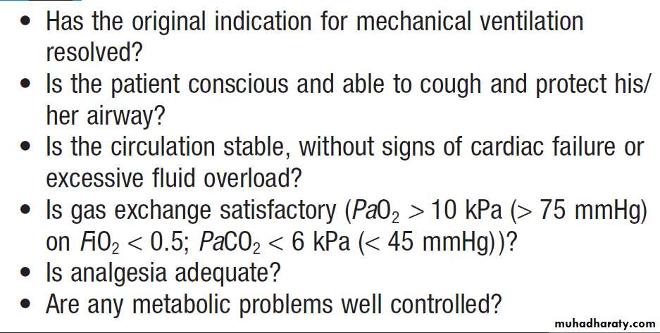
Factors to consider in deciding to wean and
extubate a ventilated patientAcute kidney injury
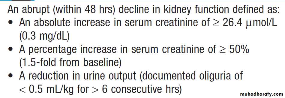
Acute kidney injury (AKI) is defined as an abrupt andsustained decrease in kidney function (Box). AKI in
the critically ill patient is often due to pre-renal problems
such as hypovolaemia, hypotension and ischaemia
resulting in reduced renal DO2. However, it may also be
due to acute tubular necrosis (ATN), which may
result from ischaemia, or nephrotoxicity caused by
chemical or bacterial toxins, or a combination of these.
Potentially nephrotoxic drugs include non-steroidal anti-inflammatory drugs (NSAIDs), angiotensin-converting
enzyme (ACE) inhibitors, angiotensin II receptor antagonists, radiological contrast media and some antibiotics.
Oliguria (< 0.5 mL/kg/hr for several hours) is an
important early sign of systemic problems in critical illness. It requires investigation and early intervention
to correct hypoxaemia, hypovolaemia, hypotension
and renal hypoperfusion. Successful resuscitation is
associated with restoration of good urine output, an
improving acid–base balance and correction of plasma
potassium, urea and creatinine.
Oliguria is an integral part of the normal stress
response to major surgery, and care should be taken not
to overfill the post-operative patient who has oliguria
but is otherwise well from a cardiovascular and biochemical point of view.
Diagnostic criteria for acute kidney injury
Renal supportSepsis is frequently implicated in the development of
AKI, and the source must be promptly identified and
adequately treated. Obstruction of the renal tract (including catheter blockage) should always be excluded and is most easily identified with abdominal ultrasound.
It must be relieved at once.
Acute glomerulonephritis and vasculitis must also be considered, and appropriate specialist referral, with investigations such as urine microscopy and immunopathological tests , carried out early. The mainstay of management is aggressive haemodynamic
resuscitation to achieve normovolaemia, normotension
and an appropriate cardiac output.
There is little evidence that specific treatments aimed at inducing a diuresis, such as low-dose dopamine, furosemide or mannitol, have any renoprotective action or other benefit in restoring renal function.
If renal function cannot be restored following resuscitation,
renal replacement therapy is indicated.
The preferred renal replacement therapy in
ICU patients is pumped venovenous haemofiltration.
This is associated with fewer osmotic fluid shifts and
hence greater haemodynamic stability than haemodialysis.
It is carried out using a double-lumen central venous
catheter placed percutaneously.
Haemofiltration should be continuous in the early phase of treatment.
Intermittent treatment may be used when the patient is recovering from the primary insult and return of normal renal function is expected. Provided the precipitating causecan be successfully treated, renal failure due to ATN
usually recovers between 5 days and several weeks later.
Survival rates from MOF, including AKI, have been
around 50% for many years, but modern haemofiltration
techniques are being shown to produce better outcomes.
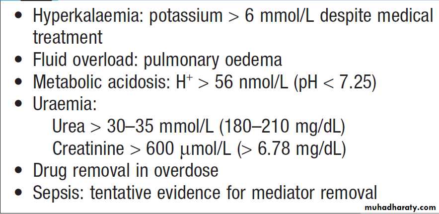
Indications for renal replacement therapy
Gastrointestinal and hepatic disturbance
Gastrointestinal symptoms, such as nausea, vomitingand large nasogastric aspirates, may be the earliest signs
of regional circulatory failure, and when associated with
a tender, distended, silent abdomen, indicate that this is
the probable site of the primary pathology. The gut has
a rapid cell turnover rate and fasting alone can produce
marked changes in mucosal structure and function. In
hypovolaemia and frank shock states, splanchnic vasoconstriction produces gut mucosal ischaemia, damaging the mucosal barrier and allowing toxins to enter the portal circulation and lymphatics.
Splanchnic ischaemia may contribute to the progression of MOF, possibly as a source of bacteraemia or systemic inflammation.
Manifestations of MOF within the gastrointestinal
tract include loss of gastric acid production, erosive
gastritis, stress ulceration, bleeding, ischaemia, pancreatitis and acalculous cholecystitis. These occur less frequently when adequate circulatory resuscitation occurs
early. Ischaemic bowel is difficult to diagnose in the
critically ill patient, but in the context of otherwise
unexplained lactic acidosis, hyperkalaemia and coagulopathy, abdominal imaging by contrast-enhanced CT and laparotomy should be considered.
Three distinctive hepatic dysfunction syndromes can
occur in the critically ill:• Shock liver or ischaemic hepatitis results from extreme
hepatic hypoxia and is characterised by hepatocellular necrosis. Transaminase levels are often massively raised (> 1000–5000 U/L) at an early stage, followed by moderate
hyperbilirubinaemia (< 100 μmol/L or < 5.8 mg/ dL). There is often associated hypoglycaemia, coagulopathy and lactic acidosis. Following successful resuscitation, hepatic function generally returns to normal.
• Hyperbilirubinaemia (‘ICU jaundice’) frequently develops following trauma or sepsis. There is a marked rise in bilirubin (predominantly conjugated), but only mild elevation of transaminase and alkaline phosphatase.
This results from failure of bilirubin transport within the liver and produces the histological appearance of intrahepatic cholestasis. Extrahepatic cholestasis must be excluded by abdominal ultrasound and potentially hepatotoxic drugs should be stopped.
Treatment is non-specific and should include early institution of enteral feeding.
Therapy that compromises splanchnic blood flow, particularly high doses of vasoconstrictor agents,
should be avoided.
• Transaminitis is most commonly due to drug
toxicity: for example, antibiotics.
Gastrointestinal and hepatic support
Early institution of enteral nutrition is the most effectivestrategy for protecting the gut mucosa and providing
nutritional support. Current evidence supports early enteral nutrition using standard feeds, with the addition of prokinetic agents such as metoclopramide or low-dose erythromycin when gastric aspirates are high.
The evidence for early supplementation with total parenteral nutrition (TPN) is weak, and it is not routinely indicated until enteral feeding attempts have been unsuccessful for approximately 7 days.
Hyperglycaemia is common during critical illness
and is associated with poor outcomes. Tight glycaemiccontrol, using insulin infusions, has been studied in
several controlled trials in the critically ill. The benefit
was greatest in surgical patients at low risk of death,
but the risk-to-benefit profile in the mixed critically ill
population is uncertain. Inadvertent hypoglycaemia
is associated with adverse patient outcomes. In most ICUs, the current target is for modestly elevated blood glucose concentrations: for example, (100–144 mg/dL).
Stress ulcer prophylaxis is best achieved with H2-
receptor antagonists (e.g. ranitidine), which are both safe
and effective. Although stress ulcer bleeding is rare with
modern resuscitation, evidence supports routine use in
mechanically ventilated patients and those with renal
failure or coagulopathy. H2-receptor antagonists are
associated with an increased incidence of nosocomial
pneumonia, and treatment should be stopped following
extubation in the absence of other indications. Withdrawal can also be considered when full enteral nutrition has been established, unless the patient has a history of peptic ulcer disease. Proton pump inhibitors are only required in upper GI bleeding due to ulceration, and continued when the patient has been taking them long-term.
Neurological failure (coma)
Impaired consciousness or coma is often an early featureof severe systemic illness (Box). Prompt assessment
of consciousness level and management of airway,
breathing and circulation are essential to prevent further
brain injury, to allow diagnosis and to permit definitive
treatment to be instituted. Any patient with confusion
or reduced conscious level should have blood sugar
measured and hypoglycaemia corrected.
Impairment of conscious level is graded using the
Glasgow Coma Scale (GCS), which is also used
to monitor progress.
A targeted neurological examination is very important. Pupil size and reaction to light, presence or absence of neck stiffness, focal neurological signs and evidence of other organ impairment should be noted.
After cardiorespiratory stability is achieved, the
cause of the coma must be sought from the history
(family, witness, general practitioner), examination and
investigation, particularly CT of brain. The possibility of
drug overdose should always be considered.
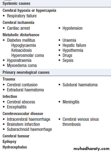
Causes of coma
Neurological supportA diverse range of neurological conditions require management in the ICU. These include not only the various causes of coma, but also spinal cord injury, peripheral
neuromuscular disease and prolonged seizures. The
goals are to:
• protect the airway, if necessary by endotracheal
intubation
• provide respiratory support to correct hypoxaemia
and hypercapnia
• treat circulatory problems, e.g. neurogenic pulmonary oedema in subarachnoid haemorrhage, autonomic disturbances in Guillain–Barré syndrome, and spinal shock following high spinal cord injuries.
• manage acute brain injury with control of raised
intracranial pressure (ICP)
• manage status epilepticus using anaesthetic agents
such as thiopental or propofol.
The aim of management in acute brain injury is to
optimise cerebral oxygen delivery by maintaining a
normal arterial oxygen content and a cerebral perfusion
pressure of more than 60 mmHg. Avoiding secondary insults to the brain, such as hypoxaemia and hypotension,
improves outcome. ICP rises in acute brain injury as a result of haematoma, contusions, oedema or ischaemic swelling.

Raised ICP causes direct damage to the cerebral cortex and, as a result of downward pressure on the brainstem, indirect damage by reducing cerebral perfusion pressure, thereby threatening cerebral blood flow and oxygen delivery:
ICP is measured by pressure transducers that are
inserted directly into the brain tissue. The normal upper
limit for ICP is 15 mmHg and management should be
directed at keeping it below 20 mmHg (Box). Sustained
pressures of more than 30 mmHg are associated
with a poor prognosis.
CPP should be maintained above 60 mmHg by
ensuring adequate fluid replacement and, if necessary,
by treating hypotension with a vasopressor such as
noradrenaline (norepinephrine).
Complex neurological monitoring must be combined
with frequent clinical assessment of GCS, pupil response
to light, and focal neurological signs. The motor response
to pain is an important prognostic sign. No response or
extension of the upper limbs is associated with severe
injury, and unless there is improvement within a few
days, prognosis is very poor. A flexor response is encouraging and indicates that a good outcome is still possible.
Neurological complications in intensive care
May occur as a result of systemic critical illness. Sepsis may be associated both with an encephalopathy characterised by delirium, cerebral oedema and loss of vasoregulation.
Hypotension and coagulopathy may provoke cerebral infarction or haemorrhage. Neurological examination is very difficult if patients are sedated or paralysed, and it is important to stop sedation regularly to re-assess their
underlying level of consciousness.
If there is evidence of a focal neurological deficit or a markedly declining level of consciousness, CT of brain should be performed.
Critical illness polyneuropathy is another potential
complication in patients with sepsis and MOF. It is due
to peripheral nerve axonal loss and can result in
areflexia, gross muscle wasting and failure to wean from
the ventilator, thus prolonging the duration of intensive
care. Recovery can take many weeks.
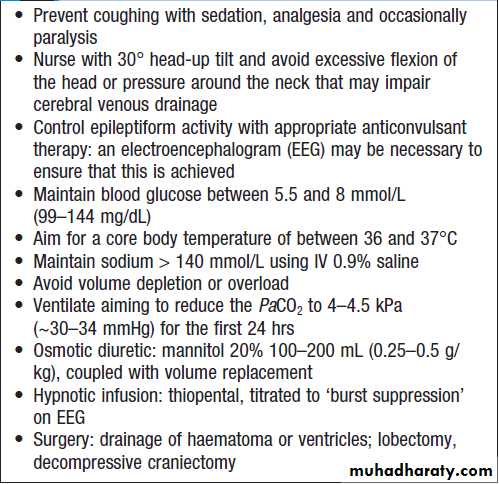
Strategies to control intracranial pressure
SepsisSepsis can occur in many clinical situations. It may be
due to a primary infection (e.g. pneumonia) or it may be
the result of clinical interventions for other conditions
(e.g. immunosuppressive drugs, chemotherapy, invasive
lines). Patients who are in hospital are at increased risk of certain specific infections, such as methicillin-resistant Staphy-aureus (MRSA).Sepsis usually originates from a localised infection that progresses to an uncontrolled systemic response. It can rapidly lead to acute physiological deterioration with the risk of MOF and death. Early identification of sepsis and appropriate intervention with oxygen, fluids, antibiotics, and more advanced resuscitation where indicated, shown to improve survival.
Important comorbidities and risk factors for sepsis are shown in Box . Any pathogen, including aerobic Gram-positive and negative bacteria, anaerobes and fungi, may cause sepsis but in nearly 45% of cases microbiological confirmation of the organism is lacking. Any or all of the features of SIRS may be present, together with an obvious focus of infection, such as purulent sputum with chest X-ray shadowing, or erythema around an intravenous line. However, severe sepsis may present with unexplained hypotension (i.e. septic shock), and the speed of onset may mimic a major pulmonary embolus or myocardial infarction.
Nosocomial infections are an increasing problem in
critical care units. Risk factors are similar to those in Box ,but also include prolonged ICU stay, invasive ventilation
and stress ulcer prophylaxis with H2 antagonists.
Cross-infection is a major concern, particularly with
MRSA, multidrug-resistant Gram-negative organisms
and Clostridium difficile. If cross-infection occurs frequently,
it should prompt a review of the unit’s infection control policies . Limiting antibiotic use helps to prevent the emergence of multidrug-resistant bacteria.
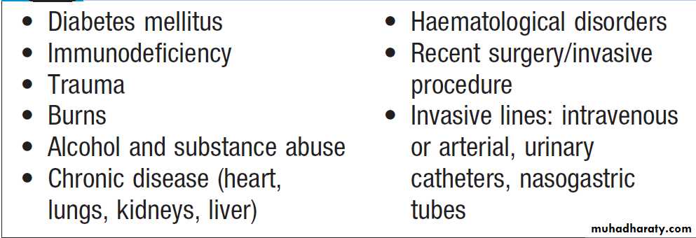
Risk factors for sepsis
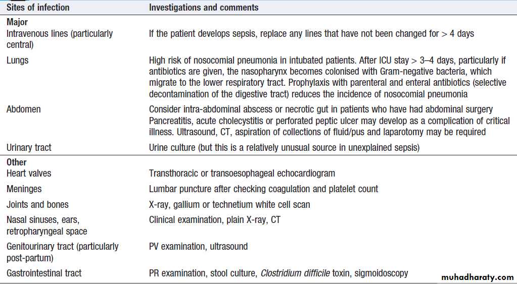
Sites of infection in critically ill patients
ManagementPrompt resuscitation, with early cultures, administration
of appropriate antibiotics and eradication of the
source of infection (if necessary by surgical drainage), is
required (Box). Antibiotics should have a spectrum
wide enough to cover probable causative organisms,
based on the likely site of infection, whether community-acquired or nosocomial, previous antibiotic therapy and known local resistance patterns.
Other investigations required include:
• cultures of sputum, intravascular lines, urine and
any wound discharge
• ABGs and coagulation profile
• urinalysis and chest X-ray.
Only 10% with a clinical diagnosis of ‘septic’ shock will have positive blood cultures, due to prior antibiotic treatment and the fact that an inflammatory state is not always due to infection. More specific investigations are driven by the history and examination. Sufficient fluid should be given to ensure that the intravascular volume is not the limiting factor in determining global oxygen delivery. Depending on haemoglobin concentration, blood or synthetic colloid should be given as 100–200 mL boluses to assess BP response to volume (see Fig.). Although ventricular function is frequently impaired, the characteristically low systemic vascular resistance (SVR) usually ensures a high cardiac output (once the patient is adequately volume-resuscitated), albeit with low BP.
The choice of the most appropriate vasoactive drug
to use should be based on a full assessment of the circulation and the different inotropic, dilating or constricting properties of these drugs (see Box). In mostcases, a vasoconstrictor such as noradrenaline is necessary to increase SVR and BP, while an inotrope (dobutamine) may be necessary to maintain cardiac output. In the later stages of severe sepsis, the fundamental problem is at the microcirculatory level.
Oxygen uptake and utilisation are impaired due to failure of the regional distribution of flow and direct cellular toxicity despite adequate global oxygen delivery.
Tissue oxygenation may be improved and aerobic
metabolism sustained by reducing demand, i.e. metabolicrate (Box). This can be achieved with sedatives
and muscle relaxants (see below).
Corticosteroids
Assessment of the pituitary–adrenal axis is difficult in
the critically ill but in some series up to 30% of patients
have adrenal insufficiency, as assessed by a short Synacthen test .
Corticosteroid replacement therapy is controversial. Recent evidence suggests that, although it is associated with earlier resolution of shock, it has no effect on survival.
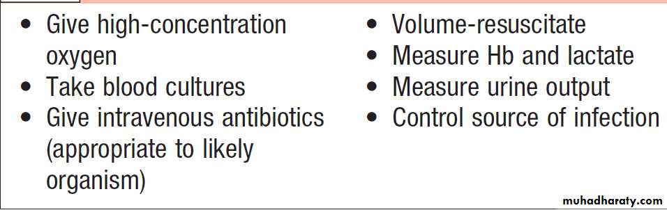
Immediate management of severe sepsis
Factors increasing the metabolic rate in
critical illnessDisseminated intravascular coagulation
Also known as consumptive coagulopathy, disseminatedintravascular coagulation (DIC) is an acquired
disorder of haemostasis , it is common in critically
ill patients and often heralds the onset of MOF. It
is characterised by an increase in prothrombin time,
partial thromboplastin time and fibrin degradation
products, and a fall in platelets and fibrinogen. The clinically dominant feature may be widespread bleeding
from vascular access points, gastrointestinal tract, bronchial tree and surgical wound sites, or widespread microvascular and even macrovascular thrombosis. Management is supportive with fresh frozen plasma and platelets, while the underlying cause is treated.
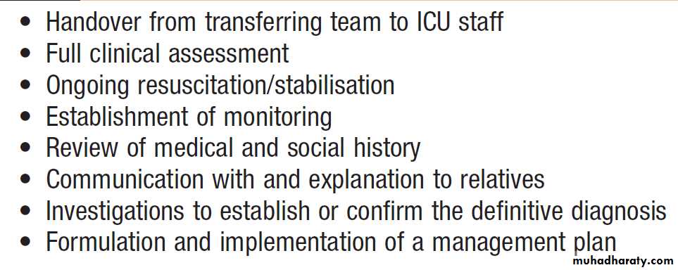
Management of patients on admission
to ICUGENERAL PRINCIPLES OF CRITICAL CARE MANAGEMENT
Daily clinical management in the ICU
Regular clinical examination is essential to identify
any changes in a patient’s condition. Detailed clinical
examination is performed at least daily, with additional
focused and systematic assessment on ward rounds at
least twice daily. Ward rounds are also an opportunity
to ensure the reliable application of evidence-based
measures to reduce complications; the mnemonic FAST
HUG provides a useful checklist of feeding, analgesia,
sedation, thromboprophylaxis, head of bed elevation,
ulcer prophylaxis, glucose control.
Patient review should include:
• Review of progress reports from nursing and medical staff, and any specialist opinions.• Review of 24-hour charts.
• Examination: general (skin, line sites, wounds etc.).
• System reviews:
Cardiovascular: haemodynamics, fluids and inotropes
Respiratory: ventilator settings and ABGs
Gastrointestinal: nutrition (calorie, protein intake,
route), nasogastric aspirate and bowel function
Renal: urine output, overall fluid balance, urea and electrolytes, and renal replacement therapy
Neurological: sedation level, GCS and pupil responses.
• Laboratory results: haematology, coagulation and
biochemistry.
• Microbiology: temperature, white blood count, line
sites and other possible sources of infection, results
of cultures, antibiotic therapy.
• Drug therapy: review with pharmacist, consider
adverse effects and interactions, and identify drugs
that can be discontinued. Medicines required for
long-term conditions should be continued in the context of the acute illness. An accurate record of the patient’s usual medicines must be obtained.
• Imaging: review X-rays and other specialist investigations with radiologists.
• Monitoring: are all measures still required? In
particular, remove central venous catheters, arteriallines and peripheral venous catheters as soon as no
longer needed, in order to avoid infection.
• Management plan: formulate an integrated plan,
with specific goals for each organ system and goals
for the patient, e.g. mobilising out of bed. Involve
the family in the patient’s care.
Sedation and analgesia
Intensive care is an extremely stressful experience forthe patient, with pain, discomfort and anxiety related to
endotracheal intubation, invasive monitoring. Most patients require sedation and analgesia to ensure comfort, relieve anxiety, and allow tolerance of an endotracheal tube, mechanical ventilation and invasive procedures.
Some conditions, especially severe neurological conditions that cause brain swelling and raised ICP, require deep sedation to reduce tissue oxygen requirements and protect organs from ischaemic damage.
Excessive sedation is common in ICU, and is associated with longer ICU stays, a higher prevalence of delirium, prolonged requirement for mechanical ventilation, and more ICU-acquired infections.
The optimally sedated patient is comfortable and tolerates
treatments, but is awake and lucid. Sedation and analgesia are usually provided via continuous infusions of sedative and/or analgesic drugs. As many critically ill patients have impaired liver and renal function, the potential for drugs to accumulate is high and the patient’s sedation must be regularly monitored.
The sedative agents used ideally have predictable short
half-lives and not affected by liver or renal impairment.
Short-acting IV agents, such as propofol, are usually employed. Analgesia can be provided using morphine infusions, but in patients with MOF, especially renal failure, active metabolites can accumulate.
Opiates such as fentanyl or alfentanil, which are not
renally metabolised or excreted, are commonly chosen.
Sedation is monitored via clinical sedation scales that record responses to voice and physical stimulation.
Regular use of these to adjust sedation is associated
with a shorter ICU stay. Many ICUs also have a daily ‘sedation break’, when all sedation is stopped in
appropriate cases for a period, in order to re-assess the
patient. This approach reduces the chance of over-sedation and shortens ICU stay.
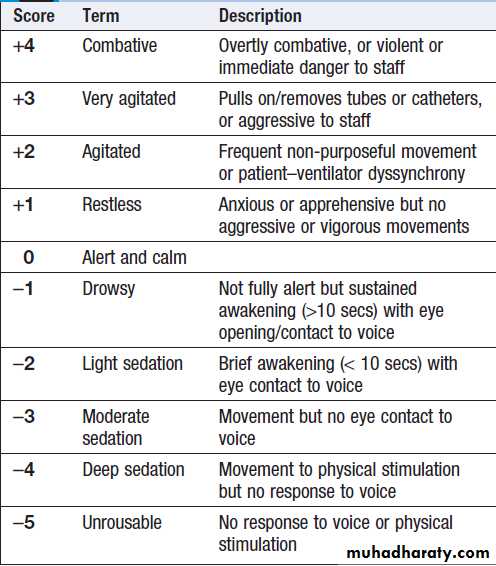
Richmond Agitation Sedation Scale (RASS)
Muscle relaxantsMuscle relaxants are avoided whenever possible in ICU
patients. Their use is associated with a higher prevalence
of critical illness neuropathy and myopathy, resulting in
muscle weakness. Sedation use also tends to be higher
when muscle relaxants are employed. They are required
to facilitate endotracheal intubation and to facilitate ventilation in patients with critical oxygenation and/or poor lung compliance. Patients with critically increased ICP
often receive intravenous infusions of muscle relaxant
drugs such as atracurium, to help control it and to
prevent coughing and high intrathoracic pressures,
which increase ICP.
Delirium
Delirium is extremely common in critically ill patients.
It often becomes apparent as sedation is reduced and
stopped. About 60–80% of patients have hypoactive
delirium, which is often missed unless formal testing is
undertaken. Between 5 and 10% of patients have agitated delirium, and 10–20% a mixed pattern. Delirium of any type is associated with poorer outcome. Management is focused on reducing or avoiding precipitating factors, such as benzodiazepines and metabolic disturbances.
Patients with agitated delirium should be managed with haloperidol in 2.5 mg increments, rather
than additional sedatives. Some sedative drugs are associated with a lower incidence of delirium, such as
α2-adrenergic agonists (clonidine and dexmedetomidine).
These have a central sedative action and can be
useful in difficult cases. There is no evidence that
pharmacological interventions are useful as prophylaxis
or in hypoactive delirium.
DISCHARGE FROM INTENSIVE CARE
Discharge is appropriate when the original indication for admission has resolved and the patient has sufficientphysiological reserve to continue recovery without the facilities of intensive care. Many ICUs and HDUs function as combined units, which allows stepdown to HDU care without a change of clinical team. Critically ill patients often have complex medical histories, multiple ongoing medical problems, and family and social issues. Many also have the emotional problems associated with survival from a life-threatening event or illness. Discharge from the ICU is stressful for patients and families, and communication with the clinical team accepting responsibility is vital.
Discharges from ICU/HDU to standard wards should take place within normal working hours to ensure adequate medical and nursing support and detailed handover, as discharge outside normal working hours (and early discharge) are associated with higher ICU re-admission rates and increased mortality.
The receiving team should be provided with a written
summary, including relevant recent investigations, and
the critical care team should remain available for advice.
Many ICU teams provide an outreach service to furnish
advice and ensure continuity. Irrespective of the reason
for admission to the ICU, many patients suffer problems
(Box). These can last from weeks to many months
and require ongoing support and rehabilitation.
Physical
• Fatigue: almost universal and can last for many weeks • Breathlessness • Muscle weakness: a combination of muscle wasting from inadequate nutritional intake, muscle breakdown associated with inflammation/infection, and damage to nerves and muscle associated with severe systemic illness (critical illness polyneuropathy and polymyopathy) • Altered taste and poor appetite: can result in weight loss • Joint stiffness • Itch • Hair loss
Psychological
• Anxiety • Depression • Traumatic memories: including delusional memories (often unpleasant), dreams, flashbacks and hyperarousal. Worse in more severe and prolonged illness, more prolonged deep sedation and after delirium. Recovery occurs over weeks/months, but can persist and affect quality of life
Common problems after ICU discharge
Withdrawal of intensive support
Withdrawal of support must be considered when it isclear that the patient has no realistic prospect of recovery or of surviving with a quality of life that he or she would
value. In these situations, intensive care will only
prolong the dying process and is therefore futile. When
intensive support is withdrawn, management remains
active and is aimed at allowing the patient to die with
dignity and as free from distress as possible .
Patients’ views are paramount and increasing use is
being made of advance directives or ‘living wills’. Communication with the patient, if possible, and with the
family, the referring clinicians, and between members of
the critical care team is crucial.
Brainstem death
Recent advances in the resuscitation and intensive caremanagement of brain-injured patients have inevitably
increased the survival of patients who remain ventilated
on ICU, in whom progression of brain injury results in
brainstem death.
When the formal criteria for brainstem death are met,
it is clearly inappropriate to continue supporting life
with mechanical ventilation, and the possibility of organ
donation should be considered.
All intensive care clinicians have a responsibility to approach relatives to seek consent for organ donation, provided there is no contraindication to their use.
This can be very difficult, but is easier if the patient carried an organ donor card or was registered with an organisation such as the UK Organ Donor Register.
In the UK, each region has a team of specialist nurses in organ donation who provide help with the process and with care of the potential organ donor.
OUTCOME OF INTENSIVE CARE
The measure used most widely to assess outcome fromintensive care is mortality. Mortality is strongly influenced
by the case mix of an ICU (the type of patients
and their illness severity at admission). Typically, about
20% of ICU patients will die during their ICU stay
despite treatment, and about 30% will die before leaving
hospital. Some factors associated with higher mortality
are shown in Box.
Mortality in patients requiring HDU care is much lower.
Many patients have preexisting illnesses prior to ICU admission, which, combined with the effect of the illness that resulted in ICU admission and the subsequent complications, means that many who survive the ICU admission have reduced life expectancy compared to those of similar age in the general population.
The long-term physical and psychological effects of critical illness can mean that surviving patients have a reduced quality of life for many months or years.
Families often carry a heavy burden of care after critical illness.
Many patients do not regain preillness health and may be unable to work, resulting in economic and social hardship.
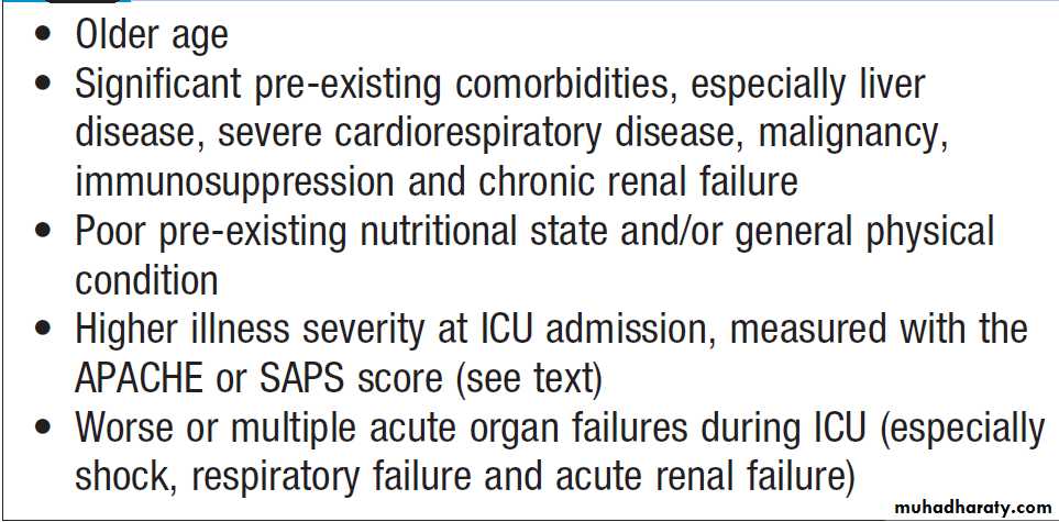
Factors associated with higher mortality
from critical illness
Scoring systems
Admission and discharge criteria vary between ICUs, soit is important to define the characteristics of the patients
admitted (case mix) in order to assess the effects of the
care provided on the outcome achieved. Two systems
are widely used to measure severity of illness:
• ‘APACHE’ II: Acute Physiology Assessment and
Chronic Health Evaluation
• ‘SAPS’ 2: Simplified Acute Physiology Score. These scores include assessment of certain admission characteristics (e.g. age and pre-existing organ dysfunction) and a variety of routine physiological measurements (e.g. temperature, BP, GCS) that reflect the response of the patient.
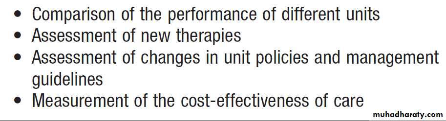
Uses of critical care scoring systems
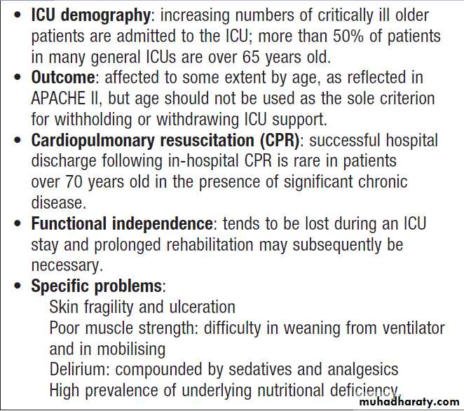
The critically ill older patient