Post Mortem Pathology / Canine and Feline Diseases / Dr. Saevan Saad Al-Mahmood
1
RABIES
Definition:
Rabies is a viral disease that affects the central nervous system of warm-
blooded animals, including humans. The causative agent is virus of lyssavirus, a
group of viruses responsible for causing encephalitis. Lyssaviruses belong to the
family Rhabdoviridae.
Clinical signs:
The first (“prodromal”) phase
occurs early during the illness. At this stage,
Behavioral changes, often in the form of a reversal of normal patterns, usually
begin to show in this phase.
In the second (“furious”) phase
, the animal becomes extremely irritable and
aggressive, often lunging at or biting anything that moves near it
The final (“dumb”) stage
is manifest by the onset of paralysis, most often in the
lower jaw and extremities.
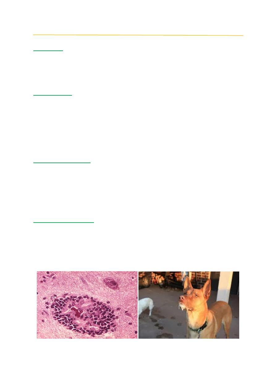
Post mortem findings
There are no characteristic gross lesions. The stomach may contain unusual
objects that were ingested. The typical histological signs, found in the CNS, are
multifocal, mild, polioencephalomyelitis and craniospinal ganglionitis with
mononuclear perivascular infiltrates, diffuse glial proliferation, regressive
changes in neuronal cells, and glial nodules. Aggregates of viral material in
neurons (Negri bodies) can be seen in some but not all cases.
Differential diagnosis:
1- Listeriosis.
2- Brain parasites.
3- Toxines.
Post Mortem Pathology / Canine and Feline Diseases / Dr. Saevan Saad Al-Mahmood
2
CANINE DISTEMPER
Definition:
is a serious viral illness that attacks a dog’s body on all fronts. The disease
may harden the paws and nose, damage the teeth, make breathing difficult and
diminish the appetite. Canine distemper virus (CDV) is a negative-stranded RNA
virus, within the family Paramyxoviridea, subfamily Paramyxovirinae, and genus
Morbillivirus.
Clinical signs:
The classic signs are depression and mucopurulent oculonasal exudates and
diarrhea frequently occur. Neurologic signs depend on the area of brain affected
and include abnormal behavior, convulsions, cerebellar and vestibular signs,
paresis or paralysis and incoordination.
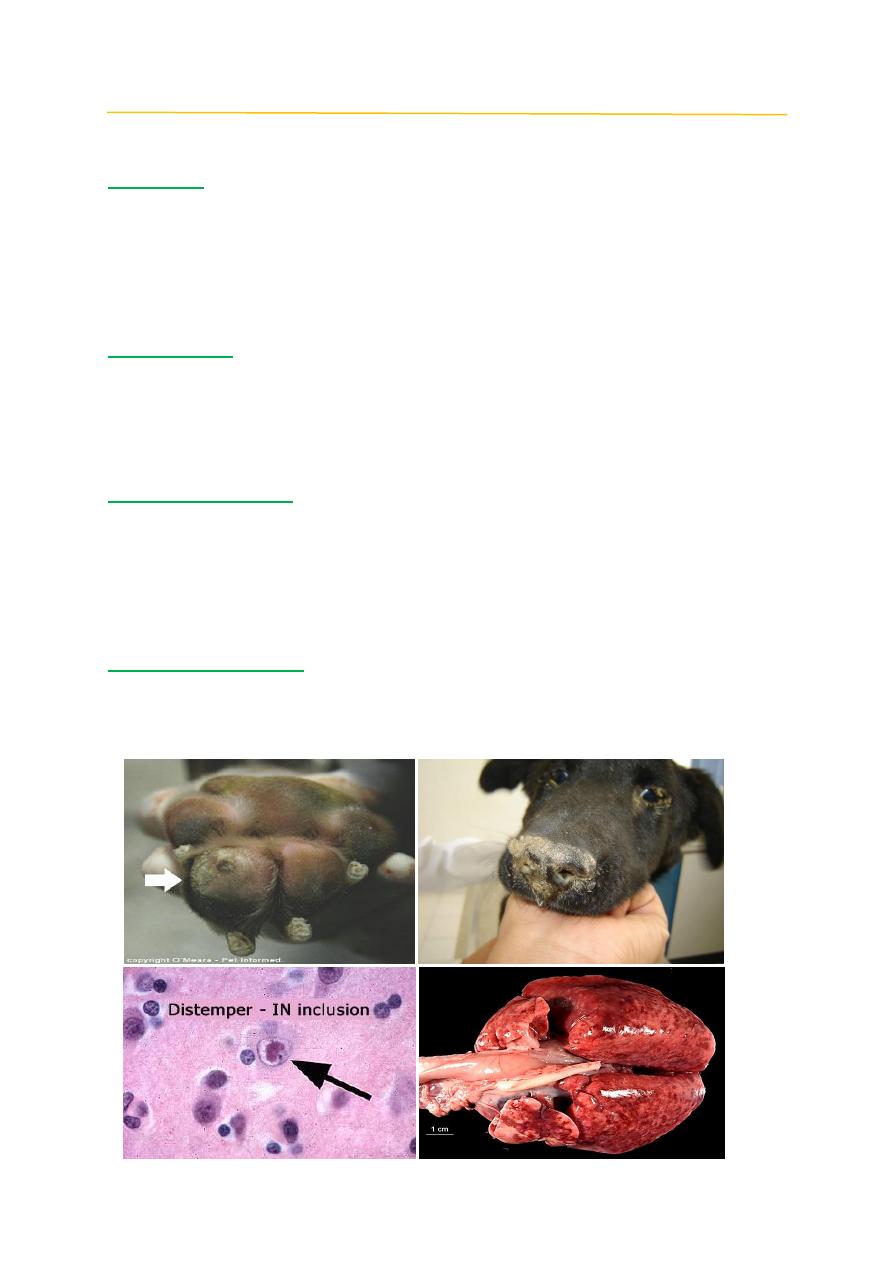
Post mortem findings
Interstitial pneumonia to bronchopneumonia, catarrhal to haemorrhagic
enteritis, and hyperkeratosis of the nose, lips, eyelids, ears, and footpads. An
important diagnostic feature of CD is the presence of intracytoplasmatic and
intranuclear eosinophilic inclusion bodies in neurons, gastric mucosa,
enterocytes, and epithelium of the respiratory and urogenital tract.
Differential diagnosis:
1- Canine Keratoconjectivitis virus.
2- Vitamin A deficiency.
3- Pulmonary Manhimosis.
Post Mortem Pathology / Canine and Feline Diseases / Dr. Saevan Saad Al-Mahmood
3
INFECTIOUS CANINE HEPATITIS (ICH)
Definition:
Worldwide, contagious disease of dogs with signs that vary from a slight
fever and congestion of the mucous membranes to severe depression, marked
leukopenia, and coagulation disorders, ICH is caused by a nonenveloped DNA
virus, canine adenovirus 1 (CAV-1),
Clinical signs:
Signs and symptoms Clinical signs develop after an incubation period of 4-
7 days and most commonly include lack of appetite, fever, conjunctivitis,
coughing, abdominal pain, vomiting, diarrhea and jaundice. In some dogs that
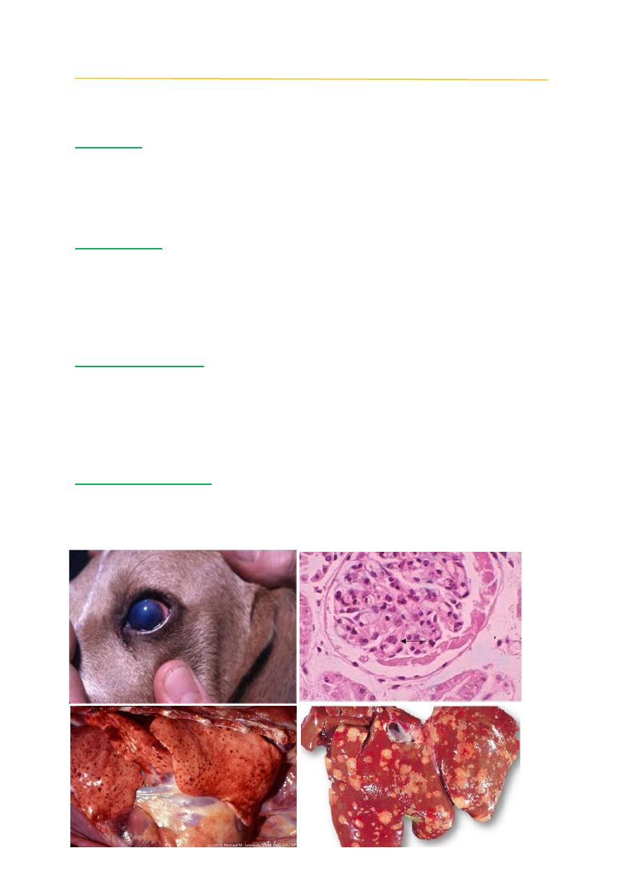
recover, a clouding of the cornea occurs, known as 'blue eye' which usually
resolves itself over time.
Post mortem findings
Endothelial damage results in “paint-brush” hemorrhages on the gastric
serosa, lymph nodes, thymus, pancreas, and subcutaneous tissues. Hepatic cell
necrosis produces a variegated color change in the liver. Histologically, there is
centrilobular necrosis, with neutrophilic and monocytic infiltration, and
hepatocellular intranuclear inclusions.
Differential diagnosis:
1- Leptospira icterohemorrhagiae infection
2- canine parvovirus.
3- Autoimmune hemolytic anemia.
Post Mortem Pathology / Canine and Feline Diseases / Dr. Saevan Saad Al-Mahmood
4
FELINE PANLEUKOPENIA (FP)
Definition:
A highly contagious viral disease of cats caused by the feline parvovirus, FP
has been known by a variety of names including
feline distemper
,
feline infectious
enteritis,
cat fever
and
cat typhoid
. The feline parvovirus infects and kills cells
that are rapidly dividing, such as those in the bone marrow, intestines, and the
developing fetus.
Clinical signs:
Infected cats usually develop bloody diarrhea, infected cats develop anemia
(due to loss of red blood cells) and are more likely to be infected with other
diseases (due to the loss of white blood cells, which play critical roles in the
immune system).
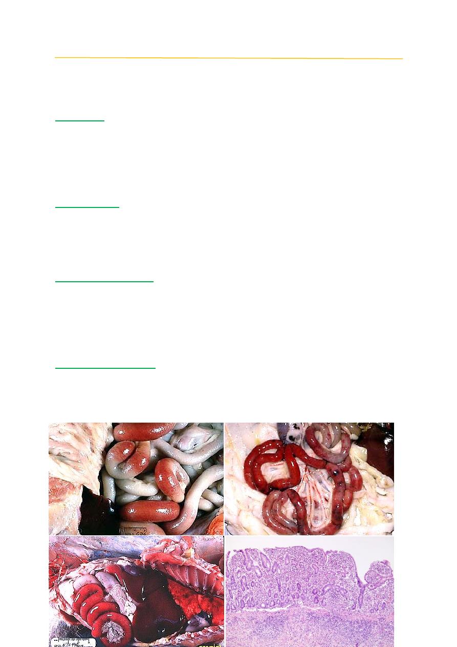
Post mortem findings:
The hallmark of FP is sever congestion of intestine and lock like sausage
also shortening of the intestinal villi due to a complete loss of epithelial cells in
the gut. The virus replicates in the rapidly dividing cells of the epithelium, the
crypts of Lieberkühn. This impairs the regeneration of the epithelium and results
in the lesions described above.
Differential diagnosis:
1- Salmonellosis.
2- Feline leukemia.
3- Cryptosporidiosis.
4- Toxoplasmosis.