JABIR IBN HAYYAN MEDICAL UNIVERSITYCOLLEGE OF MEDICINE DEPARTMENT OF HUMAN ANATOMY
Histology of Female Reproductive SystemLecture By;
Dr. Hayder ALKifaee
6/4/2017
FEMALE REPRODUCTIVE SYSTEM
HISTOLOGY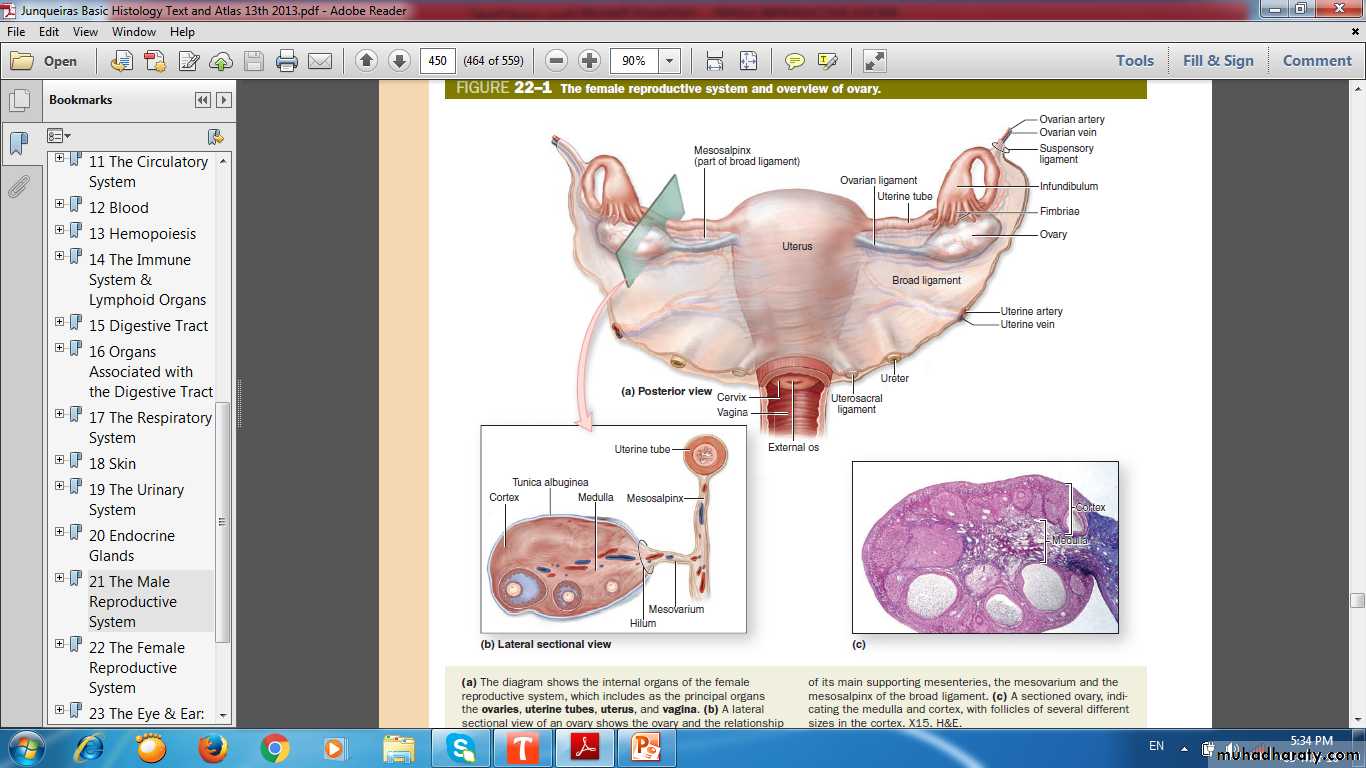
Female Reproductive System Consists of
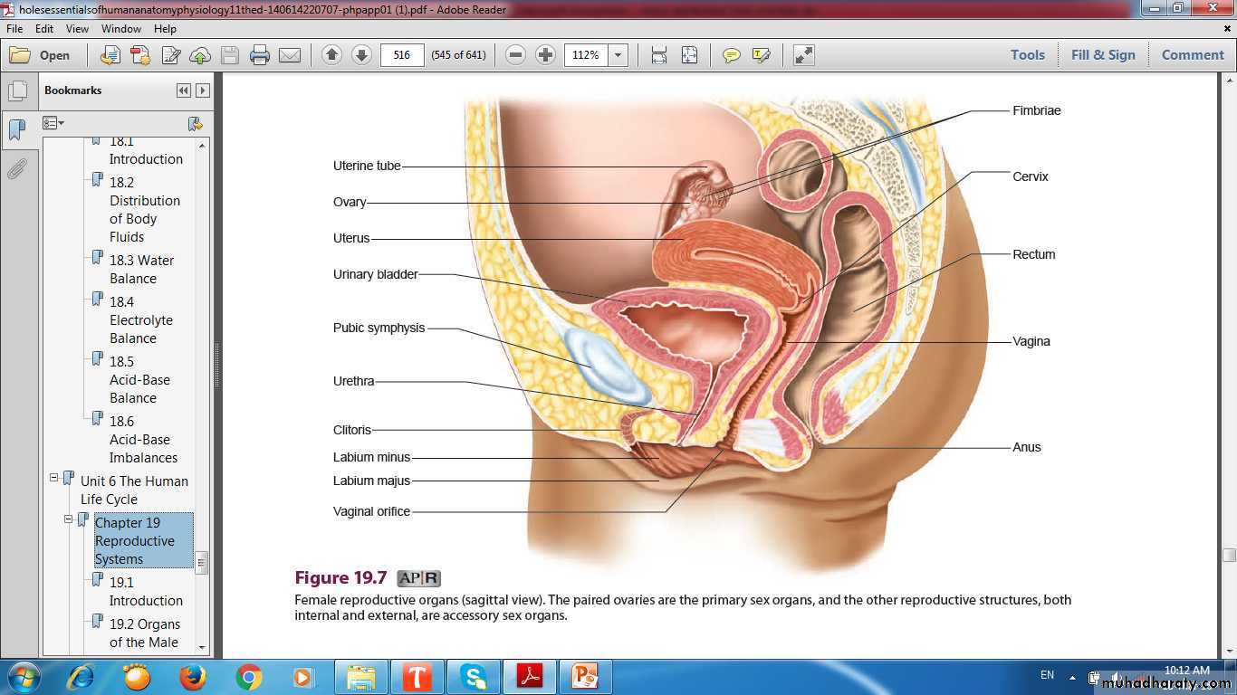
Paired ovariesUterine tubes
Uterus
Vagina
External Genitalia
Female Reproductive Organs
Cortex
MedullaFemale Reproductive Organs
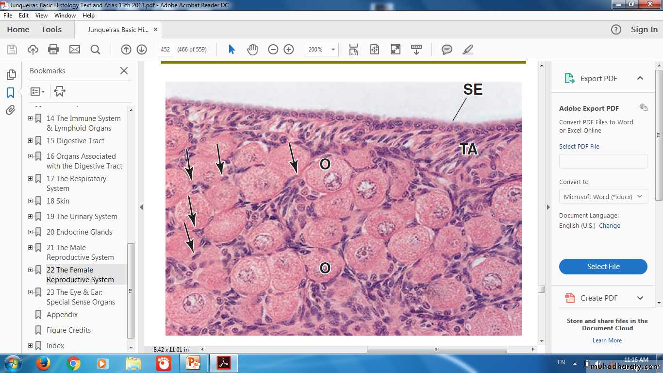
OvaryAlmond shaped (3 x 1.5 x 1 cm)
Produce Oocytes
Produce Hormones
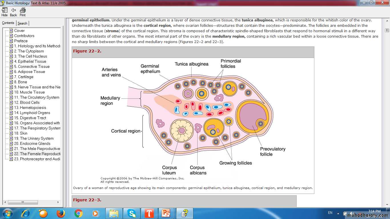
Coverings of Ovary
Coverings of Ovary
Menarche & Menopause
MenarcheMenopause period
Postmenopausal period
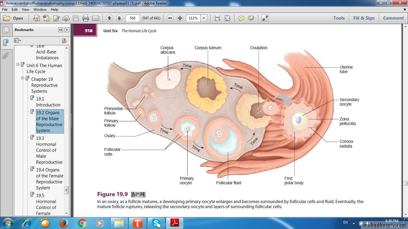
Follicular Growth & Development
Follicular Growth & Development
Beginning in Puberty with the release of FSH
Group of Primordial Follicles each month begins a process of follicular growth
Follicular Growth & Development
This involves Growth of the oocyteProliferation & changes in the Follicular cells
Proliferation & differentiation of the Stromal Fibroblasts
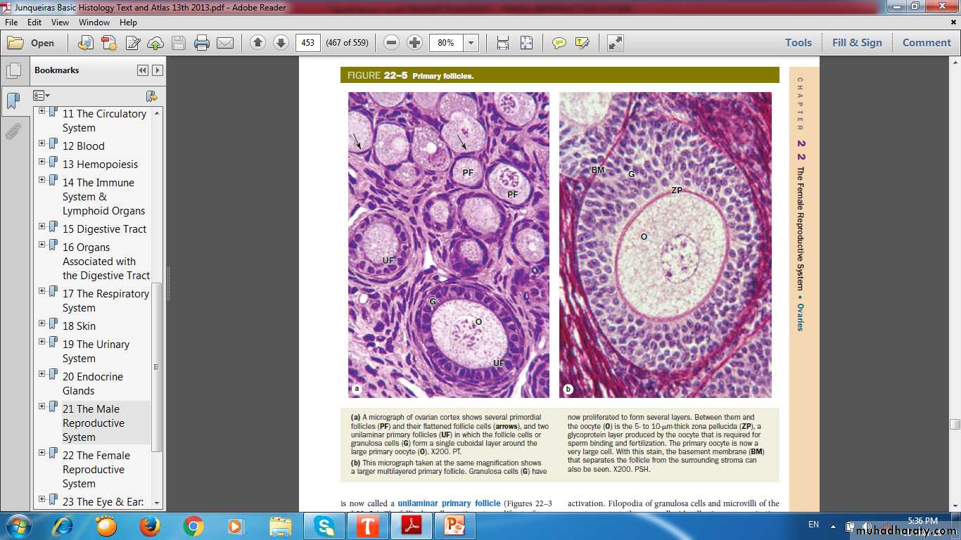
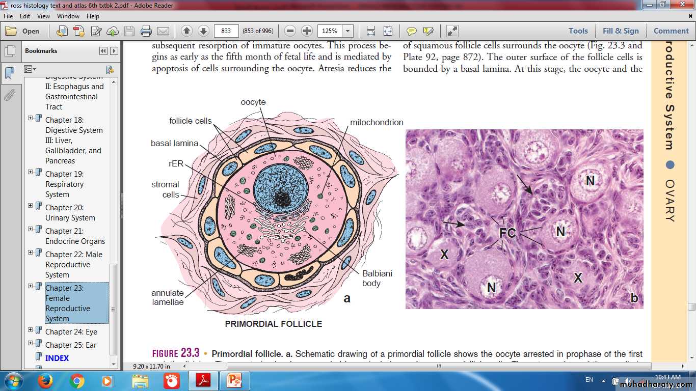
Primordial Follicle
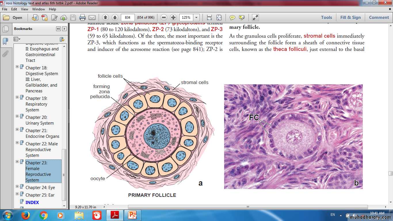
Unilaminar Primary Follicle
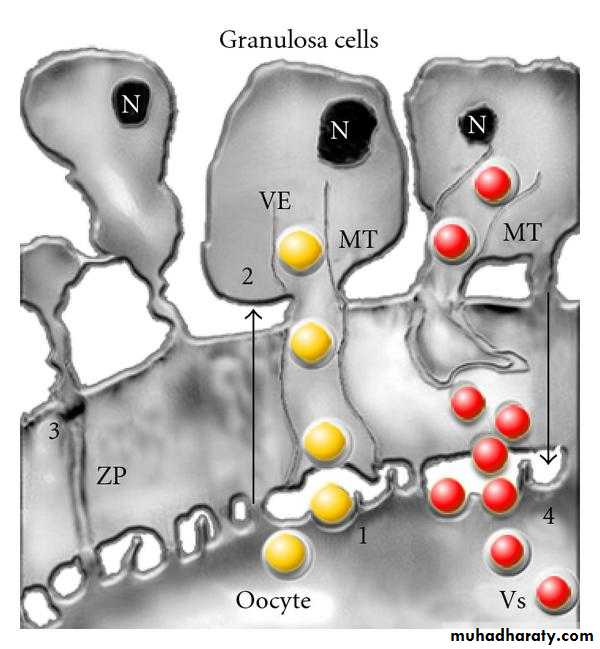
Microvilli of Oocyte & Filopodia of Granluosa cells

Microvilli
FilopodiaGranluosa cell
Microvilli of Oocyte & Filopodia of Granluosa cells
Primordial Follicle
Unilaminar FollicleMultilayered Follicle
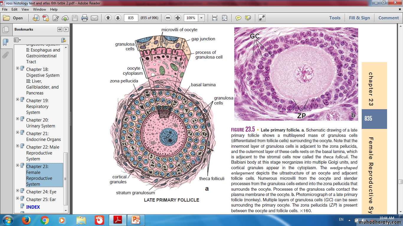
Primary Follicle
Multilaminar Primary Follicle
Antral Follicle
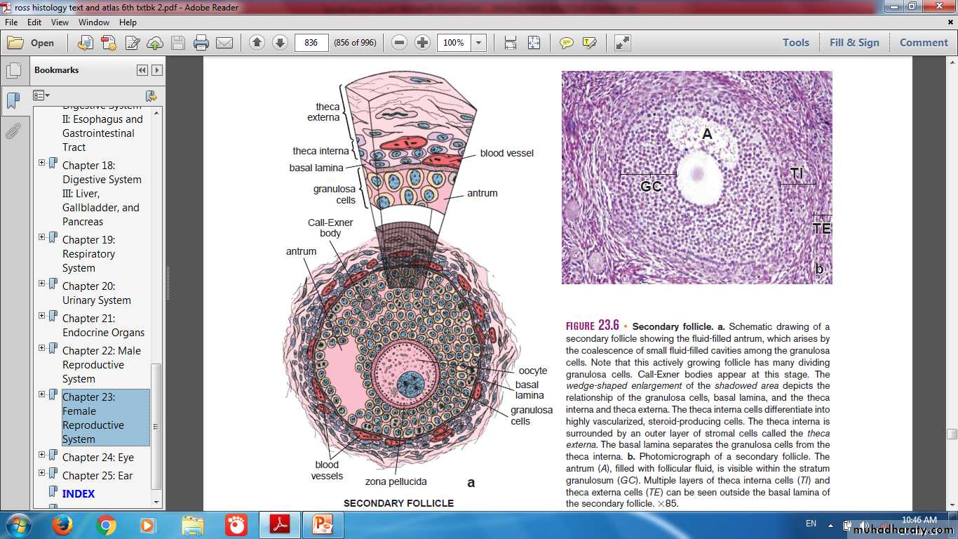
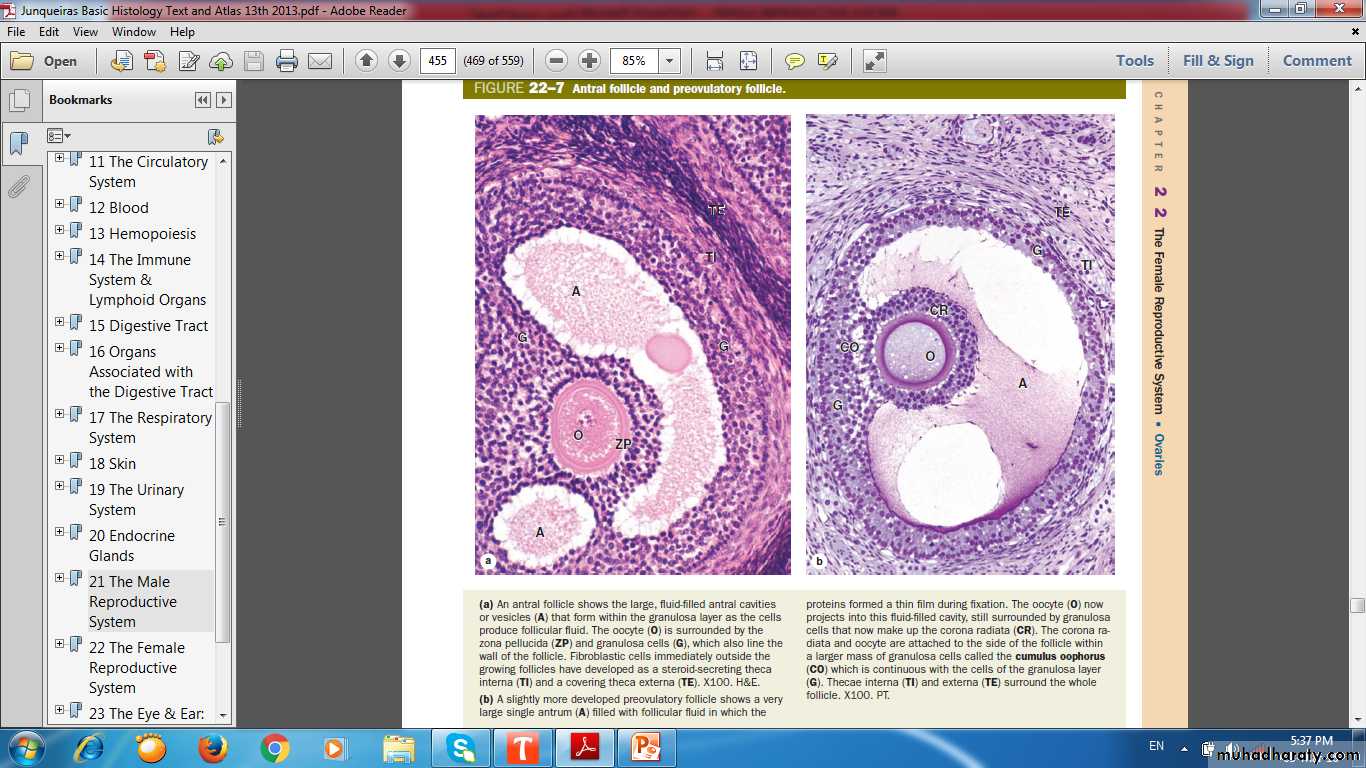
Antrum
GranulosaTheca interna & Externa
Antral Follicle
Preovulatory Follicle
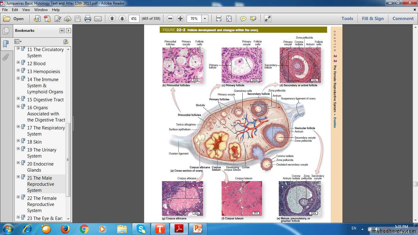
Follicular Development
Graafian Follicle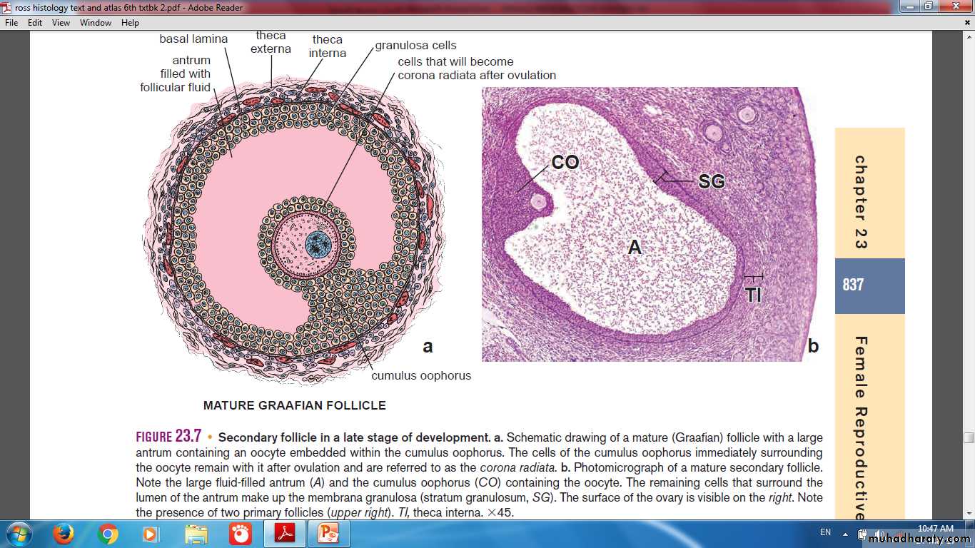
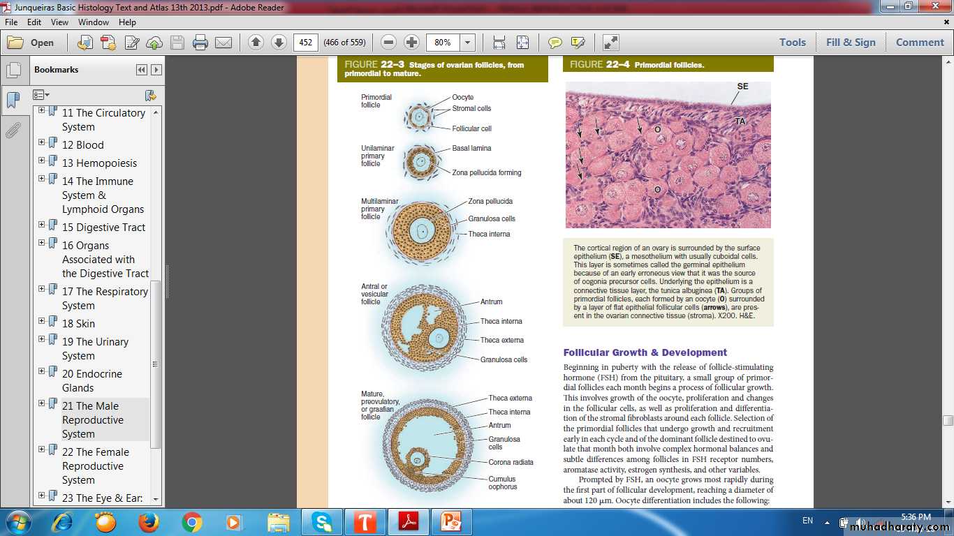
Stages of Ovarian Follicle
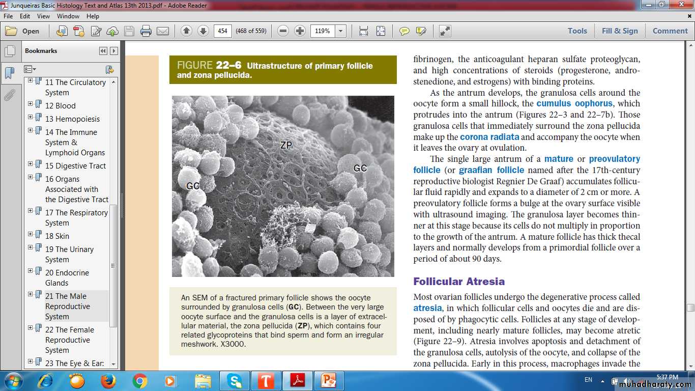
Zona Pellucida
Granluosa CellUltrastructure of Primary Follicle & Zona Pellucida
Follicular Fluid
Large GAG hyaluronic acidGrowth factors
Plasminogen
Fibrinogen
Anticoagulant heparan sulfate proteoglycan
High concentrations of steroids
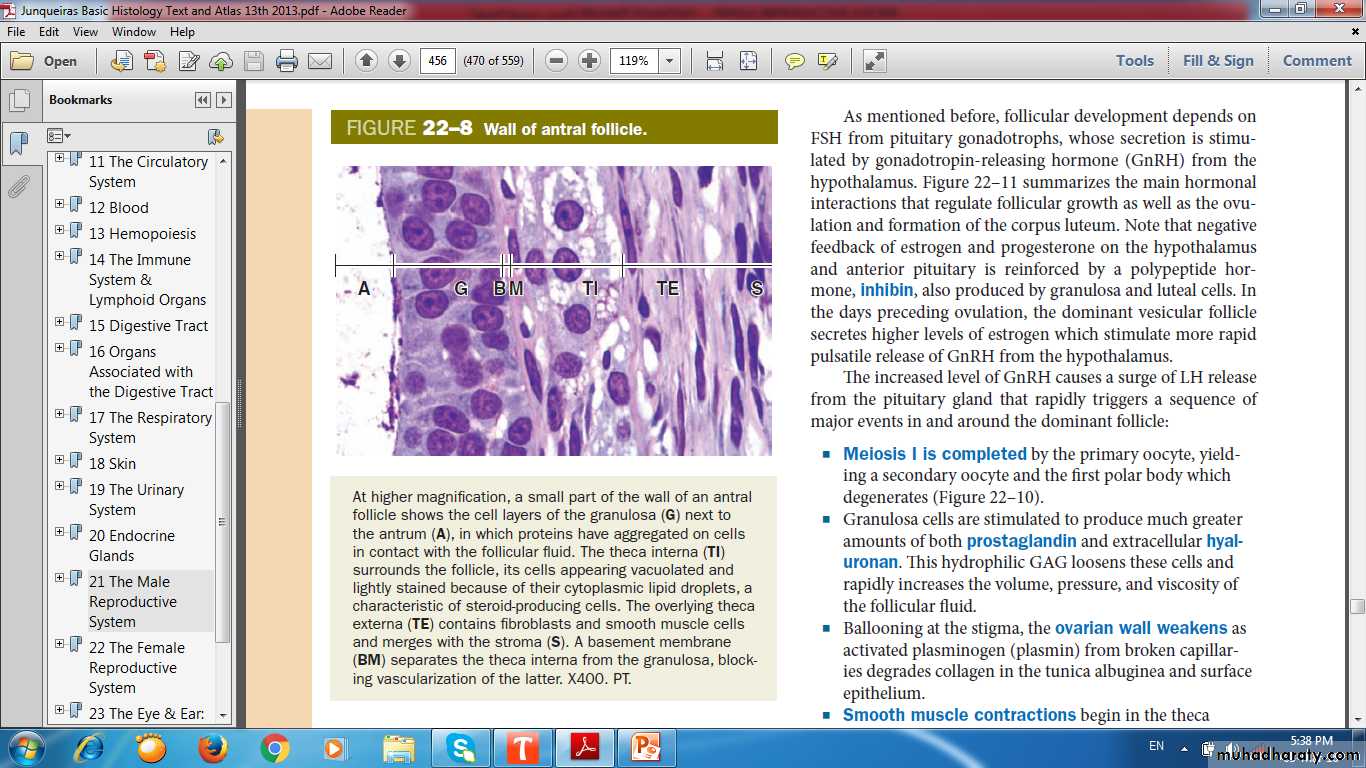
Wall of Antral Follicle
AntrumGranulosa
Theca I
Theca E
Stroma
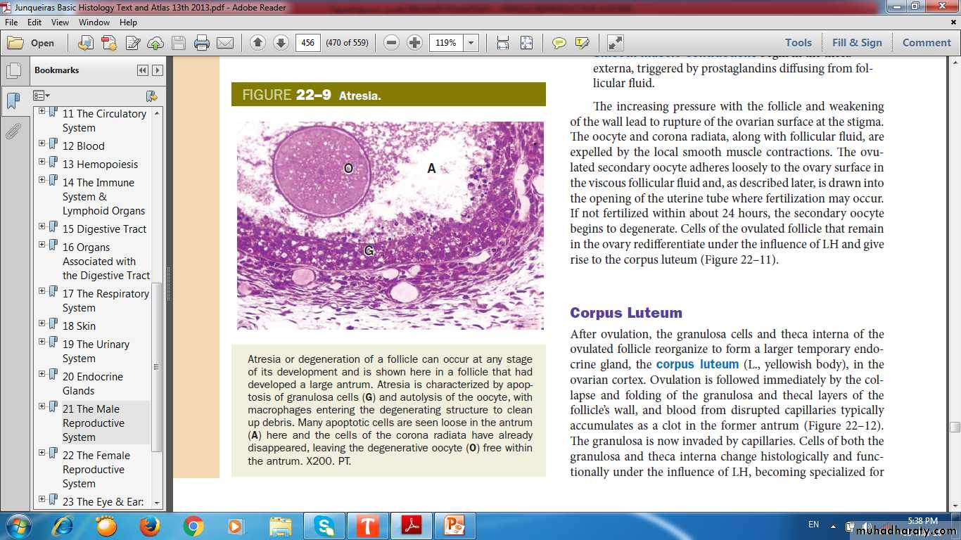
Apoptosis of G. cells
Atresia of Follicle(Apoptosis & Autolysis)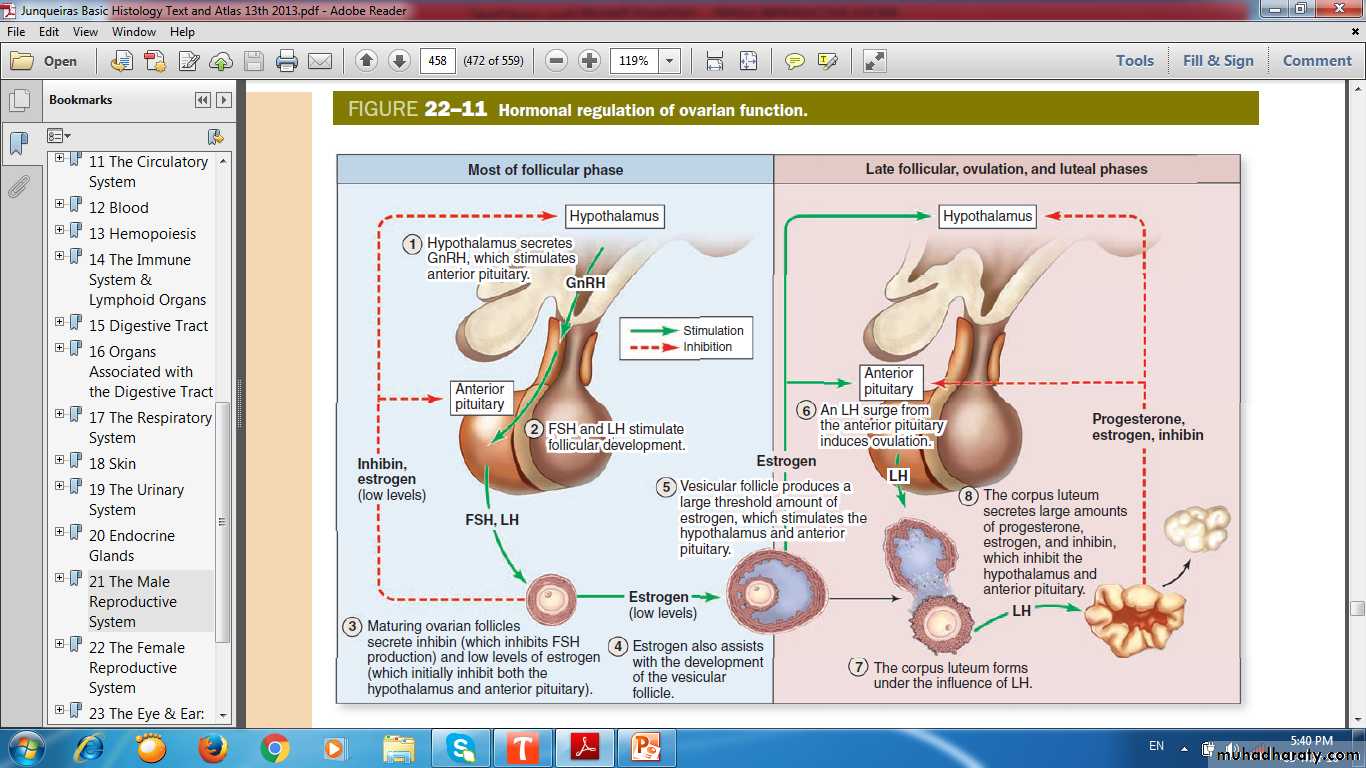
Hormonal Regulation of Ovarian Function
Corpus Luteum (yellowish body)
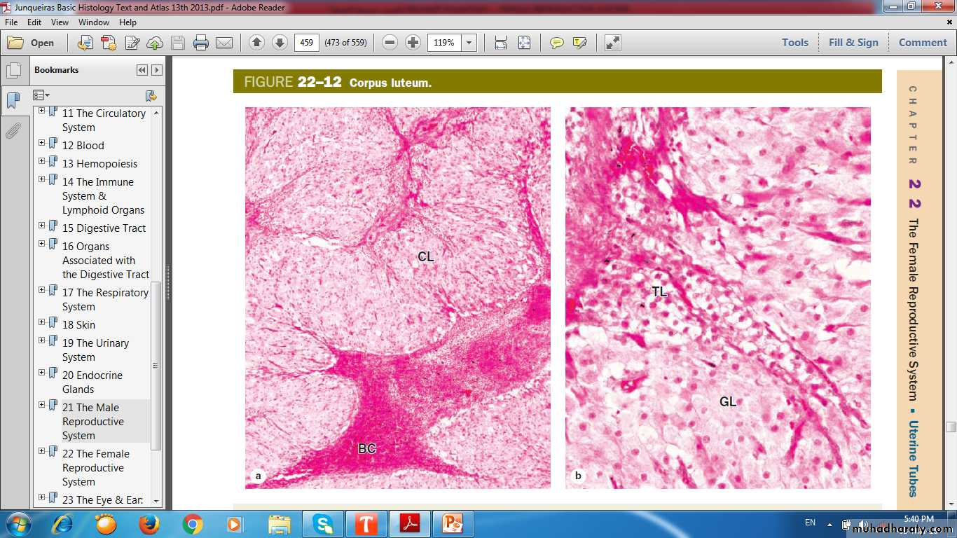
Granlosa lutein cell
Theca lutein cellCorpus LuteumGranulosa cells Granulosa lutein cells Theca interna cells Theca lutein cells
Remnants of corpus luteum are phagocytosed by macrophages, after which fibroblasts invade the area and produce a scar of dense connective tissue called a corpus albicans (L., white body).
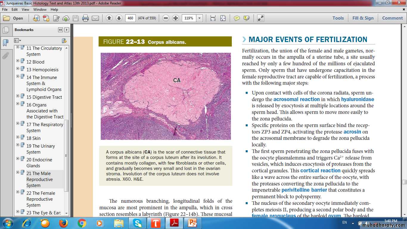
Corpus Albicans (white body)

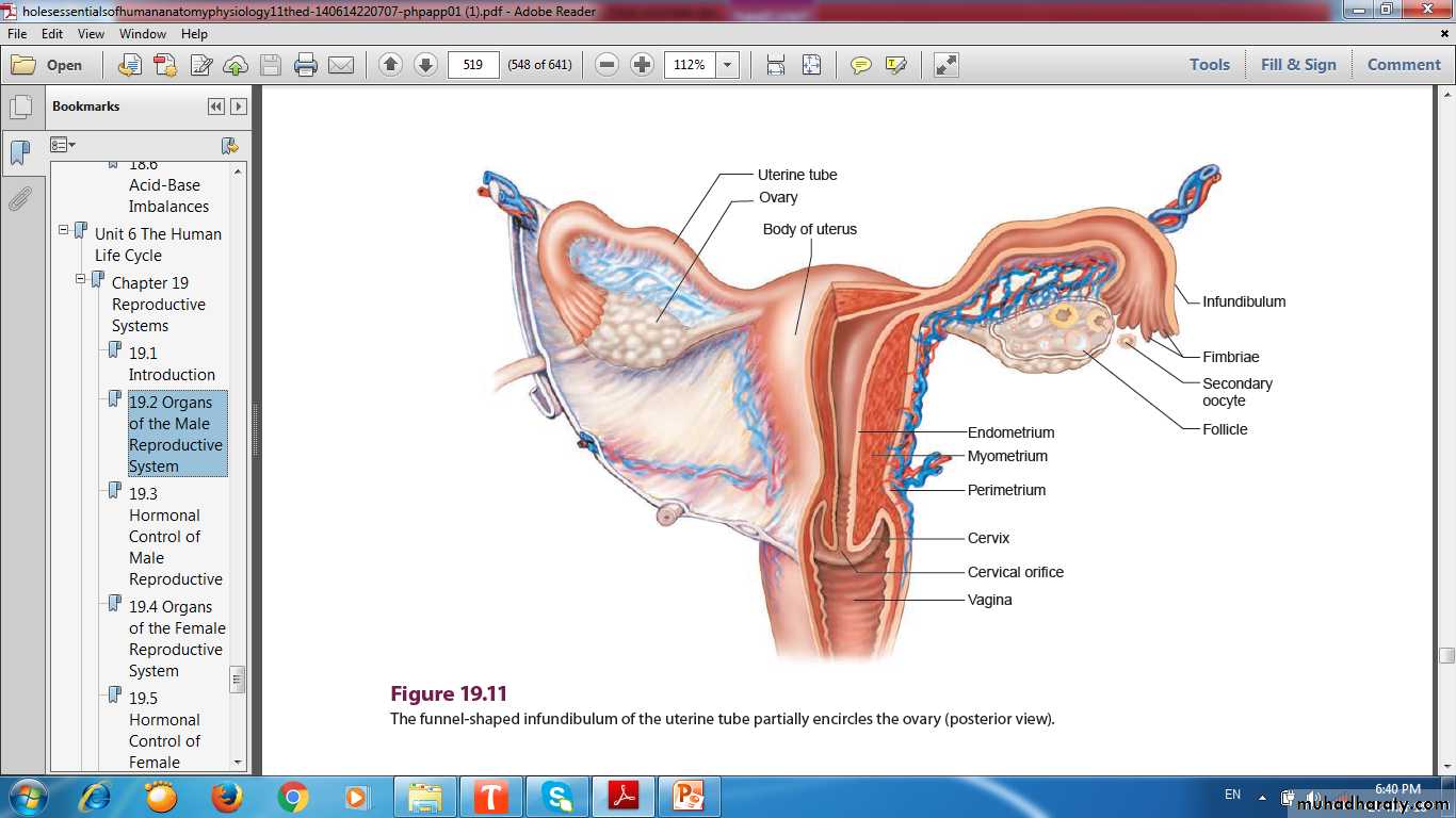
Uterine Tubes & Uterus
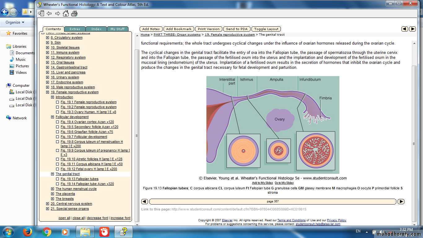
Uterine Tubes
AmpullaIsthmus
infundibulum
Intramural
Wall of the Oviduct:
MucosaMuscularis
Serosa
Cross Section in Oviduct (Ampulla)
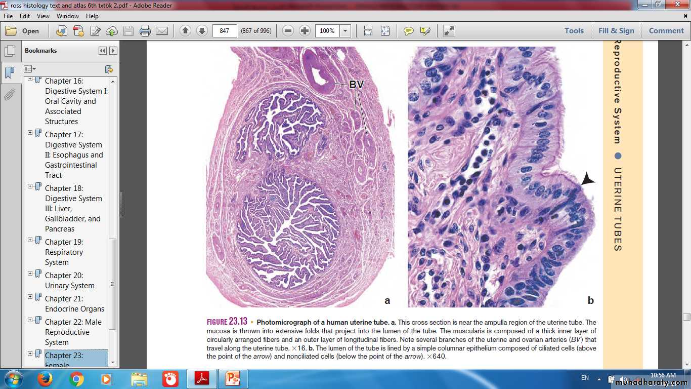
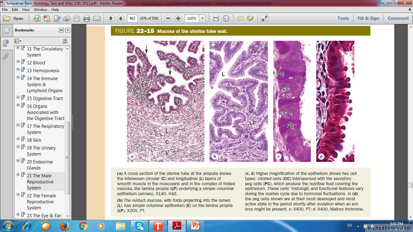
Mucosa of the Uterine Tube Wall
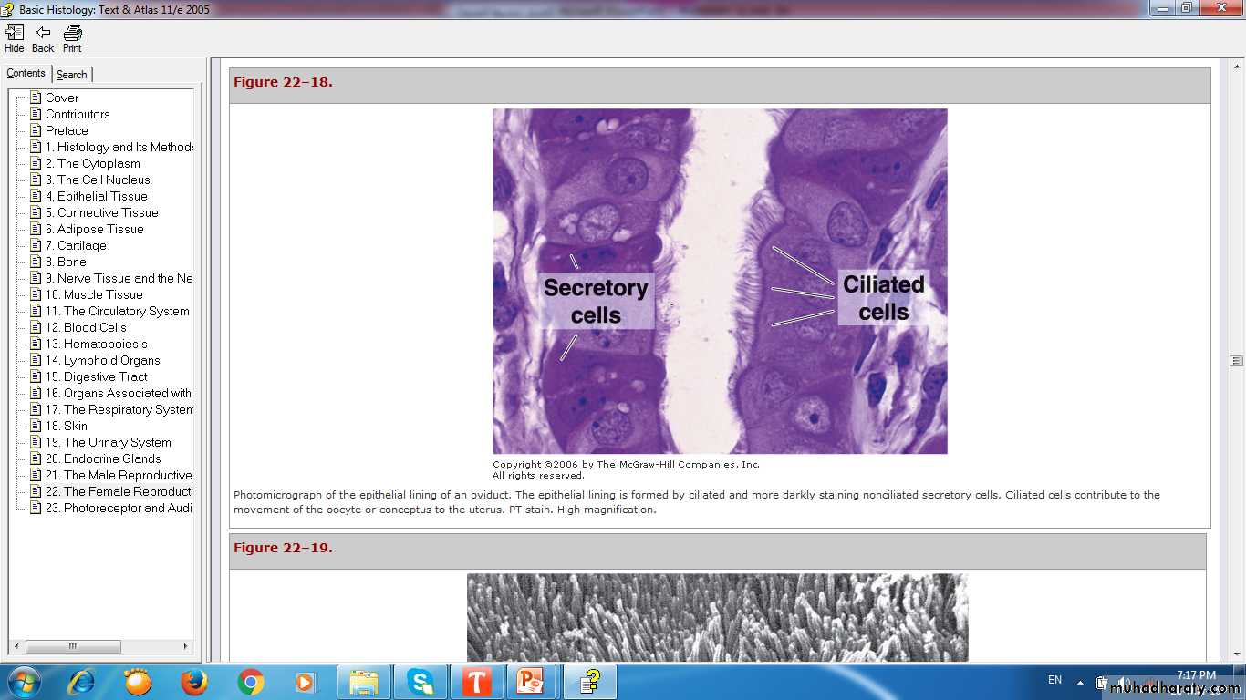
Ciliated Cell& Secretory (Peg Cell)
The Epithelium Cell Types:Ciliated Cells in which ciliary movements sweep fluid toward the uterus
Secretory Peg Cells, nonciliated & often darker staining, often with an apical bulge into the lumen, which secrete glycoproteins of a nutritive mucus film that covers the epithelium
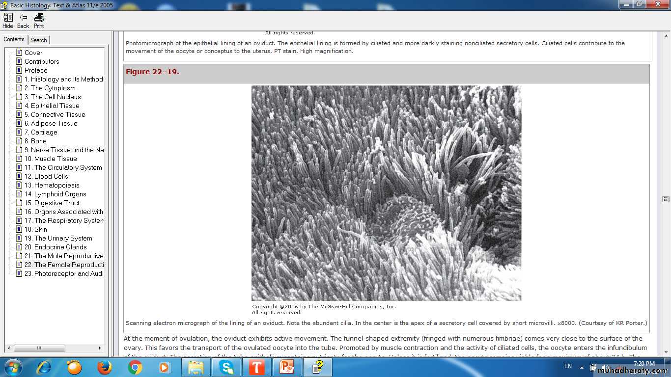
Ciliated Cell& Secretory (Peg Cell)
UTERUS Fundus, Body & Cervix
Uterine Wall Three Layers:
PerimetriumMyometrium
Endometrium
Layers of Uterine Wall
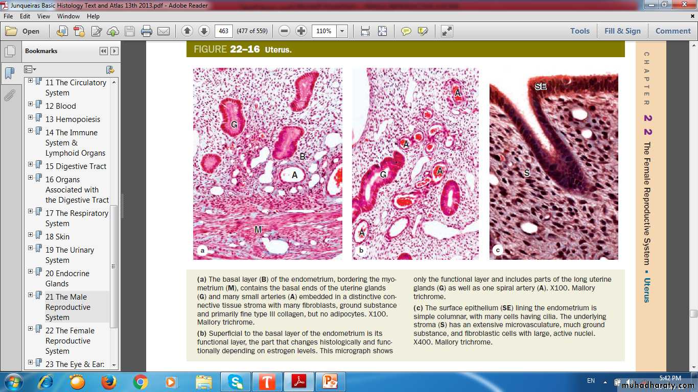
Uterus
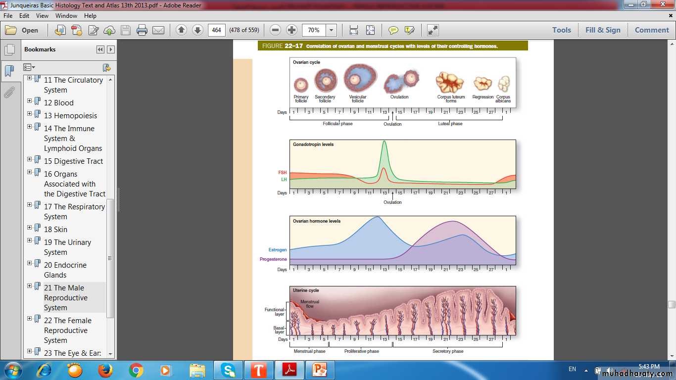
Correlation of Ovarian &Menstrual Cycle
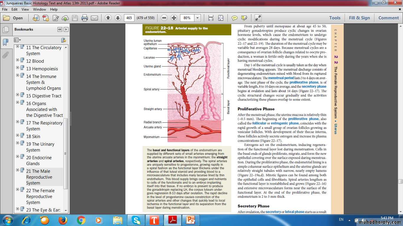
Arterial Supply of Endometrium
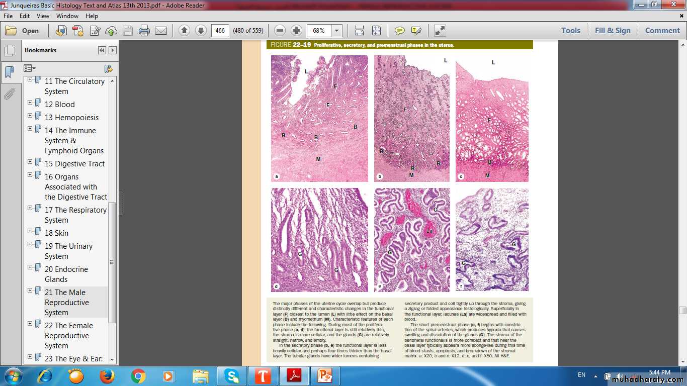
proliferative
SecretoryPremenstrual
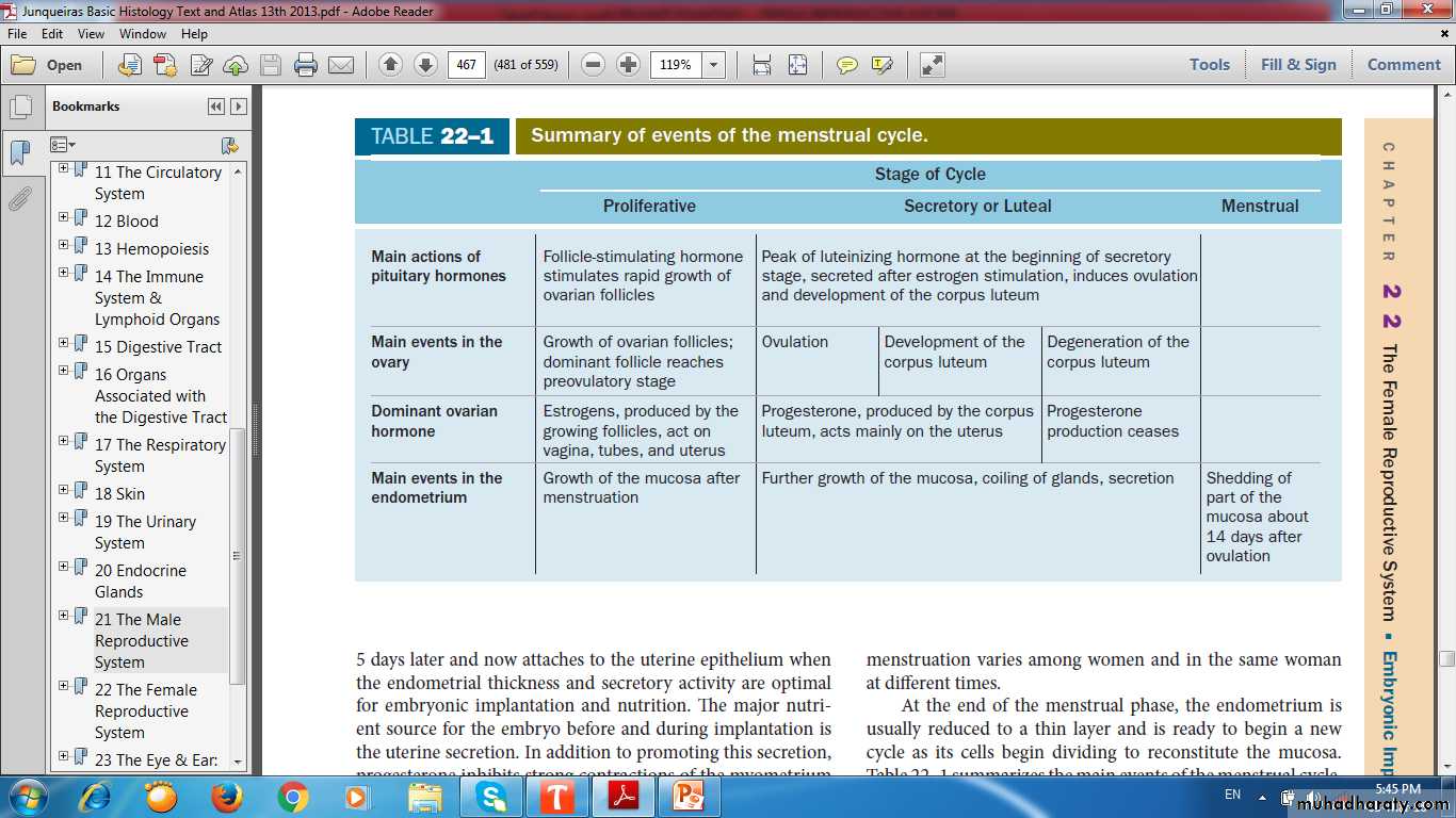
Proliferative, Secretory& Premenstrual phases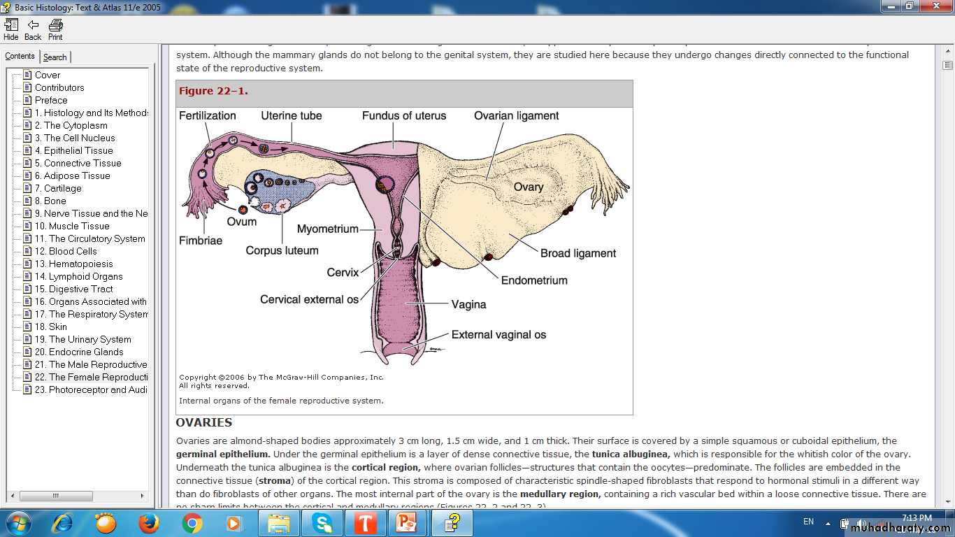
Cervix & Vagina
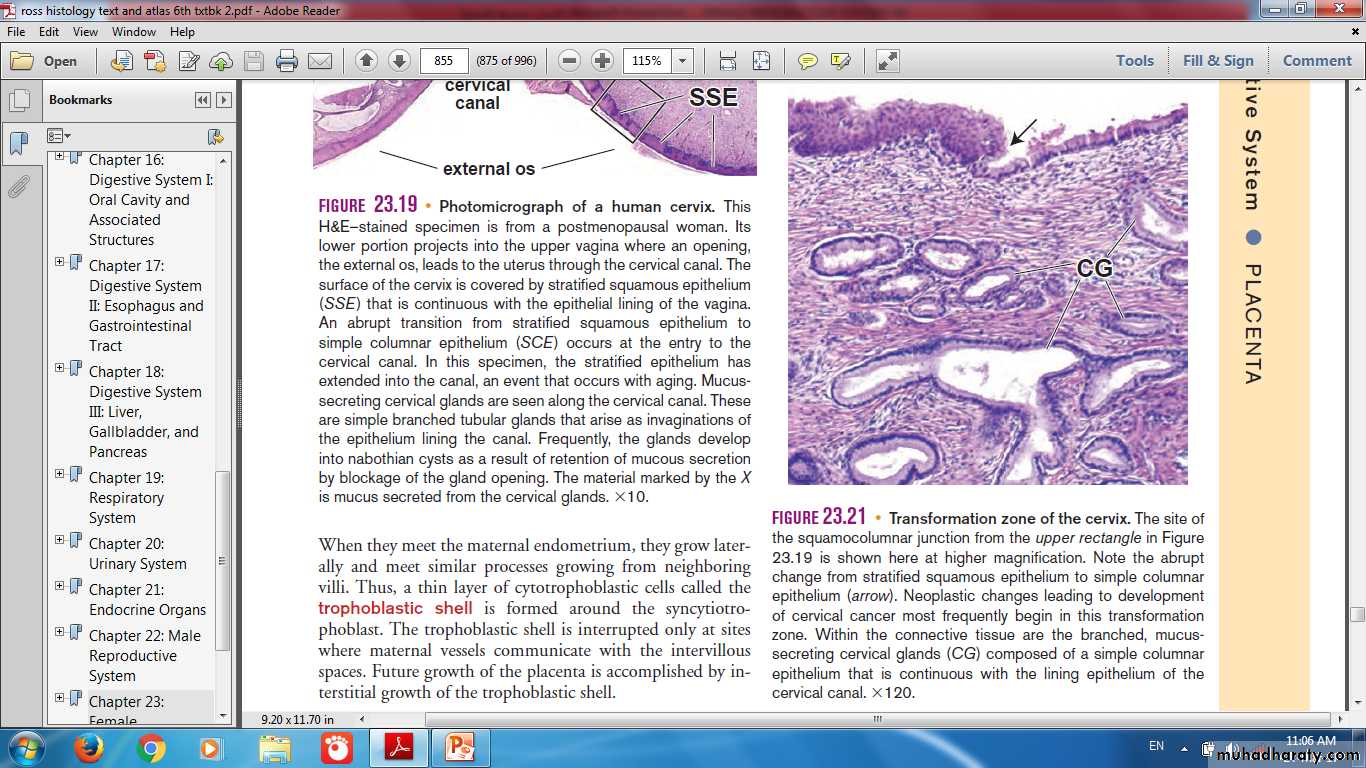
Cervix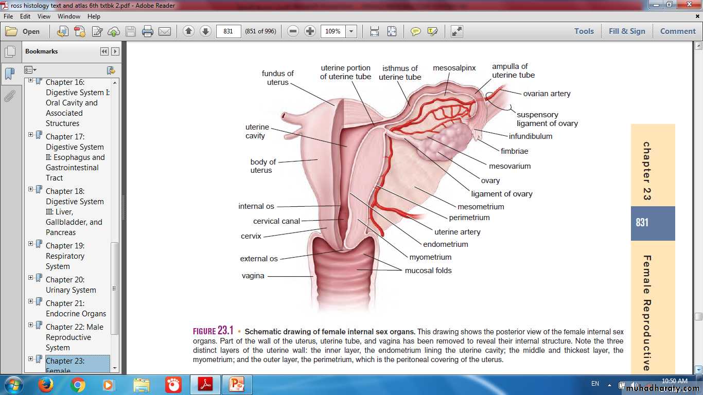
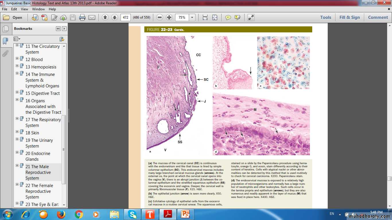
Cervical canal
Simple columnarJunction
Strat.squamous
Vagina
Exfoliative cytology of exocervix
Cervical mucosal glands
Transformation Zone(Squamo-Columnar Junction)
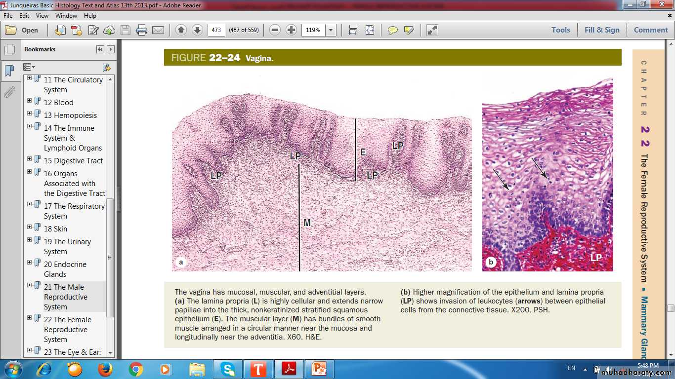
Epithelium
Muscular layerLamina propria
Vagina Mucosa, Muscular layer & Adventitia