Lec. 6 Biology
HistologyThe cartilage
Cartilage is a flexible connective tissue, including the joints between bones, the rib cage, the ear, the nose, the bronchial tubes and the intervertebral discs. It is not as hard and rigid as bone, but it is stiffer and less flexible than muscle.Characteristics of Cartilage:
Cartilage is a specialized type of connective tissue. consists, like other connective tissues, of cells and extracellular components.
A membrane of dense irregular connective tissue called the perichondrium covers the surface of most cartilage.
Unlike other connective tissue, cartilage has no blood vessels or nerves, except in the perichondrium therefore cartilage cells received their nutrition by diffusion from the vessels in the perichondrium. Since cartilage has no blood supply (avascular tissue), it heals poorly following an injury.
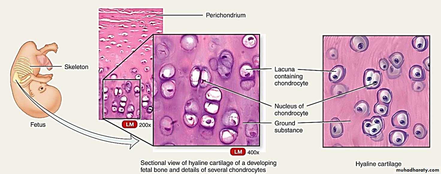
Cartilage is rather rare in the adult humans, but it is very important during development .In developing humans, most of the bones of the skeleton are preceded by a temporary cartilage "model". Cartilage is also formed very early during the repair of bone fractures.
Cartilage matrix
It consists of a dense network of collagen fibers and elastic fibers firmly embedded in chondroitin sulfate, a gel like component of the ground substance. Cartilage is nourished by diffusion of gases and nutrients through this gel.
Cartilage cells
Chondrogenic cells : Are spindle -shaped, narrow cells that derived from mesenchymal cells. These cells can differentiate into chondroblasts.Chondroblasts : immature cartilage-producing cells. They are derived from two sources; Mesenchymal cells and Chondrogenic cells.
Chondrocytes: mature cartilage cells , they occur singly or in groups within space called lacunae in the extracellular matrix.
Perichondrium
The perichondrium is a sheath of dense connective tissue that surrounds cartilage in most places, forming an interface between the cartilage and the tissue supported by the cartilage .Perichondrium contains (B.V, nerves and lymphatic vessels). It is composed of 2 layers , the outer one which is fibrous containing collagen fibers and inner layer which is cellular containing flat cells called chondrogenic cells ,this layer called chondrogenic layer, the inner portion of this layer is rich with chondroblast which differentiated into chondrocytes.Function of the perichondrium
1-Essential for growth and development of bone.
2-Nutrition .
3-Supports soft tissues.
TYPES OF CARTILAGE
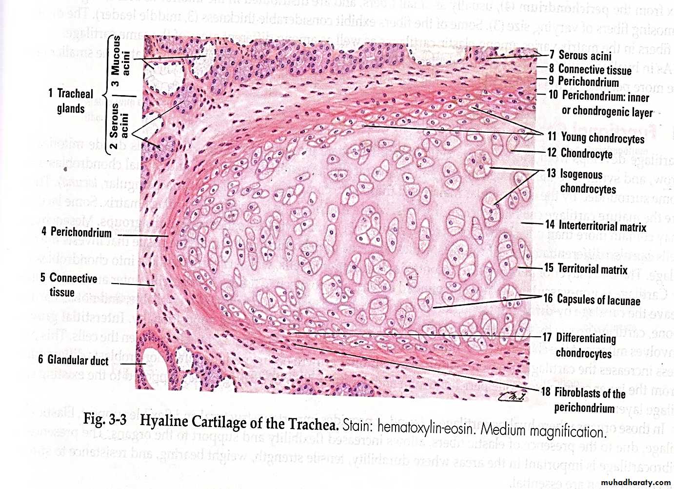
1- Hyaline CartilageHyaline cartilage (hyalos=glass):-
This type of cartilage is bluesh/wihte in the fresh state and is the most prevalent form contains a resilient homogenous ground substance. Many collagen fibers are embedding in the matrix, and prominent chondrocytes found in the spaces called lacunae. Toward the center of a mass of hyaline cartilage, the cells are large and are usually present in groups of two, four or more called cell-nests (isogenous group). Division of a single parent cell forms these groups. Toward the periphery of the cartilage, the cells are smaller. Most hyaline cartilage is surrounding by perichondrium. Hyaline cartilage functions in providing support, and precursor to bone. The locations of Hyaline cartilage include rings in the trachea, nose, articular ends of bones, and fetal skeleton .
2-Elastic cartilage
Fresh elastic cartilage has yellowish color because of the presence of elastin. It is identical to hyaline cartilage but in addition to collagen type II it contains an abundant network of fine elastic fibers. It also possesses perichondrium. Elastic cartilage possesses greater flexibility than hyaline cartilage and maintains the shape of certain structures. The locations of elastic cartilage include the external ear and the, epiglottis .
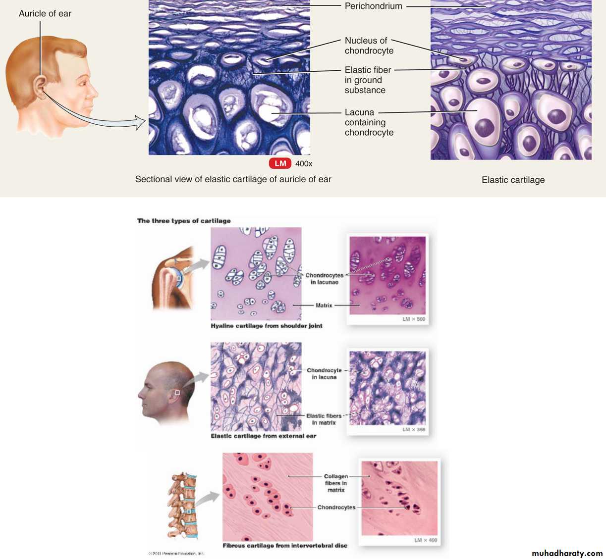
3-Fibro cartilage
It is a form of connective tissue transitional between dense connective tissue and hyaline cartilage. Chondrocytes may lie singly, but most often they form short rows between dense bundles of collagen fibers. Fibrocartilage lacks a perichondrium, combines strength and rigidity, and is the strongest of the three types of cartilage. It is typically found in relation to joints and is the main component of the intervertebral disks.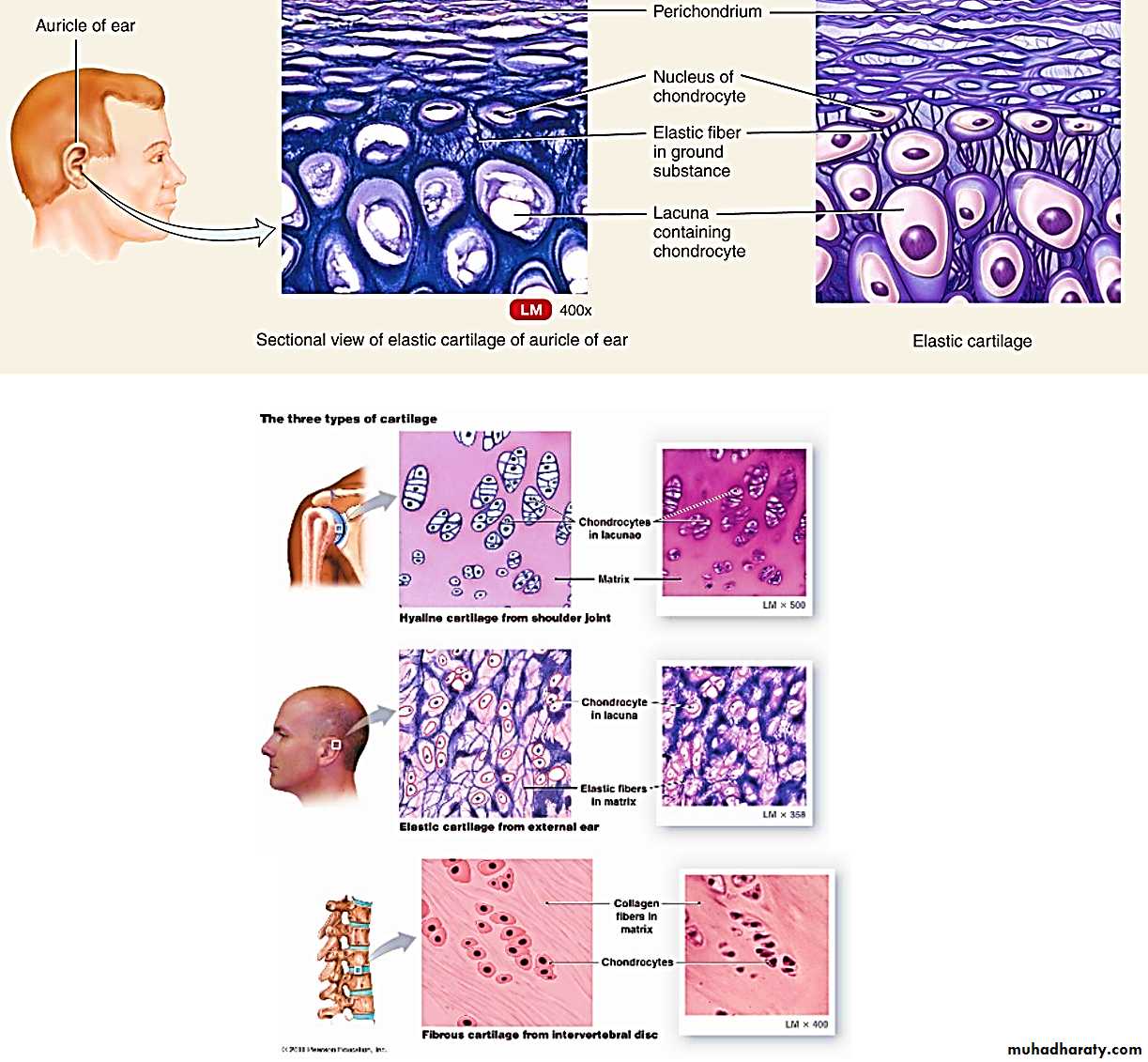
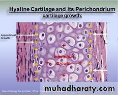
Growth of Cartilage
Growth occurs by two mechanisms:Interstitial growth (endogenous growth)
It occurs only in young cartilage (mainly in immature cartilage) in which the Chondrocytes lies in the central part of the cartilage divide and increase in number and secrete intercellular substance and causing increase of width from inside to outside.Appositional growth (exogenous growth)
Iit occurs also in mature cartilage , the chondroblasts within the inner layer of perichonderium multiply and some of them form intercellular substance and become chondrocytes where the other remain as chondroblasts , this way causing increase of width from outside to inside.Appositional growth
Appositional growth